NURS 321 Final
 Call Kai
 Learn
Learn Practice Test
 Spaced Repetition
 Match
Match Flashcards
 Knowt Play
1/65

Earn XP
Description and Tags
chest tubes
Name | Mastery | Learn | Test | Matching | Spaced | Call with Kai | Chat |
|---|
No analytics yet
Send a link to your students to track their progress
66 Terms
thoracic cavity
contains:
right and left lungs
mediastinum
heart
aorta and great vessels
esophagus
trachea
inhalation
when the diaphragm contracts it moves down, increasing the volume of the thoracic cavity
when the volume increases the pressure inside decreases
generates negative pressure
air moves from an area of higher pressure(the atmosphere) to an area of lower pressure(the lungs)
pressure within the lungs=intrapulmonary pressure
exhalation
when the phrenic nerve stimulus stops
the diaphragm relaxes and moves up in the chest→ this reduces the volume of the thoracic cavity
when the volume decreases intrapulmonary pressure increases
air flows out of the lungs to the lower atmospheric pressure
generates positive pressure
pleura
a thin continuous membrane that folds over itself
parietal pleura= lines the chest wall
visceral pleura(aka pulmonary pleura) covers the lungs'
pleural space
lies between the parietal and visceral pleural
lined with fluid to lubricate the lungs
he fluid allows the two membranes to glide smoothly against each other when you inhale and exhale.
greats a negative pressure gradient that allows the lungs to expand
mediastinum
contains the heart, aorta and great vessels, esophagus, trachea
the central compartment of the thoracic cavity located in the middle of the chest between the right and left pleural cavities that house the lungs
penumothorax
air enters the pleural space
closed pneumothorax→ chest wall is intact, rupture of the lung and visceral pleural(or airway) allows air into the pleural space
tension pneumothorax
chest wall is intact
air enters the pleural space from the lung or airway and has no way to leave
there is no vent to the atmosphere as there is in an open pneumothorax
most dangerous when patient is receiving positive pressure ventilation in which air is forced into the chest under pressure
can case a shifted mediastinum
open pneumothorax
opening in the chest wall(with or without lung puncture)
aka sucking chest wound→ air can vent through the opening in the chest wall
opening acts as a vent so pressure from the trapped air cannot build up in the chest
allows atmospheric air to enter the pleural space
penetrating trauma→ gunshot, stab, impalement, surgery
hemothorax and pleural effusion and assessment findings
accumulation of fluid(blood for hemothorax) in the pleural space
assessment findings:
SOB
decreased or absent breath sounds
pain(chest pain) on inhalation
treatment for pleural conditions
remove fluid and air ASAP
prevent drained air and fluid from returning to the pleural space
restore negative pressure in the pleural space to re-expand the lungs
What do you need for a chest tube insertion?
informed consent from MRHP
analgesic
baseline vitals(compare post-insertion)
equipment
chest tube insertion tray and sutures
bottle of sterile water
post insertion x-ray(to confirm proper placement)
bigger chest tubes used for blood/fluid
smaller tubes for air
chest tube placement
remember air rises→ chest tubes for pneumothorax are placed higher up in the lungs
remember fluid falls→ chest tubes for fluid in the pleural space are placed lower in the lungs
positive vs negative pressure
Positive Pressure: Internal pressure is higher than the outside environment. Air flows outward through any gaps or doors, which stops outside dust and germs from entering.
Negative Pressure: Internal pressure is lower than the outside environment. Air is sucked inward, which traps internal fumes, dust, or infectious particles so they cannot escape into other areas.
chest tube mechanics
expiratory positive pressure from the patient helps push air and fluid out of the chest(coughing, Valsalva)
gravity helps fluid drainage as long as the chest drainage system is below the level of the chest
suction can improve the speed at which air and fluid are pulled from the chest
keep drain below the chest for gravity drainage
monitoring an air leak
water seal is a window into the pleural space
if air is leaving the chest bubbling will be seen here
air leak meter(1-5) provides a way to measure the leak and monitor over time→ is it getting better or worse?
consistent bubblinh on inspiration and expiration means a leak is present
common leaking areas→ insertion site, connection between thoracic cavity and drainage unit or drainage system
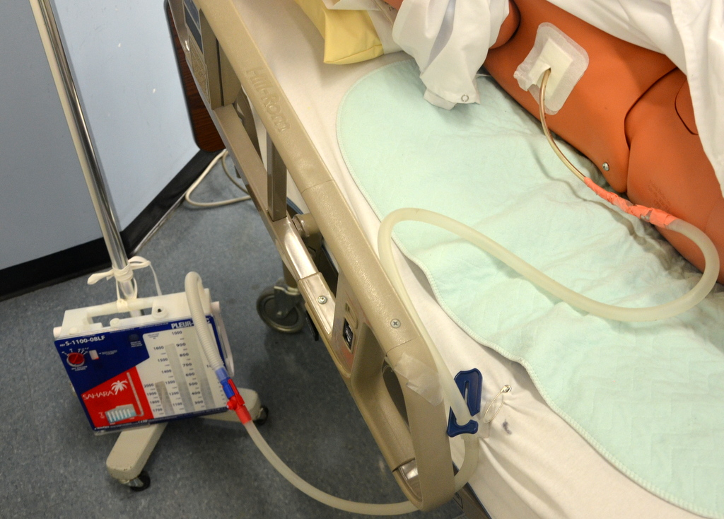
Assesment of chest tube
PATCH
PAtient
Tubing
CHamber

Assessing patient with chest tube
LOC
vital signs
Respiratory effort(breath sounds, rate, depth, ease, cough and sputum production, non-symmetrical chest excursion)
color of skin
body position
pain/restlessness/tracheal position
dressing→ dry, clean/intact
excessive bleeding at site
Subq emphysema
subcutaneous emphysema
air becomes trapped in the subcutaneous tissue
S&S:
skin has a “rice crispy”/ bubble wrap feeling
swelling of the skin
SOB
pain→ Sore throat, neck ache, or chest discomfort. [1, 2]
chest tube system
look→ sutures loosened or eyelet outside wall
listen→ hissing or air leak
feel→ Subq emphysema/mark area
collection chamber
Charting→ I/O
calibration(2cc to 200/10cc to 2000)
full/changing system/disposal
suction ay ordered level, wall regulator at 80 mmHg
air leak meter for bubblinh and fluctuating
chest tube complications
sudden increase in drainage(especially bloody drainage)
vigorous bubbling in the air leak meter
tachypnea and tachycardia with SOB
chest tube scope of practice 321
Under indirect supervision:
apply and discontinue suction
measure drainage
change dressing
MAY NOT remove
removal of chest tubes
as a nursing student or RN you cannot remove the chest tube!
physician or NP removes
provide analgesic ½ prior
purse string suture/occlusive dressing
sutures out 7-10 days
dispose of in biohazard container
monitor VS/LOC/respiratory effort
Heimlich valve
small valve attached to the catheter used for small pneumothorax
when the patient breathes in it creates negative pressure, the tube collapses on itself stopping the air from entering the chest
What safety equipment is needed at the bedside for chest tube?
Two padded/occlusive clamps — available for emergencies, such as accidental disconnection or when specifically directed by policy/provider.
Sterile occlusive dressing — for an accidental chest-tube dislodgement or emergency dressing change.
Petroleum-based gauze (e.g., Vaseline gauze) — can be used for an emergency occlusive dressing if the tube comes out.
PPE — gloves and other PPE as indicated.
Suction setup — if suction is prescribed; ensure the system is functioning and connected appropriately.
Spare drainage system/tubing — available in case the drainage system becomes damaged or contaminated.
parenteral nutrition
infusion of nutrients to sustain nutritional balance when we cannot access the GI tract
3 components→ carbohydrates, lipids, amino acids
based on the specific needs of each patient
goal of parenteral nutrition
meet caloric needs until patient can transition to enteral nutrition
transition to enteral nutrition to prevent villous atrophy(when villi shrink or disappear) and cell shrinkage→ preserve GI function
preventing complications→ GI cell shrinkage can lead to translocation of bacteria and lead to septicemia
indications for parenteral nutrition
patient is unable to digest or absorb enteral nutrition→ non-functional GI tract, extended bowel reset
patient in high stressed physiologic state→ sepsis, burns, head injury
perioperative TPN→ perioperative bowel reset
carbohydrates in parenteral nutrition
the body’s main source of energy, one of the 3 main macronutrients
dextrose is most commonly used because it is easily metabolized and stimulates the secretion of insulin
tolerated in large quantities
amino acids
building block of proteins
1 g amino acids=1 g protein
proteins maintain structure and help with wound healing
if protein is low the body will seek it out in the muscles and vita organs
lipids
aka fat emulsions are long chain fatty acids
high concentration of calories→ 9 cal/g
may contain egg yolk as an emulsifer→ check patient food allergies
maintain connective tissue integrity
fat emulsions are isotonic
available in 10, 20, 30% concentrations
non-functional GI tract causes
massive small bowel resection
GI surgery
paralytic ileus
intestinal obstruction
severe malabsorption
chemo, radiation therapy
trauma to head, neck, abdomen
Inflammatory bowel disease exacerbation
severe diarrhea
moderate to severe pancreatitis
peripheral parenteral nutrition
in the peripheral vein, for short term needs
IV is <10% dextrose in combination with amino acids and lipids→ to be less irritating to the vein and is not as calorie dense
total parenteral nutrition(TPN)
administered via central line
Iv solution are >10% dextrose→ uses a large vein for dilution
requires an infusion pump→ 40 cc-60 cc/hr for amino acid dextrose(travasol)
→ 10-20 cc/hr lipid emulsions
TPN complication: pneumothorax
air enters pleural space
causes→ catheter insertion
S&S: SOB, chest pain, coughing
peripheral IV catheter
most commonly used invasive device
fail 35-50% of the time
types: short or midline peripheral catheter
Phillips 16 steps for PIV
authorized prescriber
hand hygiene
collect your equipment and prepare the space
patient identification and psychological preparation→ explain procedure to patient
site selection, vein dilation, and visualization technology
attention to pain management
catheter selection(22-24 g= standard)
gloving
site prep
vein entry
catheter stabilization and dressing management
labeling the site
what to consider when selecting a vein?
vein condition→ soft, straight, palpable(observe vein refill)
age
patient preference→ place the IV in the patient’s non-dominant hand
patient activity: How does the patient ambulate?→ avoid placing IV on the side they ambulate
medications- anticoagulants
medical conditions→ avoid starting IV on affected site of stroke. e.g. mastectomy avoid starting IV on masectomy site
sites associated with greater risk for nerve injury
cephalic vein at wrist
antecubital fossa(AC)
palm side of wrist
superficial veins of the dorsum of the hand
cephalic vein→ 1st choice(good for first start. Avoid wrist)
metacarpal veins(not ideal for older adults)
basilic vein

superficial veins of the forearm

how to distend veins
gravity→ place arm lower than heart'
clench the fist(with ball or rolled cloth)
taping or stroking downwards on the vein
warm compress(warm blanket)→ dry heat=preferred
BP cuff→ pump cuff to 30 mmHg
tourniquet→ 10 cm above venous puncture site
IV catheter gauge
bigger number=smaller gauge
smaller number= bigger gauge
always prefer smaller gauge→ they take up less space in the vein allowing blood to flow to the catheter and cause less trauma
22-24 gauge→ used for most patients
20-24→ blood transfusion
use smaller gauge in large vein for caustic meds→ e.g. cephalic vein & 22-24 gauge
two methods to venipuncture
direct→ approach directly over vein at 15-30 degree angle
indirect→ enter the skin slightly adjacent to the vein, direct device to side of the vein
documentation for PIV insertion
date and time of insertion
site prep→ how did you prepare the iste?
needle gauge and length of device
patient response
type of dressing
what did you do with the IV→ e.g. saline lock
ongoing assessment
post-insertion assessment
atleast Q4h for alert and oriented patients
Q1-2 hours critically ill patient’s or adult’s with sensory deficits
Q1h→ pediatrics and neonatal patients
mechanical complications of parenteral nutrition
pneumothorax
air embolus
thrombosis
infection
catheter occlusion
mechanical complication of TPN- air embolus
cause: catheter insertion, during tubing/cap change, sudden dislodgement
S&S→ increased RR, apnea, hypotension, wheezing
intervention: Trendelenburg- head down, left side down (air flows up→ goal is to trap air into the right atrium and ventricle to minimize entry of air emboli to blood flow going out)
-put on oxygen and get help
prevention→ valsvala maneuver curing cap change(bearing down)
valsvala maneuver
a breathing technique where you try to exhale forcefully against a closed airway (by pinching your nose and closing your mouth or throat) for about 10 to 15 seconds
mechanical complication of TPN- pneumothorax
air enters the pleural space
cause: catheter insertion
S&S→ SOB, chest pain, coughing
mechanical complication of TPN- infection
cause: dressing changes
solution of TPN has high glucose and lipid content→ breeding ground for infection(pathogens feed off the nutrients)
S&S: high temp(fever), drainage, swelling, chills
response→ swab for culture and remove the line if needed
Prevention→ strict asepsis when caring for central line
student scope of practice for TPN
parenteral medications are a high alert med that needs an independent double check→ - this means that two health care professionals need to provide their signature. Students can participate in the process of the IDC with their primary RN or instructor but cannot sign for TPN as one of the two signatures.
anything with a central line must be done under direct supervision
mechanical complication of TPN-catheter occlusion
cause: line care and position of patient
S&S→ slow sluggish flow
prevention→ proper line care, flush and lock lines as indicated
mechanical complication of tpn thrombosis
cause: line care
S&S→ swelling, pain, redness, discoloration of skin
response→ communicate with physician
metabolic complications of TPN
hypo/hyperglycemia
hypercapnia
electrolytes
metabolic complications of TPN-hyper/hypoglycemia
hyperglycemia: TPN solution can have excess dextrose
increase in glucose can decrease neutrophils and create risk of short term infection
S&S→ thirst, headache, lethargy, high blood glucose
response→ admin insulin, monitor blood sugar
hypoglycemia:
cause→ sudden discontinue of infusion
S&S: low blood sugar, diaphoresis, shaking, confusion, loss of consciousness
Response: hang D10W
metabolic complications of TPN- hypercapnia
excessive rates of carbohydrate infusion
increase CO2 retention(>45 mmHg)
S&S: anxiety, SOB
response: oxygen
metabolic complications of TPN- electrolytes
monitor Na, Ca, K, Cl, PO4, Mg
S&S: ??
prevention→ monitor I/O, check lab values
what to monitor when giving parenteral nutrition
Intake and output→ watch for signs of fluid overload(crackles, edema, tachycardia, weight gain. do focused respiratory assessment)
Blood glucose q6h for 24 hrs
weight
lab values→ electrolytes and liver panels
complications→ metabolic and mechanical
travasol- amino acid dextrose(AAD)
maximum infusion time→ 24 hrs from priming
tubing change→ every 24 hrs
filter→ 0.22 or 0.2 micron filter
store in fridge if not immediately hung→ remove 30 mins prior to hanging(warm to room temp)
use bags in sequence!B
lipid emulsion
maximum infusion time→ 12 hours from priming
tubing changes→ when infusion is complete(12 hrs)
filter→ 1.2 micron
store at room temp
10, 20, 30% emulsions
have an indicator at the back→ yellow=safe, black=exposed to air(do not use!!)
olimel
3 in 1 parenteral solution
max infusion time→ 24 hours from priming
tubing changes→ every 24 hrs
1.2 micron filter
remove from fridge 30 minutes before hanging(warm to room temp)
only need 1 infusion pump chamber
what do you need to initiate parenteral nutrition?
physician orders
equipment: infusion set, 0.22 micron filter for AAD, 1.2 micron for lipids, 2 infusion pumps(or 1 for olimel/3 in 1) → remove AAD from fridge 30 minutes prior
name, expiry bag #
assess AAD and lipids for discolouration, leaks, crystallization
if TPN stopped for more than 8 hours, change bag
how does TPN impact a patient’s self-image
Psychosocial & Body Image Impact: Central lines, tethering pumps, and weight shifts serve as constant visual reminders of illness, disrupting physical self-concept and independence.
Social Disruption: Inability to participate in normal eating leads to loss of food-centered social connection, feelings of isolation, and potential public stigma.
Nursing Interventions: Promote autonomy through self-care education, advocate for cyclic PN (off-pump daytime hours), normalize social mealtimes, and provide sensory/oral care.
RN roles and responsibility with parenteral nutrition
assist in identifying patient who are candidates for PN→ "if the gut works use it”(" TPN should only be advocated for when Enteral Nutrition (EN) is either impossible, inadequate, or contraindicated.)
assessment→ electrolytes, nutritional and hydration status
signs and symptoms of hypo/hyperglycemia→ monitor blood glucose levels
measure weight
Ins/Outs
collaborate/communicate→ with patient/family, dietician, pharmacist, physician