Fundamentals of Photobiology & Skin Cancer
 Call Kai
 Learn
 Practice Test
 Spaced Repetition
 Match
 Flashcards
 Knowt Play
1/58

There's no tags or description
Looks like no tags are added yet.
59 Terms
Skin cancer Epi:
Skin cancer Epi:
1 in 5 Americans will develop skin cancer by the age of 70.
Skin cancer= MC in US and worldwide.
5 or more sunburns, especially early in life = 2x risk for melanoma.
Melanoma, if detected early = 99% 5-year survival rate
Melanoma + cervical lymph node metastases = 5% 5-year survival rate
Almost all skin cancers are caused by radiation from the sun or other sources such as tanning beds.
BCC = MC skin Cancer
SCC = 2nd MC
Prevention = Regular Broad Spectrum Sunscreen (UVA & UVB) SPF 30 or higher; reduces risk by 40%
Organ transplant patients are approximately 100 times more likely than the general public to develop skin cancer
Draw out the EMS

What are UV Rays?
Imp?
UVA vs B
What are UV rays?
Sunlight emits a wide range of radiation energy:
1.Visible
2.Ultraviolet (UV)
3.Infrared light.
UV = invisible + most damaging.
Importance of UV
UVA/B = main factors responsible for DNA damage -> Skin Cancers
UVA vs B:
A:
Absorbed deep into dermis
Causes TANNING, PHOTOAGING (pre-mature wrinkles).
SKIN CANCER
Penetrates through glass
B:
Absorbed primarily within the epidermis
Major cause of SUNBURNS and delayed tanning,
Key role in development of SKIN CANCER
Do not penetrate glass but reflect off reflective surfaces such as water, sand, snow, ice.
NOTE C:
Most dangerous but do not reach the earth’s surface due to the protective Ozone layer

UVA CAUSING EXTREME PHOTOAGING
Describe the multimodel pathogenesis of Skin Cancer
SKIN CANCER : Multi-Step Patho
UV mediated DNA damage
Direct damage: Thymine-dimer formation
Indirect damage: (ROS) formation
Genetic mutation formation
Inactivation of tumor suppressor genes
(p53, PTCH, CDKN2A)
Activation of oncogenes
(Ras, BRAF)
Evasion of apoptosis
(p53, Bcl-2)
Limitless replicative potential
(Telomerase)
MOA for how UV damage leads to cancer?

What is the UV Index?
Factors Affecting UV Index:
Factors Affecting UV Index:
ALTITUDE
Higher = more UV
TIME OF DAY
Sun rays are strongest from 10 am to 4 pm.
SEASON
Spring and summer experience higher levels of UV radiation than winter months.
PROXIMITY TO THE EOIJATOR
Equator = higher UV Index.
REFLECTIVE SURFACES
Water , sand, and snow

What type of UV activate Vit. D and how?

Describe the Fitzpatrick skin typing test
What is it?
Types:
Fitzpatrick skin typing test
What is it?
numerical classification (Types I-VI) for human skin shades
relates to sunburns and skin tanning.
Types:
Type I : always burns, never tans
Type II : always burns, minimal tan
Type III : burns minimally, tans moderately and gradually
Type IV : burns minimally, always tans well
Type V :rarely burns, tans easily
Type VI :never burns, tans deeply

What are the different types of Skin Cancer
Types of Skin Cancer
Non-melanoma (NMSC)
Pre-cancer: Actinic Keratosis (AK)
Squamous Cell Carcinoma (SCC)
Basal Cell Carcinoma (BCC)
Melanoma
Dysplastic nevi: precursor for melanoma
MELANOMA


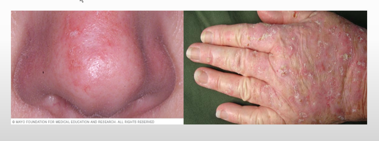
Actinic Keratosis
Describe Actinic keratosis
Clinical characteristics:
Classic presentation
Classic history:
Likely causes:
Risk factors: (3)
Precursor to
Tx (4)
Epi
Actinic keratosis = Solar keratosis= Pre-cancer
Clinical characteristics:
Skin-colored to reddish brown or yellowish ill-defined
Macule or papule w/ dry adherent scale.
Classic presentation
Fair skinned (Fitzpatrick I-III) + sand-paper feel + red/brown macule
scalp, forehead, cheek or forearm.
Classic history:
Pt has history of reoccurring red spot that they keep scratching off
Likely causes:
UV damage induced DNA mutations and subsequent cell dysplasia.
Risk factors:
Fitzpatrick I-III
Prior sunburns esp as a child,
Unprotected repeated UV exposure
Precursor to
SCC
Treatments:
Cryotherapy: Liquid Nitrogen (LNO2)
PDT (Levulan + Blue Light)
Excision with biopsy if hypertrophic
Various topical therapies
5 Flourouracil (5-FU)
Imiquimod
Epi:
majority of those diagnosed are white males over 40.
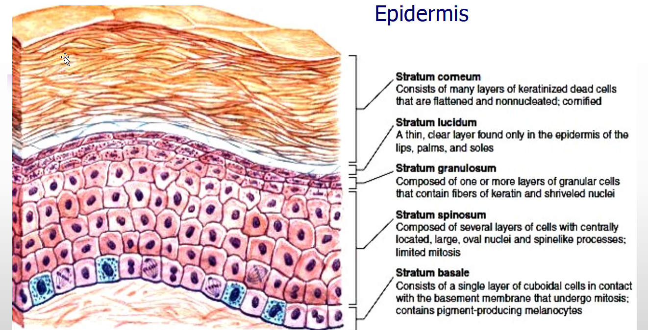

Histology of different skin cancers


Dermatopathology of an AK:
KERATINOCYTES OF THE BASAL LAYER ARE ABNORMAL: EPIDERMIS EXHIBITS HYPERKERATOSIS AND PARAKERATOSIS
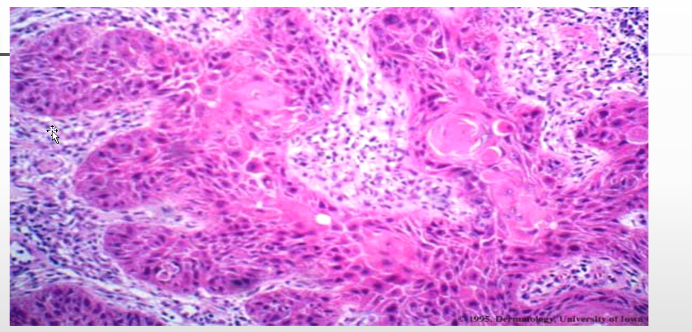
Dermatopathology of SCC
Describe SCC:
What is it?
Arises w/in:
Locations:
Importance:
SCC Types (3)
Squamous cell skin cancer (SCC)
What is it?
Common form of keratinocytic skin cancer
Arises w/in:
solar/actinic keratosis
SCC in situ
Locations:
MC = head, neck or hands of older patients
Importance:
Account for 1/3 of deaths from skin cancer among persons 65 - 84 y/o
SCC types
In-situ
Bowen’s disease
Erythroplasia of Queyrat
Invasive SCC
Keratoacanthoma

Bowen’s disease: Squamous Cell Carcinoma in-situ
Describe Bowen’s disease
Helpful descriptors: (3)
Risk Factors: (5)
Classic distribution:
Typical management: (5)
Squamous cell In-situ = Bowen’s disease
Helpful descriptors:
Red crusty or scaly patch,
Erythematous,
Ulcerated plaque + irregular borders
Risk Factors:
sun-damage,
age,
immunosuppression,
HPV 16,
arsenic exposure
Classic distribution:
sun exposed regions,
head, neck, forearms, hands, legs
Typical management:
Surgical removal,
cryotherapy,
topical chemotherapy,
5- FU,
imiquimod

Squamous cell carcinoma in-situ

SCC in-situ
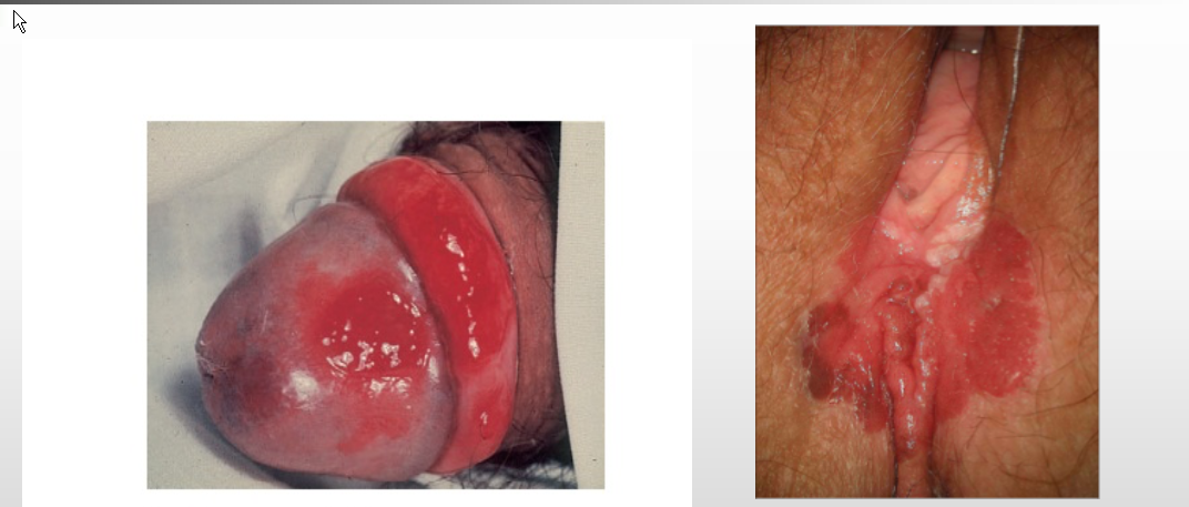
SCC in-situ:
In-situ SCC; uncircumcised men, may progress to invasive SCCErythroplasia of Queyrat

Invasive Squamous cell carcinoma
Erythematous nodule, indurated lesion, Sun exposed skin, Men> women
Slow growth

Keratoacanthoma:
Squamous Cell Carcinoma: Cutaneous Horn: Well differentiated squamous cell carcinoma
Dx/ Treatment of SCC
Prognosis
Dx/ Treatment of SCC
Biopsy = needed for Dx
Tx = complete surgical excision
Prognosis:
SCC = locally invasive and destructive
Prognosis = good for properly removed SCCs
Pt @ increased for other skin cancers
Metastases in 1-3 % of cases
To lymph nodes (50-73% survival)
Distant sites (lungs) incurable

BCC
Describe BCC
What is it?
Epi:
Course:
Risk Factors:
Types (4)
Basal Cell Carcinoma (BCC)
What is it?
Neoplasm of basal keratinocytes :
Epi:
Incidence: men>women
2.8 million/year in the US in 2010
Can occur at any age, but is more common after age 40.
Course:
Locally invasive, but rarely metastasizes.
Risk Factors:
intermittent UV exposure, ionizing radiation, indoor tanning, chemicals, arsenic, psoralen plus UVA, coal tar, Fitzpatrick I, organ transplantation and some genetic syndromes: xeroderma pigmentosa, oculocutaneous albinism, Muir-Torre syndrome, basal cell nevus syndrome (Gorlin syndrome)
Types
Nodular
Superficial
Infiltrative (Morpheaform)
Pigmented BCC
NOTE: * 7% of all BCCs are pigmented: any BCC subtypes can be pigmented, but the nodular subtypes comprises the majority of pigmented. Pigmented BCC most commonly occur on head & neck.

Nodular BCC
Describe the Characteristics of Nodular BCC (5)
Nodular BCC
Epi:
Most common subtype >50%
Characteristics:
Chronic lesion
Easy bleeding
Pearly border
Surface telangiectasias
Face, cheeks, nose, melolabial fold, forehead and eyelids most common.

Superficial BCC
Characteristics of Superficial BCC (4)
Superficial BCC Characteristics:
Erythematous scaly plaque
Slow growth
Asymptomatic
Trunk, extremities, face

Pigmented BCC
Characteristics of Pig. BCC
Pigmented BCC
Characteristics
7% of all BCC are pigmented
Any BCC can be pigmented
MC = nodular
Typical black discoloration
(Melanin deposits)
Most common area is head and neck

Infiltrative (Morpheaform) BCC
Describe Infiltrative (Morpheaform) BCC
Characteristics: (6)
MOA:
Tx:
Infiltrative (Morpheaform) BCC
Characteristics:
Uncommon variant 10% of all BCC
Aggressive with local tissue invasion
ILL defined margins,
Slightly depressed,
Flat,
Fibrotic
Firm.
Marked subclinical extension
MOA:
Tumor cells -> proliferation of fibroblasts w/in dermis + increased collagen deposition (sclerosis)
white, yellow or waxy scar.
Tx:
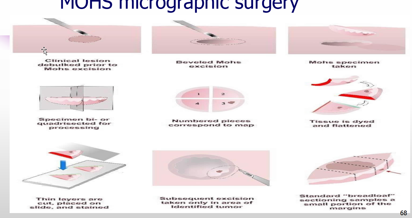
Mohs micrographic surgery is the treatment of choice

BCC; Rodent Ulcers

Dermatopathology of BCC

Basal Cell Carcinoma Tx options
Basal Cell Carcinoma Tx options
Excisional biopsy (traditional vs Mohs)
- 95% cure rate
Electrodessication and curettage (ED&C)
Radiation therapy
Imiquimod (topical
-50-75% cure rate
NOTE:
Even with low chance of metastasis, patients must still be followed after treatment.
How to do MOHS micrographic surgery
List the Risk factors for the development of non-melanoma skin cancer (NMSC)
Risk factors for the development of non-melanoma skin cancer (NMSC)
Fair Skin (Fitzpatrick types I-III)
Family history
Genetic syndromes:
Chronic sun exposure (UV rays)
Radiation therapy
Arsenic ingestion, topical tars and oils
Organ transplant patients
Viral infection (some strains of HPV)
Medications (voriconazole, B-RAF inhibitors, PUVA therapy)
Environmental pollutants

Dysplastic Nevus
Describe Dysplastic or Clark nevus
What are they?
Characteristics? (3)
Clinical Procedure: (3)
Dysplastic or Clark nevus
What are they?
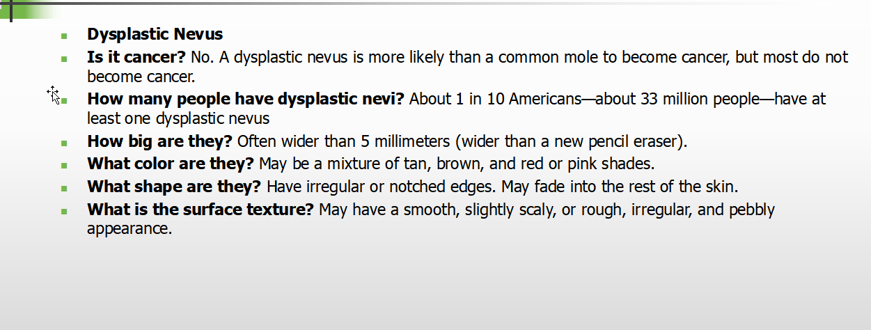
precursors for melanoma
Characteristics?
Larger + irregular shape
Uneven color/edges
Can be hereditary
Clinical Procedure:
Important to note changes in moles
common during pregnancy or with oral contraception.
Always check ABCDE changes
Always biopsy if suspicious for melanoma



Malignant Melanoma (MM)
Describe Melanoma
What is it
Epi:
Pathogenesis:
Melanoma therapies
Risk factors
Clinical types of Malignant Melanoma (5)
Melanoma
What is it
aggressive skin cancer resulting from malignant transformation of melanocytes
Epi:
< 2% global cancer diagnoses
Responsible for 80% of skin cancer deaths
MC cancer in ages 25-29
Pathogenesis:
BRAF = most frequent mutated oncogenes
V600E -> constitutive act. Of BRAF/MEK/ERK (MAPK) signaling pathway.
Melanoma therapies
Mutated BRAF = useful target for molecular therapy
BRAF kinase inhibitors (vemurafenib)
Risk factors
Fair, blonde or red hair and blue eyes
Intermittent sun exposure (Hx sunburns, tanning beds use)
Freckles and melanocytic nevi
Family history
Clinical types of Malignant Melanoma
Superficial spreading melanoma
Acral lentiginous melanoma
Lentigo maligna melanoma
Nodular melanoma
Amelanotic melanoma
Melanoma Histo Characteristics (5)
Melanoma Histo Characteristics
Consumption of epidermis,
Pagetoid spread of melanocytes,
Nests of melanocytes with variable size and shape
(which may be confluent and lack maturation),
Melanocytes w/in lymphovascular spaces,
Deep and atypical mitoses and increased apoptosis.

Histopathology of Melanoma


Acral lentiginous melanoma

Superficial spreading melanoma

Acral lentiginous melanoma- very aggressive
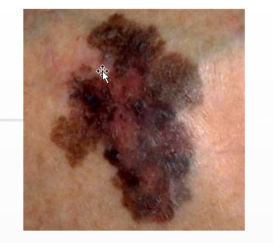
Lentigo maligna melanoma

Nodular melanoma

Melanoma
Differentiate between:
Melanoma in Situ
Invasive Melanoma
Metastatic Melanoma

Describe Breslow & Clark Level Staging System
Dif:
Clark Levels:
Breslow & Clark Level Staging System
Dif:
Breslow = thickness of melanoma
Clark = staging system that describes the layer that has been breached
Clark Levels:
Level 1:
Melanoma confined to epidermis
Level 2:
Melanoma has invaded the papillary dermis (the outermost layer of the dermis)
Level 3:
Melanoma has invaded throughout the papillary dermis and is touching on the next, deeper layer of the dermis.
Level 4:
Melanoma has reticular dermis.
Level 5:
Melanoma has invaded the fat under dermis.
Describe Melanoma Prognosis:
Describe Course of Melanoma
Melanoma Prognosis:
VERTICAL INVASION = worst prognosis
If penetrated basement membrane, dermis and entered subcutaneous fat, 5 year survival is = 49%
S-100 protein can help determine prognosis
Elevated correlates with worse prognosis.
Detection of melanoma at early stages is crucial.
Also correlated w/ Breslow:
Good prognosis Breslow < 1mm
Intermediate prognosis: Breslow 1-4 mm
Bad prognosis: Breslow > 4 mm
Course of Melanoma:
Initially, spread horizontally -> vertical growth
Can spread hematogenously or via lymphatics.
MC metastasize: brain, lung, liver and bone