7. DERMATOLOGIC DISEASES
 Call Kai
 Learn
 Practice Test
 Spaced Repetition
 Match
 Flashcards
 Knowt Play
1/36

There's no tags or description
Looks like no tags are added yet.
37 Terms
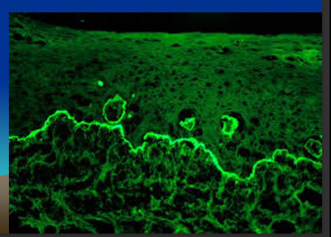
direct immunofluorescence
when you suspect autoimmune disease + you have to do biopsy from the tissue itself, where you will find autoantibodies
indirect immunofluorescence
autoimmune disease is systemic + disseminated, autoantibodies are in the blood
all know histopathology, immunofluorescence result
for this chapter
all of these are incisional biopsy
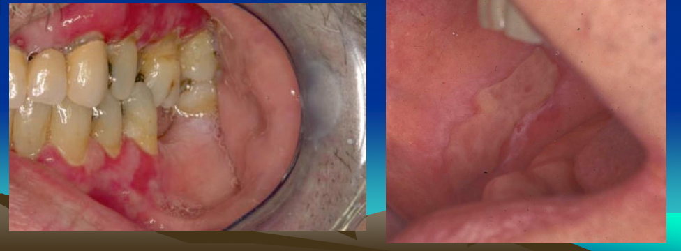
LICHEN PLANUS
Common, chronic, Immunological mucocutaneous disorder
Middle-aged female adults more commonly affected
Skin lesions: Purple, pruritic, polygonal, papules
Fine, lace-like network of white lines - Wickham's striae
Extra oral: vulvae, glans penis, nails
normally seen with stress, never goes away completely
Two forms: Reticular and Erosive
RETICULAR LICHEN PLANUS
May present as papules, asymptomatic
Waxes and wanes over weeks or months
Dorsal tongue, more keratotic plaques
Much more common than erosive form
white striae
Posterior buccal mucosa bilaterally usually
multifocal- similar lesions in multiple areas
you do not treat for this form, only inform pt

EROSIVE LICHEN PLANUS
Lesions symptomatic
also has white stria (wickhams)
Atrophic, erythematous areas with central ulceration
Atrophy/ulceration confined to gingiva = Desquamative gingivitis
very low malignant potential
lichen planus HISTOPATHOLOGIC FEATURES
Hyper-ortho and parakeratosis
Thickness of spinous layer varies
Rete ridges absent or hyperplastic, pointed or "saw-toothed”
Destruction of basal layer (hydropic degeneration)
Intense, band-like infiltrate of lymphocytes subjacent to epithelium
Degenerating keratinocytes - Civatte bodies
½ in formalin, ½ in michalins solution (live vs fixed)

d/d for desquamative gingiva
erosive lichen planus
mucous membrane pemphigoid
pemphigoid vulgaris
lichen planus DIAGNOSIS
Reticular: Clinical basis alone especially if classic lesions buccal mucosa bilaterally
Erosive: More difficult to diagnose clinically
Direct immunofluorescent – Fibrinogen positivity (fluorescent green area)
IgG , IgA, IgM, c3 + fibrinogen tested for DIF)
lichen planus TREATMENT AND PROGNOSIS
Reticular - No treatment
Erosive - Corticosteroids
LICHENOID LESIONS: GRAFT-VERSUS-HOST DISEASE
Recipients of allogeneic bone marrow transplants bc HLA match not always exact
GRAFT-VERSUS-HOST DISEASE CLINICAL FEATURES
Tongue, labial/buccal mucosa affected
Burning - Candidiasis should be ruled out
Xerostomia - Due to immune response to salivary gland tissue
GRAFT-VERSUS-HOST DISEASE TREATMENT AND PROGNOSIS
Careful histocompatibility matching, immunosuppressant drugs
Topical corticosteroids
acute GRAFT-VERSUS-HOST DISEASE
Within first few weeks after bone marrow transplantation
Affects 50% of BM patients, 33-75% oral involvement
chronic GRAFT-VERSUS-HOST DISEASE
Continuation of acute or develops >100 days after bone marrow transplantation
May appear several years after procedure, >80% oral lesions
Reticular network of white striae resembling lichen planus
LICHENOID LESIONS: LUPUS ERYTHEMATOSUS
Systemic Lupus Erythematosus (SLE) - Multisystem disease
Genetic factors play a role in pathogenesis but precise cause unknown
Chronic Cutaneous Lupus Erythematosus: Primarily skin / oral
F>M, Average age – 31
Fever, weight loss, arthritis, general malaise
40-50% butterfly rash over malar area and nose
Sunlight often makes lesions worse
Kidneys and heart frequently affected (40-50%) serious sequelae
LUPUS ERYTHEMATOSUS ORAL MANIFESTATIONS
Oral lesions 5-25%, as high as 40%
Palate, buccal mucosa, lips, gingiva
May appear as lichenoid areas
Ulceration, pain, erythema, hyperkeratosis
LUPUS ERYTHEMATOSUS DIAGNOSIS & TREATMENT
Clinical, microscopic & immunologic studies
Direct immunofluorescence - IgM, IgG, or C3 in a shaggy or granular band at basement membrane zone
direct + indirect possible
95% - Antinuclear antibodies (ANAs)
Acute episodes- Systemic corticosteroids, may be combined with other immunosuppressive agents

PEMPHIGUS VULGARIS
*not lichenoid
Most common of pemphigus disorders (Vulgaris is Latin for "common")
Oral lesions first sign of disease, most difficult to treat
Blistering - Autoantibodies against component of desmosomes
Immunologic attack causes intra-epithelial split
acantholysis: spinous layer cells are destroyed → blood fills
vesiculobullous disease
Untreated often results in death

PEMPHIGUS VULGARIS CLINICAL FEATURES
60-70% initial lesions in oral cavity
Adults average age at diagnosis 50
Superficial, ragged ulcerations
Palate, labial / buccal mucosa, gingiva
Flaccid vesicles / bullae (blister) rupture quickly, leaving erythematous, denuded surface
Bulla induced on normal-appearing skin or oral mucosa by firm lateral pressure (+ve Nikolsky sign)
systemic disorder
PEMPHIGUS VULGARIS HISTOPATHOLOGIC FEATURES
Intraepithelial separation just above basal cell layer
Spinous cells fall apart, (acantholysis- not exclusive to this), cells become rounded "Tzanck cells"
Biopsy ulcerated mucosa- results inconclusive, lack of intact epithelium- connective tissue interface
Confirmed by direct immunofluorescence
Perilesional tissue placed in Michel’s solution
Antibodies between involved epithelial cells - IgG & C3
Indirect immunofluorescence- positive 80-90%, circulating autoantibodies in serum

PEMPHIGUS VULGARIS TREATMENT AND PROGNOSIS
Systemic corticosteroids - Often in combination with other immunosuppressive drugs (so-called "steroid-sparing" agents)
Success of therapy- Measure titers of circulating autoantibodies (indirect immunofluorescence)
Mortality rate 5 to 10% - Due to complications of long term systemic corticosteroid use
Mucous Membrane Pemphigoid
looks like pemphagus but not it
Chronic, blistering, mucocutaneous autoimmune disease (cicatrical pemphigoid) “Cicatricial”, meaning "scar."
Tissue-bound autoantibodies against components of basement membrane- Hemidesmosomes
SUBEPITHELIAL
Oral lesions do not scar
Similar to pemphigus, differs in prognosis / microscopic features
Gingival involvement - Desquamative gingivitis
Desquamative gingivitis seen in other conditions- Erosive LP, pemphigus vulgaris
Mucous Membrane Pemphigoid CLINICAL FEATURES
Older adults average age 60
Females > males by a 2:1 ratio.
Oral lesions in most patients
Conjunctiva, nose, esophagus, larynx, vagina / skin
Oral lesions begin as vesicles or bullae
Blister forms in sub epithelial location, thicker, stronger roof than pemphigus
Blisters rupture - large, superficial, ulcerated mucosa
Ulcers painful, persist for weeks to months
Most significant complication- ocular involvement - 14-25%
Conjunctiva becomes inflamed and eroded
Scarring between bulbar and palpebral conjunctiva
Adhesions called symblepharons result
Blindness occurs if not treated

Mucous Membrane Pemphigoid HISTOPATHOLOGIC FEATURES
Biopsy of perilesional mucosa - split between epithelium and connective tissue
Direct immunofluorescence testing to confirm diagnosis
Immunoglobulins at basement membrane zone – IgG & C3
Indirect immunofluorescence not helpful

nikolsky's sign
Bulla induced on normal-appearing skin or oral mucosa by firm lateral pressure (+ve Nikolsky sign)
seen in pemphigus vulgaris , mucous membrane pemphigoid + erosive lichen planus
Mucous Membrane Pemphigoid TREATMENT AND PROGNOSIS
Referral to an ophthalmologist
If only oral lesions, potent topical corticosteroids
Gingival lesions respond to oral hygiene measures
If topical steroids fail, systemic steroids or other immunosuppressive agents used
Aggressive therapy - Indicated with advancing ocular disease
ERYTHEMA MIGRANS (Geographic Tongue)
psoriasiform mucositis (another name)
Common condition primarily of tongue, Increased frequency in atopic individuals, patients with psoriasis
Usually asymptomatic, may be burning with spicy foods
If symptomatic - Topical corticosteroids
If fissured - Antifungals (yeast can grow in fissures)
Well-demarcated zones of erythema - Atrophy of filiform papillae
Atrophic areas surrounded by white serpentine borders
Lesions heal in a few days or weeks, then develop in another area
Infrequently occurs on other sites: (ectopic geographic tongue) can also been seen on palate
etiology unknown but ppl w allergies are more likely (atopic)
strong familial
no malignant potential

SYSTEMIC SCLEROSIS (SCLERODERMA)
Rare immunologically mediated condition
Dense collagen deposited in large amounts
body gets stiff
Women three times men
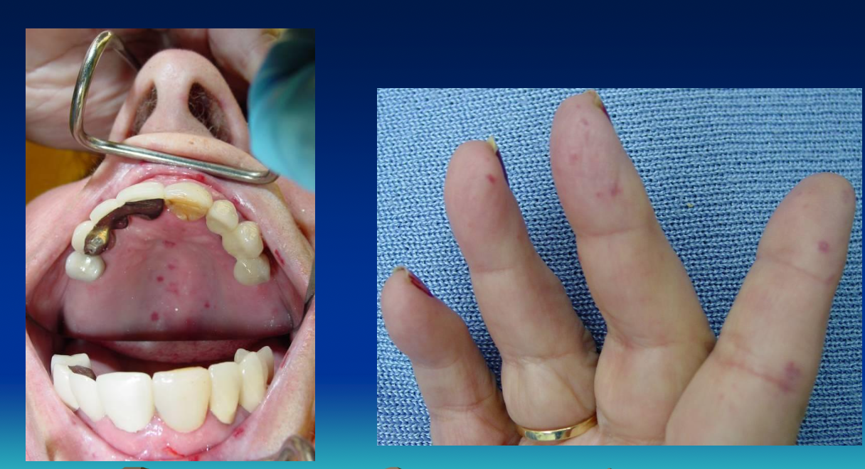
First sign Raynaud's phenomenon, vasoconstriction
Vascular events and abnormal collagen deposition produce ulcers on fingertips
Flexion contractures produce shortened claw-like fingers

SYSTEMIC SCLEROSIS (SCLERODERMA) CLINICAL AND RADIOGRAPHIC FEATURES
Furrows radiating from mouth - "purse-string" appearance
Tongue stiff, board-like
Deposition of collagen in esophagus, difficulty in swallowing (dysphagia)
Microstomia (cant open mouth) from collagen deposition in perioral tissues
Subcutaneous collagen deposition in facial skin - "mask-like" facies
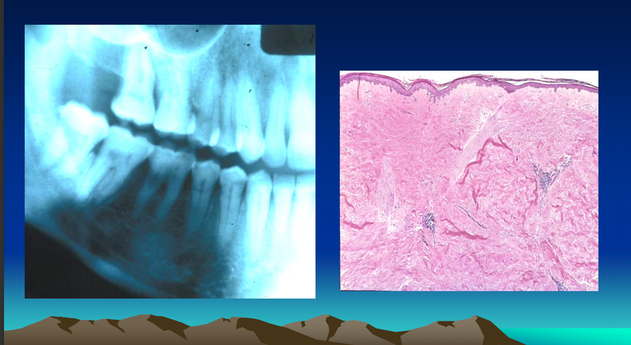
Diffuse widening of pdl space
Resorption of posterior ramus, coronoid process and condyle (20%)
SYSTEMIC SCLEROSIS (SCLERODERMA) Diagnosis & Treatment
Skin biopsy supportive of diagnosis
Anti-Scl-70 (Anti-Topoisomerase I)
D - penicillamine to inhibit collagen production
Prognosis is poor
CREST SYNDROME
Relatively mild variant of systemic sclerosis
Anti-Centromere Antibody (ACA)
Acronym for:
Calcinosis cutis
Raynaud's phenomenon
Esophageal dysfunction
Sclerodactyly (claw fingers, bc restricted movement)
Telangiectasia (dialated blood vessels)
HEREDITARY HEMORRHAGIC TELANGIECTASIA (RENDU-OSLER-WEBER DISEASE)
Autosomal dominant disorder
Multiple vascular defects – Telengiectasias
Epistaxis – Presenting symptom, begins in childhood
Bleeds of nose, GI, persistent anemia
Cutaneous & oral lesions – Mostly on tongue, lips

EPIDERMOLYSIS BULLOSA
Genetic mutations encoding structural proteins, lack of hemidesmosomes, defects in anchoring fibrils
Defect in attachment of epithelial cells to each other or to the connective tissue
23 forms of EB, mostly hereditary
Blisters in areas of minor trauma
EB Acquisita – Autoimmune

EPIDERMOLYSIS BULLOSA Oral Manifestations
Highly variable depending on EB type
Increased fragility
Ulceration of mucosa, lips, cheeks, tongue, palate
Obliteration of oral vestibule
Enamel hypoplasia, rampant caries
REACTIVE ARTHRITIS (Reiter’s Syndrome)
Immune mediated, triggered by infection
1-4 weeks after episode of dysentery / venereal disease
Non gonococcal urethritis, arthritis, conjunctivitis (cant see cant pee cant climb a tree)
Prevalent in young adult men
Glans penis - Balanitis circinata (geographic tongue lesion but on the penis)
Oral lesions - <20% of pts,similar to geographic tongue
Painless, erythematous papules
Skin lesions – Erythematous erosions with white scalloped boundary

if the skin is sloughed off
you do perilesional biopsy (around lesion) to compare where autoantibodies are