Anatomy II - Cranial Cavity
 Call Kai
 Learn
 Practice Test
 Spaced Repetition
 Match
 Flashcards
 Knowt Play
1/87

There's no tags or description
Looks like no tags are added yet.
88 Terms
calvaria
part of skull that houses the brain and brainstem
pia mater
subarachnoid space
arachnoid mater
dura mater
bone
What are the structure surrounding the brain from deep to superficial?
cerebrospinal fluid
Subarachnoid space contains
dura mater
As in the vertebral canal, arachnoid mater adheres to
bone
Dura mater adheres to
Sulcus
groove in cerebral cortex
Lambdoid suture

Sutural bones

Coronal Suture
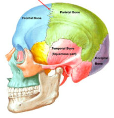
Bregma
junction of coronal and sagittal sutures
Bregma

Squamosal suture

pterion
junction of frontal, parietal, temporal and sphenoid bones
Pterion

middle meningeal artery
ascends deep to the pterion is
cranium and dura mater
middle meningeal artery is between
middle meningeal artery
Epidural Hematoma is the rupture of what artery
lateral head trauma
An epidural hematoma is due to
convex
An epidural hematoma is what shape
Epidural Hematoma

dura mater and arachnoid
bridging vein is between
2
The dura mater has how many layers
dural venous sinuses
At various locations, the 2 layers of the dura mater split to form
periosteal layer
meningeal layer
2 layers of dura mater
inner surface of cranium
The periosteal layer of the dura mater adheres to
dural venous sinus
the meningeal layer separates from the periosteal layer to form a space called
bridging veins
Blood from cerebral veins drains to the dural venous sinuses via
arachnoid granulations
CSF drains to dural venous sinuses via
bridging veins
Subdural hematoma is the rupture of
head trauma
A subdural hematoma often follows
mix of concave and convex
What is the shape of a subdural hematoma
Subdural hematoma

cerebral arteries
What is ruptured for subarachnoid hematoma
ruptured aneurysm
A subarachnoid hematoma is often caused by
headache
A patient with a subarachnoid hematoma often describes it as
Blood fills the Subarachnoid Space
what happens in subarachnoid hematoma
star
What is the shape of a subarachnoid hematoma
Subarachnoid Hematoma

scarred over
If a patient survives a subarachnoid hemorrhage, what may happen to the arachnoid granulations?
communicating hydrocephalus
Blockage of reabsorption of CSF can lead to
Communicating Hydrocephalus
enlargement of ventricles

emissary veins
provides communications between dural venous sinuses and the scalp
infection
Emissary veins are a potential route for
calva
What can be seen where emissary veins pass through?
parietal foramen
entry point for the emissary vein?
tentorium cerebelli
falx cerebri
infoldings of the dura mater
Tentorium Cerebelli
separates cerebrum and cerebellum
cerebellum
inferior to the tentorium cerebelli
cerebral hemispheres
falx cerebri separates
crista galli
anterior attachment for falx cerebri
straight sinus
runs between the falx cerebri and tentorium cerebri
inferior sagittal sinus
confluence of sinuses
What does the straight sinus connect
superior sagittal sinus
straight sinus
Confluence of sinuses receives venous blood from
transversus sinus
runs along lateral edge of tentorium cerebelli
internal jugular vein
The sigmoid sinus drains the venous sinus system to
anterior
middle
posterior
What are the 3 cranial fossas
frontal
What lobe is in the anterior cranial fossa
temporal
What lobe is in the middle cranial fossa
cerebellum
What is in the posterior cranial fossa
sphenoid crest
separates the anterior and middle cranial fossae
petrous ridge of temporal lobe
separates the middle and posterior cranial fossae
anterior clinoid process
posterior clinoid process
petrous ridge of temporal bone
groove for transverse sinus
What are the four attachments for tentorium cerebelli
olfactory foramina
Olfactory Nerve (CN I) passes through
optic canal
Optic Nerve (CN II) passes through
optic canal
Ophthalmic artery passes through
foramen spinosum
Middle meningeal artery passes through
superior orbital fissure
Oculomotor nerve (CN III) passes through
superior orbital fissure
Trochlear nerve (CN IV) passes through
superior orbital fissure
The ophthalmic branch of the trigeminal nerve (CN V1) passes through
superior orbital fissure
Abducens nerve (CN VI) passes through
foramen rotundum
The maxillary branch of the trigeminal nerve (CN V2) passes through
forame ovale
The mandibular branch of the trigeminal nerve (CN V3) passes through
foramen magnum
The spinal cord passes through
groove for sigmoid sinus
Sinuses drain to the internal jugular vein through
internal acoustic meatus
The facial nerve ( CN VII) passes through
internal acoustic meatus
The vestibulocochlear nerve (CN VIII) passes through
petrous temporal
occipital bone
The jugular foramen is between what bones
jugular foramen
The glossopharyngeal nerve (CN IX) passes through
jugular foramen
The vagus nerve (CN X) passes through
jugular foramen
The accessory nerve (CN XI) passes through
hypoglossal canal
The hypoglossal nerve (CN XII) passes through
jugular foramen
Accessory nerve (CN XI) exits through
Sella Turcica
depression in sphenoid bone
hypoglossal fossa
The sella turcica includes
pituitary gland
The hypoglossal fossa is the location of
olfactory foramen
olfactory nerve (CN 1) passes through
foramen ovale
CN V3 passes into
foramen spinosum
middle meningeal artery passes through