embryology of palate and related craniofacial/ oral structures
 Call Kai
 Learn
Learn Practice Test
 Spaced Repetition
 Match
Match Flashcards
 Knowt Play
1/30

There's no tags or description
Looks like no tags are added yet.
Name | Mastery | Learn | Test | Matching | Spaced | Call with Kai | Chat |
|---|
No analytics yet
Send a link to your students to track their progress
31 Terms
early embryonic development: bastulaiton
2 cells → 4 → 8 → morula (mass of cells) → early blastula → late blastula
refer to image

implantation

early embryonic development: gastrulation (D 14-17)
key things
epi+hypoblast
invaginating mesoderm cells
ecto/meso/endoderm

human gastrula
b = above
c = below

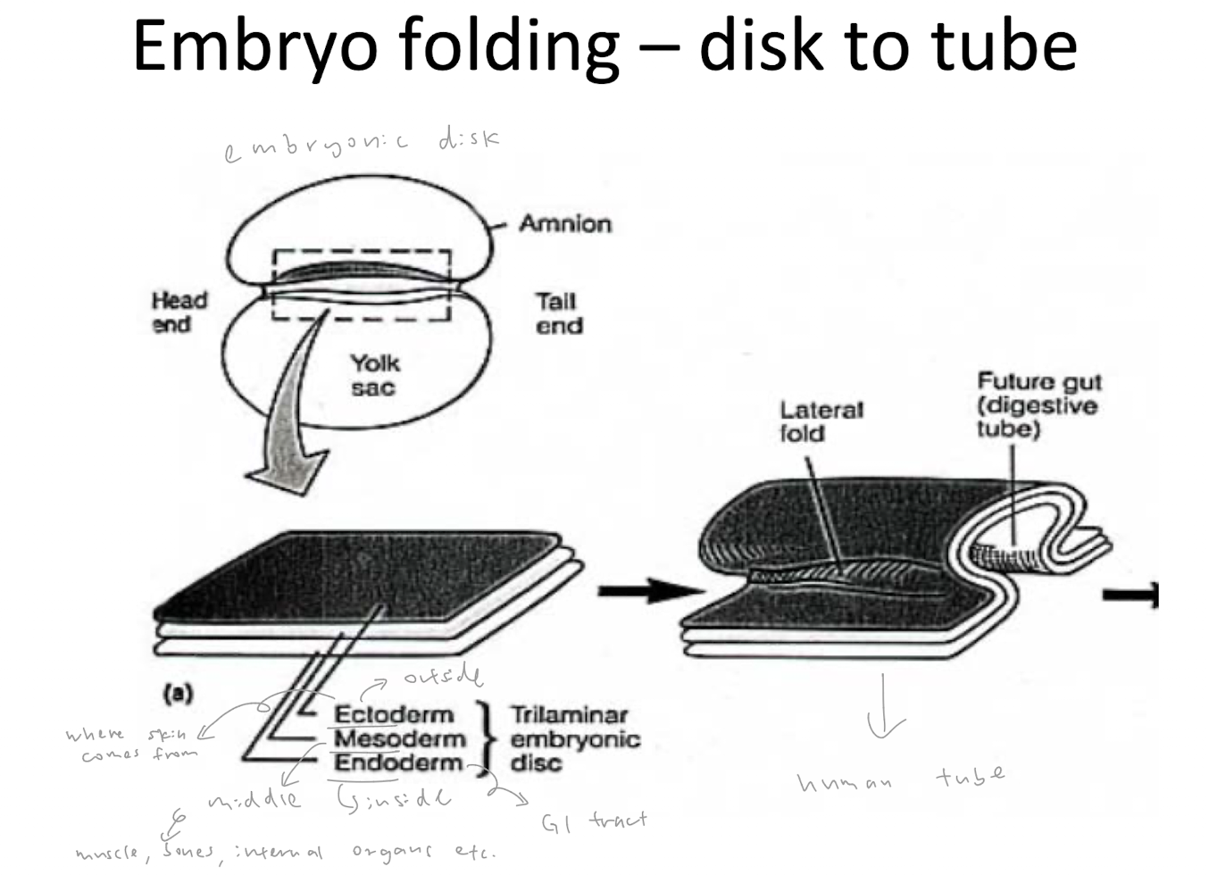
embryonic folding - disk to tube
ectoderm = outside
mesoderm = middle
endoderm = inside
embryonic disk folds → human tube
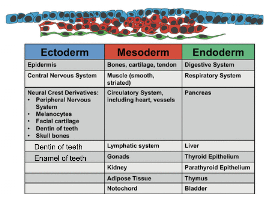
3 germ layers give rise to diff tissues
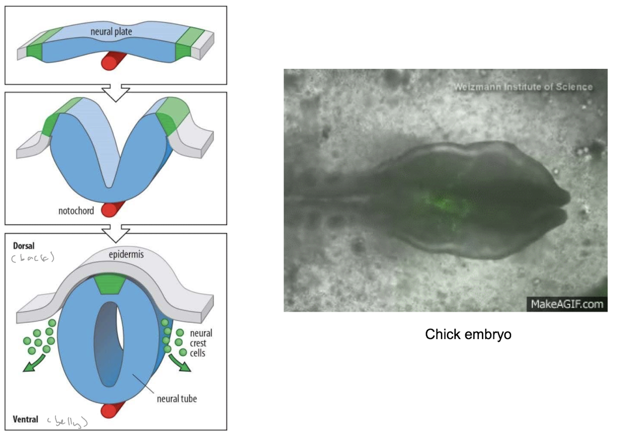
neurulation (W 3-4)
*formation of neural tube
neural crest
leaves to from most of major structures of face (come from back)
form peripheral nerves
(R = just ectoderm)

neural tube closure

neural tube defects
often due to folic acid deficiencies
pregnant women advised to take folic acid supplements

neural crest formation and migration
cranial neural crest cells
form dentin and most of craniofacial skeletal tissues
skeletal tissue of craniofacial region: from neural crest (comes from ectoderm)
anywhere else = from mesoderm
migrate to head and face

neurocristopathy
abnormal development of neural crest cells

contributions of neural crest cells in head
nearly ½ of head and face skeleton comes from migration of cells from back to front of embryo
significant contribution to cranial ganglia

pharyngeal arches
(skeletal derivatives?)
post-migratory neural crest cells populate future face
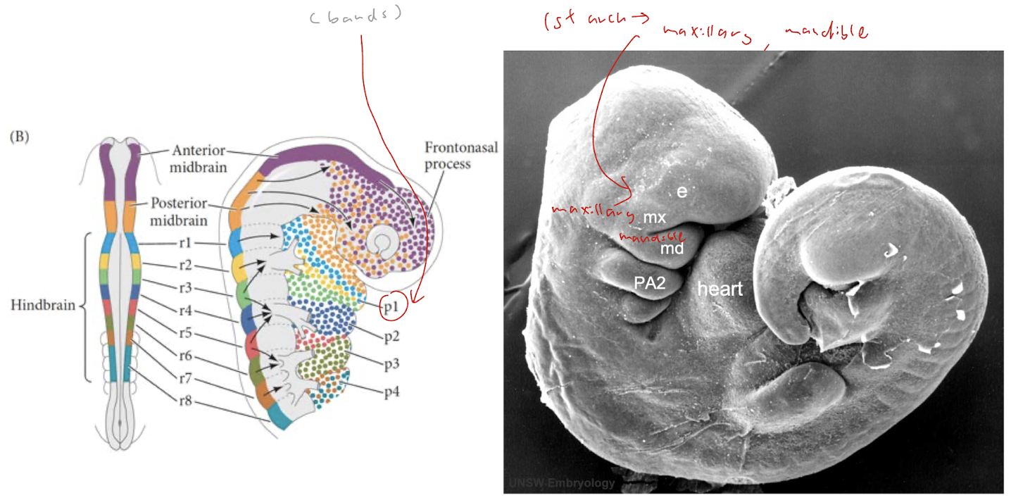
development of face
maxillary process
mandibular arch → mandible

MNP and inter maxillary segment (primary palate)
yellow = MNP (medial nasal prominences)
2 MNPs merge at midline to form intermaxillary segment
orange = maxillary prominence/ process

development of palate
primary palate develops at end of W5 from intermaxillary segment
secondary includes hard and soft palate

hard palate
comes from fusion between inter maxillary and a pair of maxillary processes
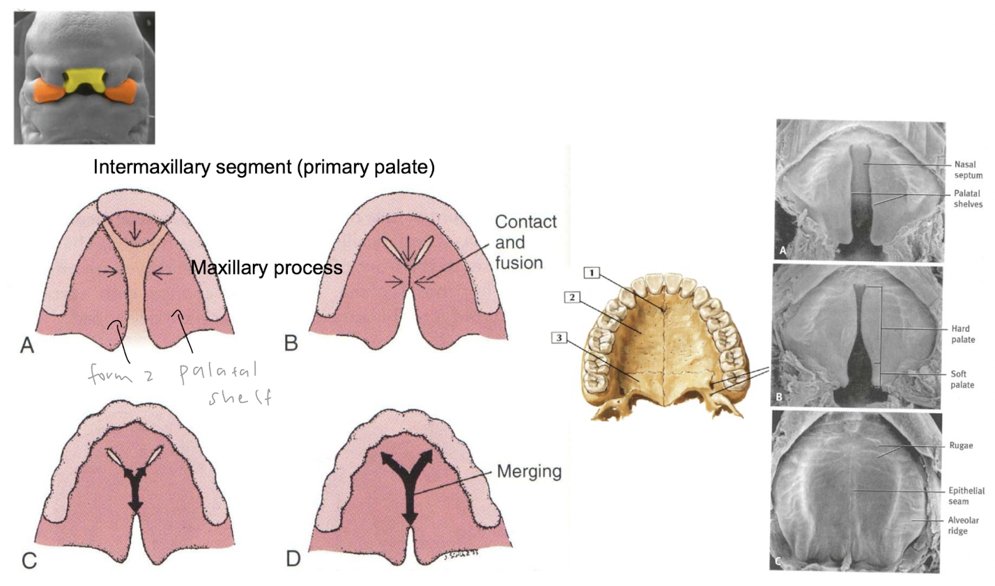
elevation and fusion of palatal shelves
2 lateral palatine processes project from internal of maxillary prominences and grow downward on either side of tongue
lateral palatal shelves grow and move to horizontal position (to tongue)
(fusion of primary palate with lateral palatine processes and nasal septum)

fusion of palatal shells and naval septum
naval septum divide nasal cavity into 2 separate cavities

when fusion goes wrong

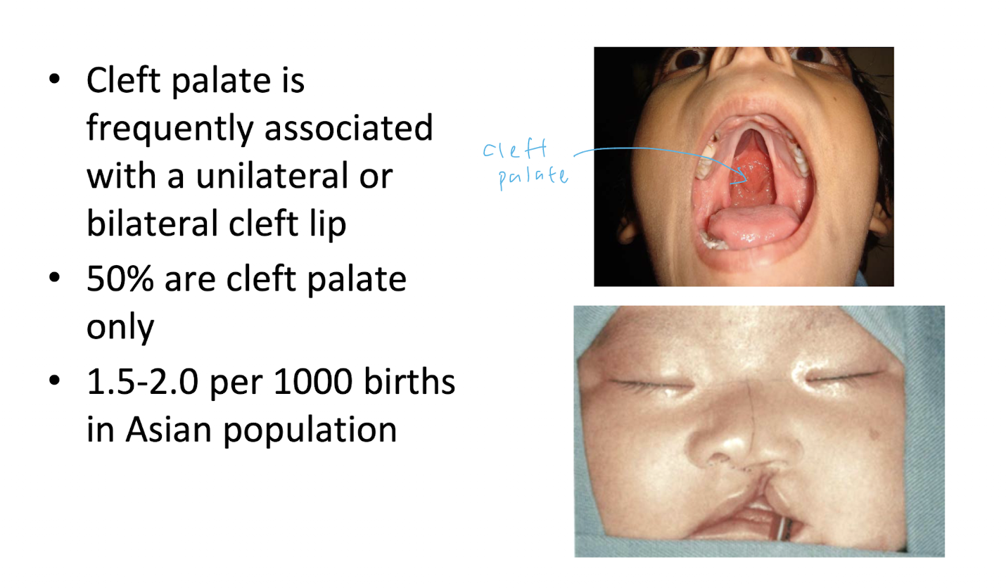
cleft lip and cleft palate
CP freq associated with uni/bilateral CL
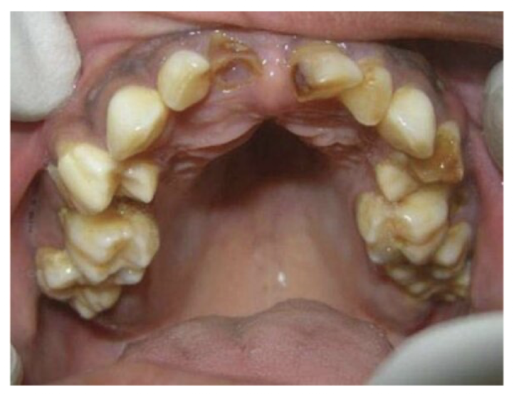
high-arched palate
tall and narrow U-shaped palate (arc instead of flat)
similar pathogenic mechanism of CP
feeding difficulties
can cause tooth crowding
cleft malformation
anterior
incl CL with/without cleft of alveolar part of maxilla
BCD
posterior
incl. clefts of posterior palate
EF

developmental mechanisms of CL &/ P
improper growth of primordia involved in palate development
small jaw → tongue fall backwards and block secondary palate closure
e.g. Pierre-Robin Seq
failure of fusion between spatial shelves/ inter maxillary segment and palatal shelves

(palate fusions)
shelf to shelf (the midline): forms the secondary palate;
shelves to the intermaxillary segment (at the incisive foramen): joins the front and back of the palate;
shelves to the nasal septum (from above): separates the nose from the mouth.
etiology - causes of CL/P
genetic
syndromic (part of wider spectrum of disease), non-syndromic (>common) (otherwise healthy), many genes associated
gene and environmental interaction
gene → > susceptible
eg. smoking (GSTT1 and NOS3), alcohol consumption (ADH1C)
drug
excessive retinoid acid (vit A)
craniofacial microsomia (CFM)

Treacher Collins Syndrome

DiGeorge Syndrome

Pierre-Robin sequence
small jaw → tongue fall back → high-arched palate etc.
