Lecture 5-8: Protein Sequence and Shape

0.0(0)
Studied by 0 people
call kaiCall Kai
learnLearn
examPractice Test
spaced repetitionSpaced Repetition
heart puzzleMatch
flashcardsFlashcards
GameKnowt Play
Card Sorting

1/58

encourage image

There's no tags or description

Looks like no tags are added yet.

59 Terms

1
New cards

What is the basic structure of a protein, and what is myoglobin?

  • Proteins consist of long, linear chains of amino acids connected by peptide bonds.

  • The specific sequence of amino acids forms the polypeptide chain.

  • Myoglobin is an oxygen-binding protein found in muscle tissue.

  • Myoglobin contains 153 amino acids in its polypeptide chain.


<ul><li><p>Proteins consist of long, linear chains of amino acids connected by peptide bonds.</p></li><li><p>The specific sequence of amino acids forms the polypeptide chain.</p></li><li><p><strong>Myoglobin</strong> is an oxygen-binding protein found in muscle tissue.</p></li><li><p>Myoglobin contains 153 amino acids in its polypeptide chain.</p></li></ul><p></p>
2
New cards

How does myoglobin's linear polypeptide chain form its three-dimensional structure?

  • Myoglobin's linear chain of 153 amino acids folds into a compact, three-dimensional globular structure.

  • A space-filling model provides a detailed representation of its structure.

  • However, the high level of detail makes it difficult to visualize the protein's folding patterns.

  • Simplified structural representations can make these patterns easier to understand.


<ul><li><p>Myoglobin's linear chain of 153 amino acids folds into a compact, three-dimensional globular structure.</p></li><li><p>A space-filling model provides a detailed representation of its structure.</p></li><li><p>However, the high level of detail makes it difficult to visualize the protein's folding patterns.</p></li><li><p>Simplified structural representations can make these patterns easier to understand.</p></li></ul><p></p>
3
New cards

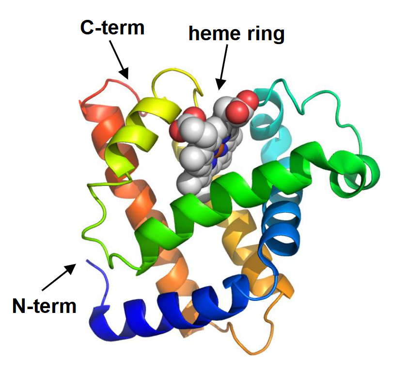
What does the ribbon representation of myoglobin reveal about its structure?

  • The ribbon traces the path of the polypeptide backbone without showing amino acid side chains (peptide bonds only!).

  • The ribbon is colour-coded from blue at the N-terminus to red at the C-terminus.

  • Myoglobin contains eight spiral-shaped (helical) segments.

  • Amino acid side chains occupy the spaces surrounding the backbone, even though they are not shown.

  • The structure also contains a heme ring.

<ul><li><p>The ribbon traces the path of the polypeptide backbone without showing amino acid side chains (peptide bonds only!).</p></li><li><p>The ribbon is colour-coded from blue at the N-terminus to red at the C-terminus.</p></li><li><p>Myoglobin contains eight spiral-shaped (helical) segments.</p></li><li><p>Amino acid side chains occupy the spaces surrounding the backbone, even though they are not shown.</p></li><li><p>The structure also contains a heme ring.</p></li></ul><p></p>
4
New cards

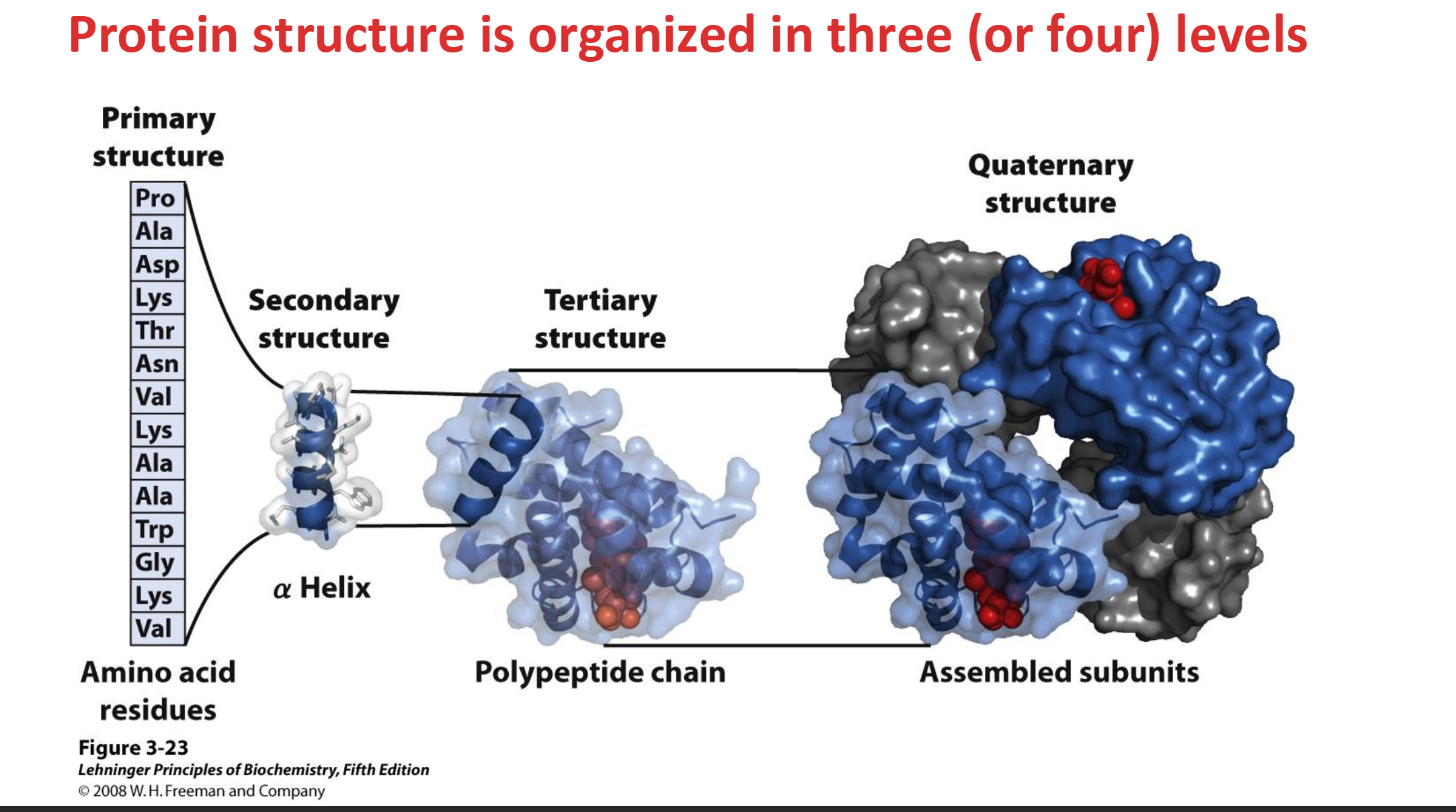
What are the four levels of protein structure?

  • Primary: Linear sequence of amino acids in a polypeptide chain.

  • Secondary: Regular, repetitive folding patterns along short sections of the polypeptide (5–20 amino acids), such as helical sections in myoglobin.

  • Tertiary: Overall three-dimensional folding of an entire polypeptide chain.

    • Ex. In myoglobin, 8 helical sections enclose a central cavity

  • Quaternary: Arrangement of multiple polypeptide chains into a protein complex (e.g., 4 globin units in hemoglobin).

  • Not all proteins possess quaternary structure.

<ul><li><p><strong>Primary:</strong> Linear sequence of amino acids in a polypeptide chain.</p></li><li><p><strong>Secondary:</strong> Regular, repetitive folding patterns along short sections of the polypeptide (5–20 amino acids), such as <span>helical sections in myoglobin.</span></p></li><li><p><strong>Tertiary:</strong> Overall three-dimensional folding of an entire polypeptide chain.</p><ul><li><p><span>Ex. In myoglobin, 8 helical sections enclose a central cavity</span></p></li></ul></li><li><p><strong>Quaternary:</strong> Arrangement of multiple polypeptide chains into a protein complex (e.g., 4 globin units in hemoglobin).</p></li><li><p>Not all proteins possess quaternary structure.</p></li></ul><p></p>
5
New cards

Why is the three-dimensional folding of proteins important for their function?

  • Protein function depends on the precise three-dimensional positioning of key amino acids.

  • Amino acids that are far apart in the primary sequence may become close together through folding.

  • The exact folding pattern is critical for proper protein function.


6
New cards

What is protein hydrolysis, and what methods can be used to hydrolyze peptide bonds?

  • Protein hydrolysis breaks peptide bonds to release individual amino acids.

  • Acid hydrolysis: 6 M HCl at 110°C for 24–72 hours.

  • Base hydrolysis: 4 M NaOH at 110°C for 16 hours.

  • Enzymatic hydrolysis: Uses proteases, which catalyze peptide bond hydrolysis.

  • Some amino acids may be destroyed during acid or base hydrolysis.


<ul><li><p>Protein hydrolysis breaks peptide bonds to release individual amino acids.</p></li><li><p><strong>Acid hydrolysis:</strong> 6 M HCl at 110°C for 24–72 hours.</p></li><li><p><strong>Base hydrolysis:</strong> 4 M NaOH at 110°C for 16 hours.</p></li><li><p><strong>Enzymatic hydrolysis:</strong> Uses proteases, which catalyze peptide bond hydrolysis.</p></li><li><p>Some amino acids may be destroyed during acid or base hydrolysis.</p></li></ul><p></p>
7
New cards

How can acid or base hydrolysis alter amino acids, and how are amino acids analyzed afterward?

  • Acid and base hydrolysis can destroy certain amino acids.

  • Hydrolysis of amide groups converts Asn → Asp and Gln → Glu, releasing ammonia.

  • After protein hydrolysis, chromatography can determine the amount of each amino acid present.

  • This reveals amino acid composition, but not their original sequence.


8
New cards

What causes chemical reactivity, and what are nucleophiles and electrophiles?

  • Chemical reactivity arises from an uneven distribution of valence electrons.

  • C–C and C–H bonds share electrons relatively equally, making them nonpolar and relatively unreactive.

  • Nucleophiles: Atoms with available lone pairs that donate electrons to form new bonds.

  • Electrophiles: Electron-deficient atoms that accept electron pairs.

  • Many biochemical reactions are initiated by nucleophiles

  • Negatively charged oxygen is a stronger nucleophile than neutral oxygen


<ul><li><p>Chemical reactivity arises from an uneven distribution of valence electrons.</p></li><li><p>C–C and C–H bonds share electrons relatively equally, making them nonpolar and relatively unreactive.</p></li><li><p><strong>Nucleophiles:</strong> Atoms with available lone pairs that donate electrons to form new bonds.</p></li><li><p><strong>Electrophiles:</strong> Electron-deficient atoms that accept electron pairs.</p></li><li><p><span>Many biochemical reactions are initiated by nucleophiles</span></p></li><li><p>Negatively charged oxygen is a stronger nucleophile than neutral oxygen</p></li></ul><p></p>
9
New cards

What are the three ways an atom can use its lone pair of electrons?

  • Hydrogen bond acceptor: Uses its lone pair to attract an O–H or N–H group through hydrogen bonding.

  • Base: Uses its lone pair to capture a proton (H⁺).

  • Nucleophile: Shares its lone pair with an electron-deficient atom to form a new covalent bond.

  • Key distinction: A base accepts H⁺, while a nucleophile forms a bond with an electrophile.


<ul><li><p><strong>Hydrogen bond acceptor:</strong> Uses its lone pair to attract an O–H or N–H group through hydrogen bonding.</p></li><li><p><strong>Base:</strong> Uses its lone pair to capture a proton (H⁺).</p></li><li><p><strong>Nucleophile:</strong> Shares its lone pair with an electron-deficient atom to form a new covalent bond.</p></li><li><p><strong>Key distinction:</strong> A base accepts H⁺, while a nucleophile forms a bond with an electrophile.</p></li></ul><p></p>
10
New cards

What is the difference between nucleophilic substitution and nucleophilic addition?

  • Nucleophilic substitution: An incoming nucleophile attacks an electron-deficient atom and displaces a leaving group.

  • The leaving group takes its bonding electrons when it departs.

  • Nucleophilic addition: A nucleophile attacks an atom involved in a double bond.

  • One bond of the double bond breaks, but the original group remains attached.

  • Key distinction: Substitution replaces a group; addition does not remove a group.


<ul><li><p><strong>Nucleophilic substitution:</strong> An incoming nucleophile attacks an electron-deficient atom and displaces a leaving group.</p></li><li><p>The leaving group takes its bonding electrons when it departs.</p></li><li><p><strong>Nucleophilic addition:</strong> A nucleophile attacks an atom involved in a double bond.</p></li><li><p>One bond of the double bond breaks, but the original group remains attached.</p></li><li><p><strong>Key distinction:</strong> Substitution replaces a group; addition does not remove a group.</p></li></ul><p></p>
11
New cards

How does peptide bond hydrolysis occur through nucleophilic displacement?

  • The carbonyl carbon of the peptide bond is electron-deficient (electrophile) because oxygen attracts electrons.

  • OH⁻ acts as a nucleophile and attacks the carbonyl carbon, forming a new C–O bond.

  • Electrons from the C=O double bond move toward oxygen, forming a negatively charged intermediate (transition state).

  • The C=O double bond reforms, and the C–N peptide bond breaks.

  • The amino nitrogen acts as the leaving group.

  • Curly arrows represent the movement of electron pairs during the reaction.


<ul><li><p>The carbonyl carbon of the peptide bond is electron-deficient (electrophile) because oxygen attracts electrons.</p></li><li><p>OH⁻ acts as a nucleophile and attacks the carbonyl carbon, forming a new C–O bond.</p></li><li><p>Electrons from the C=O double bond move toward oxygen, forming a negatively charged intermediate (transition state).</p></li><li><p>The C=O double bond reforms, and the C–N peptide bond breaks.</p></li><li><p>The amino nitrogen acts as the leaving group.</p></li><li><p><strong>Curly arrows represent the movement of electron pairs during the reaction.</strong></p></li></ul><p></p>
12
New cards

How does Sanger's method identify the N-terminal amino acid of a protein?

  • At high pH, the free α-amino group at the N-terminus deprotonates and acts as a nucleophile (NH2-).

  • The N-terminal amino acid reacts with fluorodinitrobenzene (FDNB), attaching a bright yellow tag.

  • Complete hydrolysis releases the individual amino acids.

  • The yellow-labelled N-terminal amino acid is identified using chromatography.

  • Limitation: Hydrolysis destroys the remaining polypeptide chain, so only the first amino acid can be identified.


<ul><li><p>At high pH, the free α-amino group at the N-terminus deprotonates and acts as a nucleophile (NH2-).</p></li><li><p>The N-terminal amino acid reacts with fluorodinitrobenzene (FDNB), attaching a bright yellow tag.</p></li><li><p>Complete hydrolysis releases the individual amino acids.</p></li><li><p>The yellow-labelled N-terminal amino acid is identified using chromatography.</p></li><li><p><strong>Limitation:</strong> Hydrolysis destroys the remaining polypeptide chain, so only the first amino acid can be identified.</p></li></ul><p></p>
13
New cards

What is Edman degradation, and how does it improve upon Sanger's method?

  • Edman degradation determines the amino acid sequence of a protein, starting at the N-terminus.

  • The N-terminal amino acid is chemically labelled, removed, and identified.

  • Unlike Sanger's method, the remaining polypeptide chain is not destroyed.

  • The process can be repeated to identify successive amino acids.

  • Key advantage: Multiple amino acids can be sequenced rather than identifying only the first residue.


14
New cards

What are the two steps of Edman degradation?

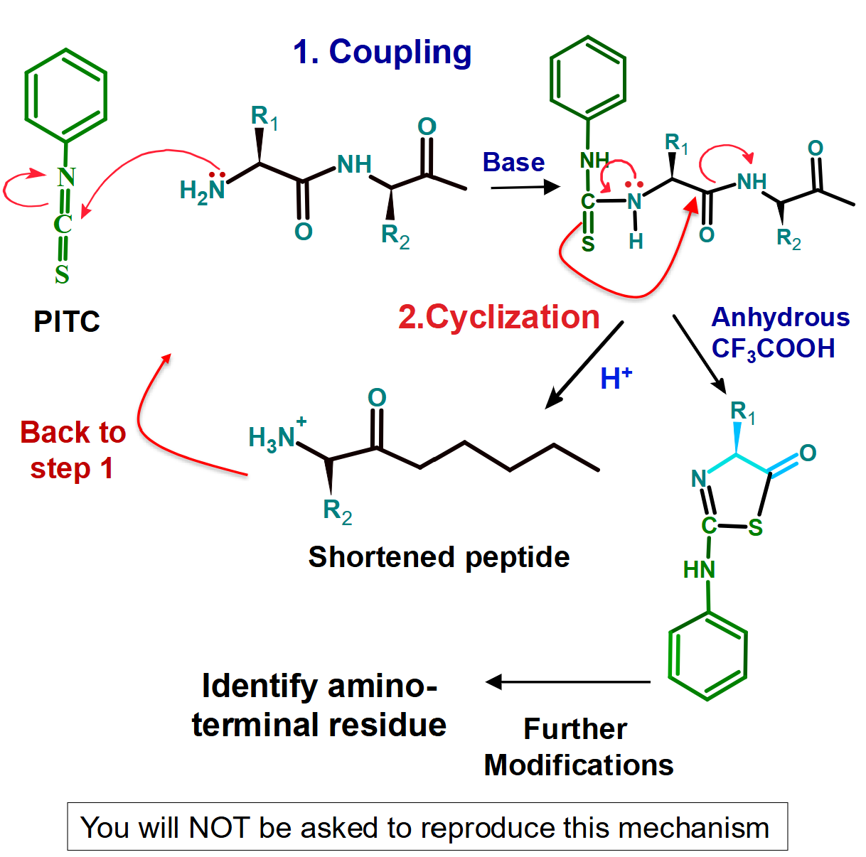
  • Coupling (basic conditions/requires base): Phenyl isothiocyanate (PITC) labels the N-terminal amino acid.

    • The coupling reaction must finish before cyclization begins.

  • Cyclization (acidic conditions/requires acid): The first peptide bond is cleaved, removing the labelled N-terminal amino acid.

    • The removed amino acid is identified, leaving a shortened polypeptide.

  • The cycle can be repeated up to approximately 50 times using automated sequencers.

<ul><li><p><strong>Coupling (basic conditions/requires base):</strong> Phenyl isothiocyanate (PITC) labels the N-terminal amino acid.</p><ul><li><p>The coupling reaction must finish before cyclization begins.</p></li></ul></li><li><p><strong>Cyclization (acidic conditions/requires acid):</strong> The first peptide bond is cleaved, removing the labelled N-terminal amino acid.</p><ul><li><p>The removed amino acid is identified, leaving a shortened polypeptide.</p></li></ul></li><li><p>The cycle can be repeated up to approximately 50 times using automated sequencers.</p></li></ul><p></p>
15
New cards

What are the cleavage specificities of trypsin, chymotrypsin, and cyanogen bromide?

  • Trypsin: Cleaves peptide bonds on the C-terminal side of Lys (K) and Arg (R).

  • Chymotrypsin: Primarily cleaves on the C-terminal side of aromatic amino acids Phe (F), Tyr (Y), and Trp (W).

  • These reagents produce smaller peptide fragments that can be individually sequenced.

  • Key concept: Cleavage occurs AFTER the recognized amino acid, toward its C-terminus.

  • Selective hydrolysis cuts the polypeptide at specific locations, to yield a limited number of oligopeptides of definite size


16
New cards

How does proline affect peptide cleavage by trypsin and chymotrypsin?

  • Trypsin: Normally cleaves after Lys (K) and Arg (R).

  • Exception: No cleavage occurs if Lys or Arg is immediately followed by Pro (P).

  • Proline has the wrong shape to fit into the enzyme's binding position.

  • Chymotrypsin: The lecture diagram also shows no cleavage when Phe is followed by Pro.

  • Key concept: Always check whether Pro follows a potential cleavage site!


<ul><li><p><strong>Trypsin:</strong> Normally cleaves after Lys (K) and Arg (R).</p></li><li><p><strong>Exception:</strong> No cleavage occurs if Lys or Arg is immediately followed by Pro (P).</p></li><li><p>Proline has the wrong shape to fit into the enzyme's binding position.</p></li><li><p><strong>Chymotrypsin:</strong> The lecture diagram also shows no cleavage when Phe is followed by Pro.</p></li><li><p><strong>Key concept:</strong> Always check whether Pro follows a potential cleavage site!</p></li></ul><p></p>
17
New cards

How does cyanogen bromide (CNBr) cleave polypeptides?

  • CNBr is a chemical cleavage reagent, not an enzyme.

  • Specifically cleaves peptide bonds on the C-terminal side of methionine (Met, M).

  • Unlike trypsin and chymotrypsin, CNBr can cleave even when the next amino acid is proline (Met–Pro).

  • During cleavage, methionine is converted into homoserine (Hse).

  • Key concept: CNBr cleaves AFTER Met, including Met–Pro bonds.

  • For a linear peptide, the number of fragments equals the number of cleavage sites + 1.

  • Exception: A Met at the very end of the peptide does not create an additional fragment because no peptide bond follows it.


<ul><li><p>CNBr is a chemical cleavage reagent, not an enzyme.</p></li><li><p>Specifically cleaves peptide bonds on the C-terminal side of methionine (Met, M).</p></li><li><p>Unlike trypsin and chymotrypsin, CNBr can cleave even when the next amino acid is proline (Met–Pro).</p></li><li><p>During cleavage, methionine is converted into homoserine (Hse).</p></li><li><p><strong>Key concept:</strong> CNBr cleaves AFTER Met, including Met–Pro bonds.</p></li><li><p>For a linear peptide, the number of fragments equals the number of cleavage sites + 1.</p></li><li><p><strong>Exception:</strong> A Met at the very end of the peptide does not create an additional fragment because no peptide bond follows it.</p></li></ul><p></p>
18
New cards

What is peptide mass fingerprinting, and how is it used to identify proteins?

  • Proteins digested with enzymes like trypsin yield characteristic patterns of
    fragments of different molar mass (fingerprint)

  • Mass spectrometry determines the masses of the resulting fragments.

  • The fingerprint is compared with those of known proteins to identify the original protein.

  • For unknown proteins: Oligopeptide Fragments can be separated using chromatography and sequenced using Edman degradation.


<ul><li><p><span>Proteins digested with enzymes like trypsin yield characteristic patterns of</span><br><span>fragments of different molar mass (fingerprint)</span></p></li><li><p>Mass spectrometry determines the masses of the resulting fragments.</p></li><li><p>The fingerprint is compared with those of known proteins to identify the original protein.</p></li><li><p><strong>For unknown proteins:</strong> <span>Oligopeptide </span>Fragments can be separated using chromatography and sequenced using Edman degradation.</p></li></ul><p></p>
19
New cards

How are overlapping peptide fragments used to reconstruct a protein's amino acid sequence?

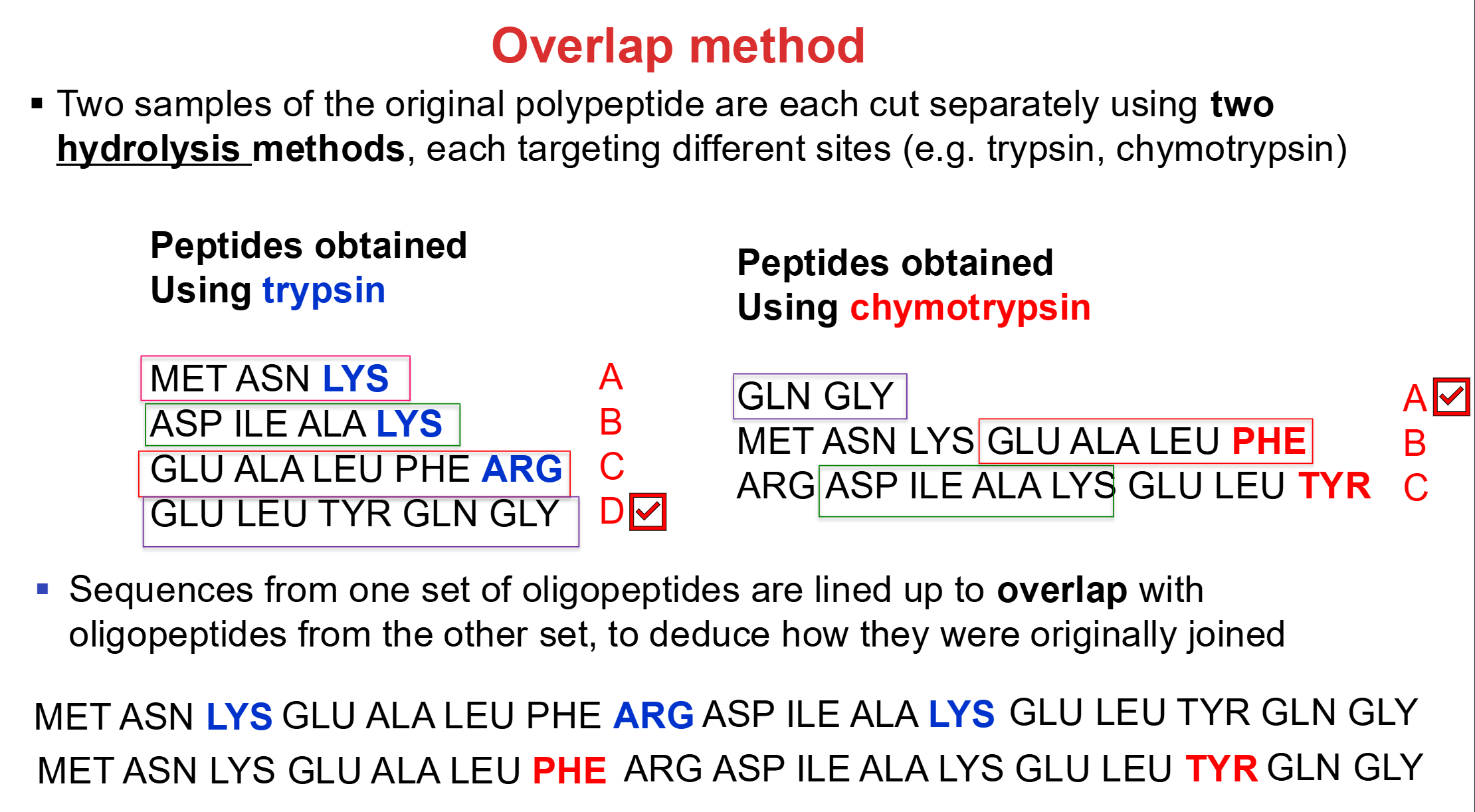
  • Two samples of the same protein are digested using different cleavage reagents (hydrolysis methods).

  • Each reagent produces peptide fragments with different cleavage sites.

  • The fragments are separated and individually sequenced.

  • Overlapping amino acid sequences are identified between fragments from the two digestions.

  • These overlapping regions establish the correct order of fragments.

  • Key concept: Different cleavage patterns allow the original protein sequence to be reconstructed.

<ul><li><p>Two samples of the same protein are digested using different cleavage reagents (hydrolysis methods).</p></li><li><p>Each reagent produces peptide fragments with different cleavage sites.</p></li><li><p>The fragments are separated and individually sequenced.</p></li><li><p>Overlapping amino acid sequences are identified between fragments from the two digestions.</p></li><li><p>These overlapping regions establish the correct order of fragments.</p></li><li><p><strong>Key concept:</strong> Different cleavage patterns allow the original protein sequence to be reconstructed.</p></li></ul><p></p>
20
New cards

How are most protein amino acid sequences determined today?

  • Most protein sequences are predicted from DNA sequences stored in genome databases.

  • Scientists determine the DNA sequence of a protein-coding gene.

  • The genetic code is used to predict the corresponding amino acid sequence.

  • This approach is relatively cheap and easy.

  • Key advantage: It does not require direct protein sequencing experiments.


21
New cards

How does tandem mass spectrometry (MS/MS) determine protein sequences?

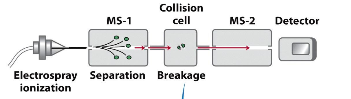
  • 1. Digestion: A protease breaks the protein into smaller peptides (hydrolysis!).

  • 2. MS-1: Separates the peptides and selects one peptide type to
    go into the collision cell

  • 3. Collision cell: Fragment each peptide molecule once (usually
    the peptide bond) in a random fashion
    .

  • 4. MS-2: Measures the masses of the resulting fragments.

  • Differences between fragment masses help identify the amino acid sequence.

  • Key advantage: Only tiny amounts of protein are required.

<ul><li><p><strong>1. Digestion:</strong> A protease breaks the protein into smaller peptides (hydrolysis!).</p></li><li><p><strong>2. MS-1:</strong> Separates the peptides and selects one peptide <span>type to</span><br><span>go into the collision cell</span></p></li><li><p><strong>3. Collision cell:</strong><span> </span><span>Fragment each peptide molecule once (usually<br>the peptide bond) in a random fashion</span><span>.</span></p></li><li><p><strong>4. MS-2:</strong> Measures the masses of the resulting fragments.</p></li><li><p>Differences between fragment masses help identify the amino acid sequence.</p></li><li><p><strong>Key advantage:</strong> Only tiny amounts of protein are required.</p></li></ul><p></p>
22
New cards

Why is trypsin the preferred protease for tandem mass spectrometry?

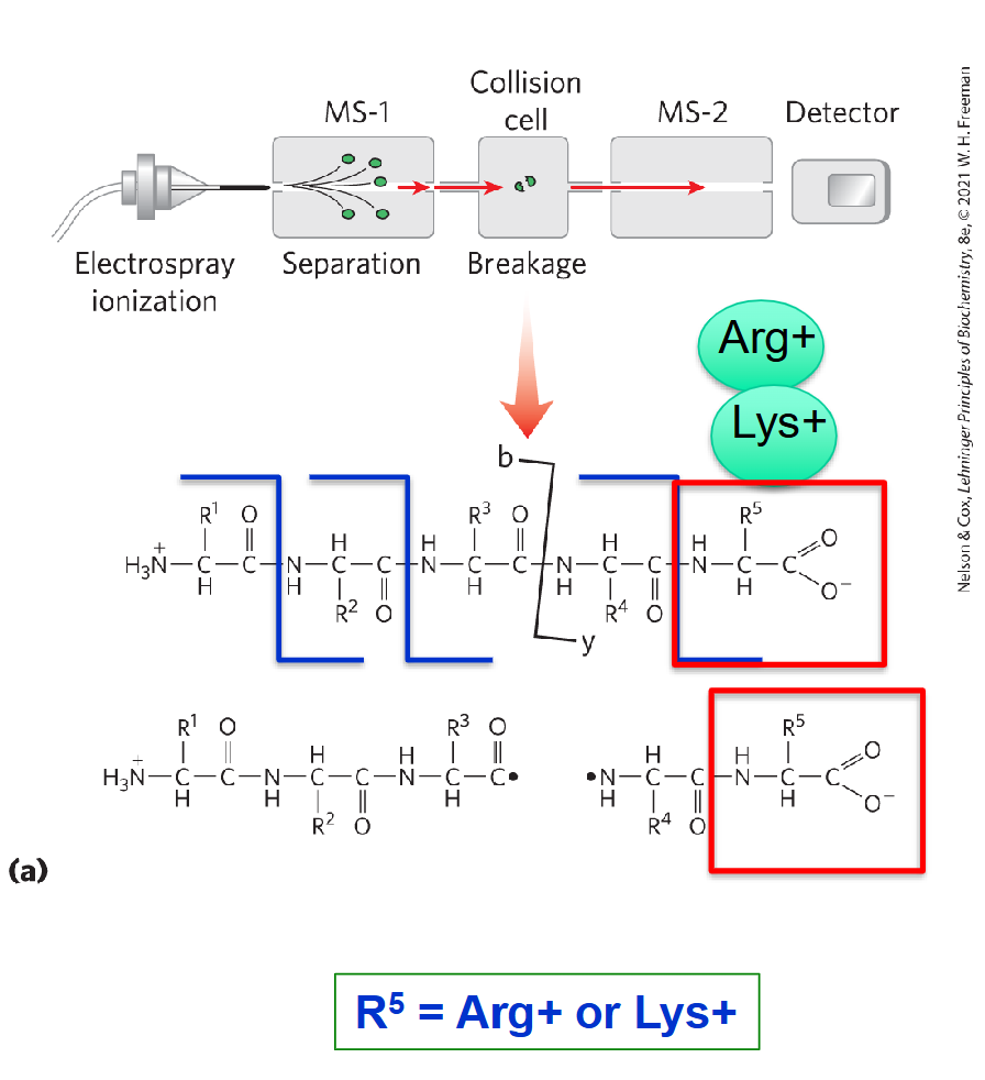
  • Trypsin first cleaves proteins after Lys (K) or Arg (R), producing peptides that generally end with these residues.

  • These peptides are then fragmented further inside the collision cell.

  • b-type fragments: Retain the N-terminus of the trypsin-generated peptide.

  • y-type fragments: Retain the C-terminus, including the terminal Lys or Arg.

  • Lys and Arg can carry positive charges, helping y-type fragments produce the highest mass spectrometry signals.

  • Key concept: Trypsin facilitates the detection and analysis of y-type fragments.

<ul><li><p>Trypsin first cleaves proteins after Lys (K) or Arg (R), producing peptides that generally end with these residues.</p></li><li><p>These peptides are then fragmented further inside the collision cell.</p></li><li><p><strong>b-type fragments:</strong> Retain the N-terminus of the trypsin-generated peptide.</p></li><li><p><strong>y-type fragments:</strong> Retain the C-terminus, including the terminal Lys or Arg.</p></li><li><p>Lys and Arg can carry positive charges, helping y-type fragments produce the highest mass spectrometry signals.</p></li><li><p><strong>Key concept:</strong> Trypsin facilitates the detection and analysis of y-type fragments.</p></li></ul><p></p>
23
New cards

How can amino acid sequences be determined from y-type fragment masses in tandem mass spectrometry?

  • MS-2 measures the masses of y-type fragments containing the peptide's C-terminus.

  • Chromatogram - Each peak corresponds to a y-type fragment formed by
    breakage of a peptide bond at a different point in the peptide chain

  • Going from the largest to the smallest, each successive peak has one less amino acid than the peak before

  • Subtract consecutive fragment masses to determine the mass of the missing amino acid.

  • Match each mass difference to the corresponding amino acid residue mass.

  • Moving from largest to smallest fragments reveals the sequence from N-terminus to C-terminus.

  • Example: 989 − 932 = 57 Da, corresponding to Gly.
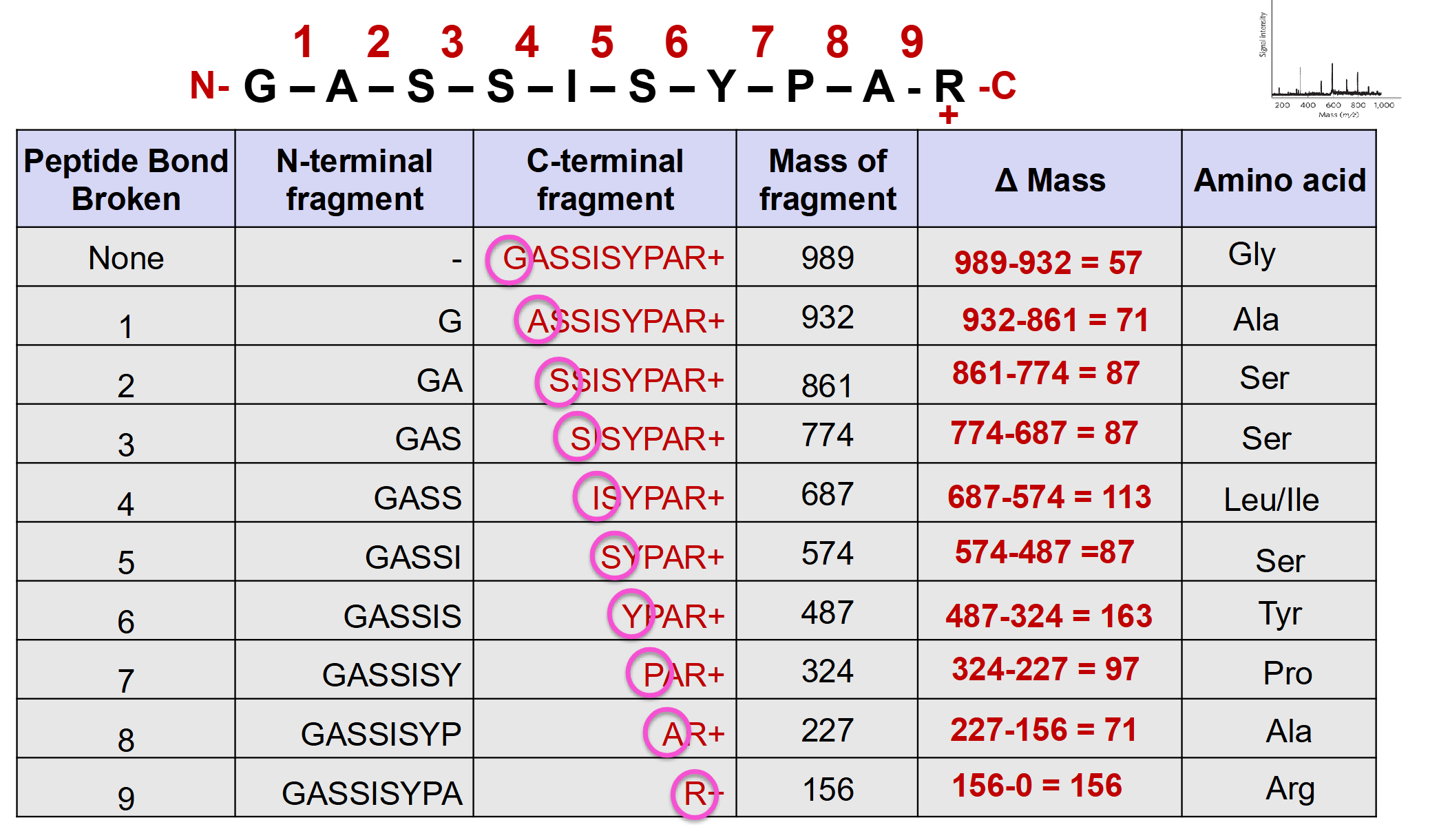

<ul><li><p>MS-2 measures the masses of y-type fragments containing the peptide's C-terminus.</p></li><li><p>Chromatogram - Each peak corresponds to a y-type fragment formed by<br>breakage of a peptide bond at a different point in the peptide chain</p></li><li><p>Going from the largest to the smallest, each successive peak has one less amino acid than the peak before</p></li><li><p>Subtract consecutive fragment masses to determine the mass of the missing amino acid.</p></li><li><p>Match each mass difference to the corresponding amino acid residue mass.</p></li><li><p>Moving from largest to smallest fragments reveals the sequence from N-terminus to C-terminus.</p></li><li><p><strong>Example:</strong> 989 − 932 = 57 Da, corresponding to Gly.</p></li></ul><p></p>
24
New cards

Useful Slide

Important: Because we're moving from the largest y-type fragment to the smallest, we're identifying amino acids from the N-terminus toward the C-terminus.

Imagine the original peptide is:

Gly–Ser–Ala–Arg

The y-type fragments would be:

  • Gly–Ser–Ala–Arg = 371 Da

  • Ser–Ala–Arg = 314 Da

  • Ala–Arg = 227 Da

  • Arg = 156 Da

Notice how each smaller fragment loses one amino acid from the N-terminal end?

<p>Important: Because we're moving from the largest y-type fragment to the smallest, we're identifying amino acids from the <strong>N-terminus toward the C-terminus</strong>.</p><p>Imagine the original peptide is:</p><p><strong>Gly–Ser–Ala–Arg</strong></p><p>The y-type fragments would be:</p><ul><li><p>Gly–Ser–Ala–Arg = 371 Da</p></li><li><p>Ser–Ala–Arg = 314 Da</p></li><li><p>Ala–Arg = 227 Da</p></li><li><p>Arg = 156 Da</p></li></ul><p>Notice how each smaller fragment loses <strong>one amino acid from the N-terminal end</strong>?</p>
25
New cards

What are NCBI and BLAST, and how are BLAST results interpreted?

  • NCBI (National Center for Biotechnology Information): Provides biological databases containing DNA, RNA, protein sequences, genomes, health-related mutations, and access to PubMed.

  • BLAST (Basic Local Alignment Search Tool): Compares an input sequence against known sequences in biological databases.

  • BLAST produces a list of the best matches, called hits.

    • 100% identity: Positive match with a known sequence.

    • Good identity: Suggests a homolog (evolutionarily related protein).

    • No identity: May indicate a new protein; biochemical tests are needed to investigate its identity.


26
New cards

Review: What are the four levels of protein structure?

  • Primary: Linear amino acid sequence.

  • Secondary: Local backbone folding patterns (α-helices and β-sheets).

  • Tertiary: Overall 3D structure of a single polypeptide.

  • Quaternary: Arrangement of multiple polypeptide subunits.


<ul><li><p><strong>Primary:</strong> Linear amino acid sequence.</p></li><li><p><strong>Secondary:</strong> Local backbone folding patterns (α-helices and β-sheets).</p></li><li><p><strong>Tertiary:</strong> Overall 3D structure of a single polypeptide.</p></li><li><p><strong>Quaternary:</strong> Arrangement of multiple polypeptide subunits.</p></li></ul><p></p>
27
New cards

How can rotation around covalent bonds change a protein's three-dimensional structure?

  • Rotation around certain single covalent bonds changes the spatial arrangement of atoms.

  • Groups connected by single bonds can rotate about bond axis

  • Chain flexibility arises from bond rotation, not bond bending

  • Normal 109* tetrahedral or 120* trigonal planar bond angles are present

  • Bond rotation allows the peptide chain to adopt a variety of shapes


<ul><li><p>Rotation around certain single covalent bonds changes the spatial arrangement of atoms.</p></li><li><p><span>Groups connected by single bonds can rotate about bond axis</span></p></li><li><p><span>Chain flexibility arises from bond rotation, not bond bending</span></p></li><li><p><span>Normal 109* tetrahedral or 120* trigonal planar bond angles are present</span></p></li><li><p><span>Bond rotation allows the peptide chain to adopt a variety of shapes</span></p></li></ul><p></p>
28
New cards

What is the difference between conformation and configuration in proteins?

  • Conformation: Spatial arrangement of atoms that can change through rotation around single bonds.

  • Conformational changes do not require breaking covalent bonds.

    • For macromolecules such as proteins, we are usually concerned
      with conformations

  • Configuration: Spatial arrangement that cannot change through simple single-bond rotation.

  • Changing configurations generally requires breaking and reforming covalent bonds.

  • Examples of configuration: Cis/trans double-bond isomers and D/L stereoisomers.


<ul><li><p><strong>Conformation:</strong> Spatial arrangement of atoms that can change through rotation around single bonds.</p></li><li><p>Conformational changes do not require breaking covalent bonds.</p><ul><li><p><span>For macromolecules such as proteins, we are usually concerned</span><br><span>with conformations</span></p></li></ul></li><li><p><strong>Configuration:</strong> Spatial arrangement that cannot change through simple single-bond rotation.</p></li><li><p>Changing configurations generally requires breaking and reforming covalent bonds.</p></li><li><p><strong>Examples of configuration:</strong> Cis/trans double-bond isomers and D/L stereoisomers.</p></li></ul><p></p>
29
New cards

How is X-ray diffraction used to study protein structure?

  • Crystals are ordered arrays of molecules

  • Atoms and molecules have similar dimensions to the wavelength of X-rays (0.7-1.5 Å)

  • When X-rays reflect off a regular repetitive structure, e.g., molecular crystal or fibre, they are deflected by an angle dependent on wavelength of X-ray and spacing of pattern

  • X-rays scatter when they pass through an ordered sample, producing a diffraction pattern.

  • If X-ray wavelength is known, dimensions of the repeating pattern of molecules can be calculated.

  • Some important structural parameters were identified from early studies of diffraction patterns of fibrous proteins such as α-keratin and β-keratin.

    • α-Keratin: Major repeat of 5.4 Å and minor repeat of 1.5 Å.

    • β-Keratin (fibroin): Major repeat of 7.0 Å and minor repeat of 3.5 Å.
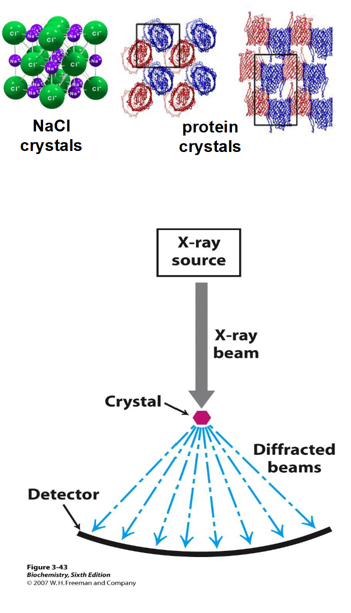

<ul><li><p><span>Crystals are ordered arrays of molecules</span></p></li><li><p><span>Atoms and molecules have similar dimensions to the wavelength of X-rays (0.7-1.5 Å)</span></p></li><li><p><span>When X-rays reflect off a regular repetitive structure, e.g., molecular crystal or fibre, they are deflected by an angle dependent on wavelength of X-ray and spacing of pattern</span></p></li><li><p>X-rays scatter when they pass through an ordered sample, producing a diffraction pattern.</p></li><li><p><span>If X-ray wavelength is known, dimensions of the repeating pattern of molecules can be calculated.</span></p></li><li><p><span>Some important structural parameters were identified from early studies of diffraction patterns of fibrous proteins such as α-keratin and β-keratin.</span></p><ul><li><p><strong>α-Keratin:</strong> Major repeat of 5.4 Å and minor repeat of 1.5 Å.</p></li><li><p><strong>β-Keratin (fibroin):</strong> Major repeat of 7.0 Å and minor repeat of 3.5 Å.</p></li></ul></li></ul><p></p>
30
New cards

Why are peptide bonds rigid and planar?

  • The peptide bond has two resonance forms, one with a double bond

  • Resonance restricts rotation around the peptide C–N bond.

  • The peptide group is therefore rigid and planar.

    Bond type

    Length

    Typical C–N single bond

    1.49 Å

    Peptide C–N bond

    1.32 Å

    Typical C=N double bond

    1.27 Å

  • Note: C-N bond between a single bond at 1.49 Å and a double bond at 1.27 Å

  • Peptide bonds generally adopt a trans configuration (behaves more like a double bond than a single bond).


<ul><li><p><span>The peptide bond has two resonance forms, one with a double bond</span></p></li><li><p>Resonance restricts rotation around the peptide C–N bond.</p></li><li><p>The peptide group is therefore rigid and planar.</p><table style="min-width: 50px;"><colgroup><col style="min-width: 25px;"><col style="min-width: 25px;"></colgroup><tbody><tr><th colspan="1" rowspan="1"><p>Bond type</p></th><th colspan="1" rowspan="1"><p>Length</p></th></tr><tr><td colspan="1" rowspan="1"><p>Typical C–N single bond</p></td><td colspan="1" rowspan="1"><p>1.49 Å</p></td></tr><tr><td colspan="1" rowspan="1"><p><strong>Peptide C–N bond</strong></p></td><td colspan="1" rowspan="1"><p><strong>1.32 Å</strong></p></td></tr><tr><td colspan="1" rowspan="1"><p>Typical C=N double bond</p></td><td colspan="1" rowspan="1"><p>1.27 Å</p></td></tr></tbody></table></li><li><p>Note: C-N bond between a single bond at 1.49 Å and a double bond at 1.27 Å</p></li><li><p>Peptide bonds generally adopt a trans configuration (behaves more like a double bond than a single bond).</p></li></ul><p></p>
31
New cards

Which bonds in a polypeptide backbone can rotate, and what are the phi and psi angles?

  • Phi (φ): Rotation around the N–Cα bond.

  • Psi (ψ): Rotation around the Cα–C bond.

  • These rotations allow the polypeptide backbone to adopt different conformations.

  • Peptide C–N bonds (α-amino and α-carboxylate): Rotation is restricted because of resonance.

  • The backbone consists of relatively rigid peptide planes connected by rotatable bonds (only the two bonds to α-C can rotate freely)


<ul><li><p><strong>Phi (φ):</strong> Rotation around the N–Cα bond.</p></li><li><p><strong>Psi (ψ):</strong> Rotation around the Cα–C bond.</p></li><li><p>These rotations allow the polypeptide backbone to adopt different conformations.</p></li><li><p><strong>Peptide C–N bonds</strong> (α<span>-amino and </span>α<span>-carboxylate)</span><strong>:</strong> Rotation is restricted because of resonance.</p></li><li><p>The backbone consists of relatively rigid peptide planes connected by rotatable bonds (<span>only the two bonds to </span>α<span>-C can rotate freely)</span></p></li></ul><p></p>
32
New cards

How do phi and psi rotations contribute to protein secondary structure?

  • Restricted bond rotation limits freedom of motion, so that only a few regular structures can form

  • The peptide backbone changes direction by 109* at each tetrahedral α-C, defining two possible regular repeating patterns:

  • In a helical shape, every α-C bond down the peptide chain turns in same direction (e.g. clockwise)

  • In an extended shape, the α-C bonds turn in alternate directions down the peptide chain

  • If there is no regular, repeating structure, get random coil

    • α-Helix: Coiled backbone arrangement.

    • β-Strand: Extended backbone arrangement.

    • Random coil: Irregular backbone arrangement.


<ul><li><p><span>Restricted bond rotation limits freedom of motion, so that only a few regular structures can form</span></p></li><li><p><span>The peptide backbone changes direction by 109* at each tetrahedral </span>α<span>-C, defining two possible regular repeating patterns:</span></p></li><li><p><span>In a helical shape, every </span>α<span>-C bond down the peptide chain turns in same direction (e.g. clockwise)</span></p></li><li><p><span>In an extended shape, the </span>α<span>-C bonds turn in alternate directions down the peptide chain</span></p></li><li><p><span>If there is no regular, repeating structure, get random coil</span></p><ul><li><p><strong>α-Helix:</strong> Coiled backbone arrangement.</p></li><li><p><strong>β-Strand:</strong> Extended backbone arrangement.</p></li><li><p><strong>Random coil:</strong> Irregular backbone arrangement.</p></li></ul></li></ul><p></p>
33
New cards

What are the structural characteristics of an α-helix?

  • Helix forms when amino acids all have same orientation, and α-C bonds turn in same direction (i.e. all clockwise)

  • Pauling’s models showed a very stable structure with 3.6 amino acids per turn of helix

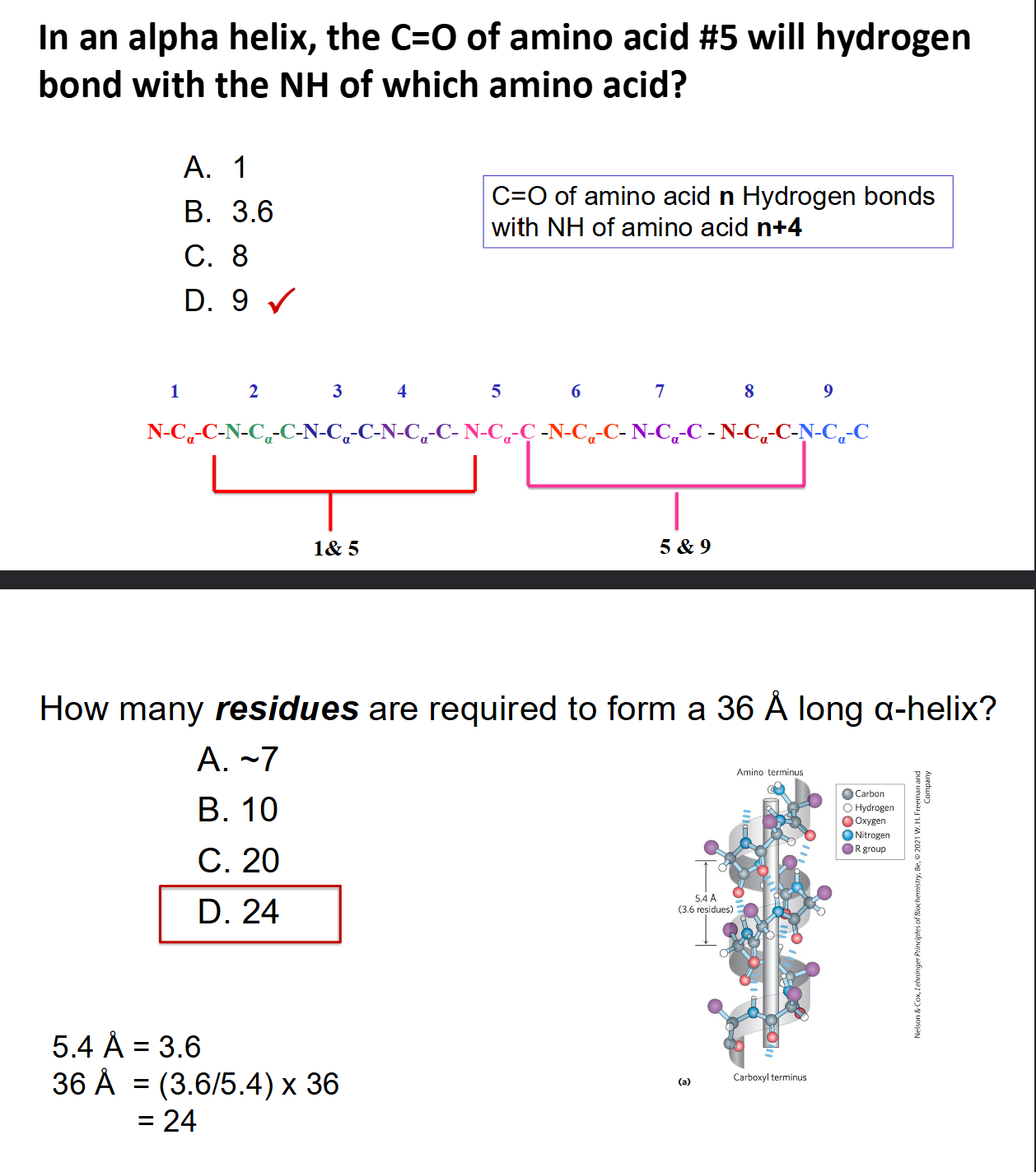
  • #1 C=O lines up with H–N #5 to form hydrogen bonds that make α-helix stable

  • Distance between each turn of helix is 5.4 Å

  • 5.4 ÷ 3.6 = 1.5 Å is distance along the helix per amino acid

  • These distances match α-keratin patterns exactly, hence the name α-helix


<ul><li><p><span>Helix forms when amino acids all have same orientation, and </span>α<span>-C bonds turn in same direction (i.e. all clockwise)</span></p></li><li><p><span>Pauling’s models showed a very stable structure with 3.6 amino acids per turn of helix</span></p></li><li><p><span>#1 C=O lines up with H–N #5 to form hydrogen bonds that make </span>α<span>-helix stable</span></p></li><li><p><span>Distance between each turn of helix is 5.4 Å</span></p></li><li><p><span>5.4 ÷ 3.6 = 1.5 Å is distance along the helix per amino acid</span></p></li><li><p><span>These distances match </span>α<span>-keratin patterns exactly, hence the name </span>α<span>-helix</span></p></li></ul><p></p>
34
New cards

Hydrogen Bonding Calculation and Helix Length Calculation

  • Remember: Hydrogen bonding: C=O of residue n bonds with N–H of residue n+4.

  • Length = 1.5 x Number of Residues


<ul><li><p>Remember: <strong>Hydrogen bonding:</strong> C=O of residue n bonds with N–H of residue n+4.</p></li><li><p>Length = 1.5 x Number of Residues </p></li></ul><p></p>
35
New cards

What is the difference between parallel and antiparallel β-sheets?

  • β-Sheets consist of extended β-strands stabilized by backbone hydrogen bonds.

  • Parallel β-sheet: Adjacent strands run in the same N→C direction.

  • Antiparallel β-sheet: Adjacent strands run in opposite N→C directions.

  • Hydrogen bonds are generally more directly aligned in antiparallel β-sheets.


<ul><li><p>β-Sheets consist of extended β-strands stabilized by backbone hydrogen bonds.</p></li><li><p><strong>Parallel β-sheet:</strong> Adjacent strands run in the same N→C direction.</p></li><li><p><strong>Antiparallel β-sheet:</strong> Adjacent strands run in opposite N→C directions.</p></li><li><p>Hydrogen bonds are generally more directly aligned in antiparallel β-sheets.</p></li></ul><p></p>
36
New cards

Which amino acids prefer β-sheet structures, and why?

  • β-Sheets provide more space for bulky or awkwardly shaped side chains.

  • Trp, Tyr, Phe (WYF): Have bulky aromatic side chains.

  • Val, Ile, Thr (VIT): Have branching at the β-carbon.

  • Cys (C): Contains a relatively large sulfur atom.

  • Memory trick: WYF, VIT, C.

  • These amino acids tend to favour β-sheet structures.
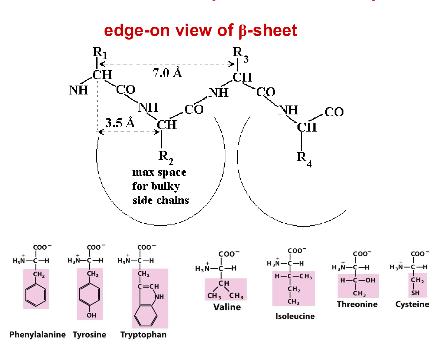

<ul><li><p>β-Sheets provide more space for bulky or awkwardly shaped side chains.</p></li><li><p><strong>Trp, Tyr, Phe (WYF):</strong> Have bulky aromatic side chains.</p></li><li><p><strong>Val, Ile, Thr (VIT):</strong> Have branching at the β-carbon.</p></li><li><p><strong>Cys (C):</strong> Contains a relatively large sulfur atom.</p></li><li><p><strong>Memory trick:</strong> WYF, VIT, C.</p></li><li><p>These amino acids tend to favour β-sheet structures.</p></li></ul><p></p>
37
New cards

Review: What X-ray diffraction patterns are associated with α-keratin and β-keratin?

knowt flashcard image
38
New cards

How does amino acid composition influence secondary structure formation?

  • Secondary structure depends on the local majority of amino acid preferences.

  • α-Helix formers: Ala, Arg, Gln, Glu, His, Leu, Lys, Met, (Phe).

  • β-Sheet formers: Trp, Tyr, (Phe), Val, Ile, Thr, Cys.

  • Phe: Can favour either structure.

  • The local majority helps predict whether a segment forms an α-helix or β-sheet.


39
New cards

What are secondary structure breakers, and what is their role in protein folding?

  • Breakers: Gly, Pro, Asn, Asp, Ser (GPNDS).

  • Can interfere with regular secondary structure formation.

  • Professor's rule: Two breakers within four amino acids can interrupt secondary structure.

  • Breaker clusters promote turns and flexible loops.

  • These regions allow the polypeptide backbone to change direction, sometimes by approximately 180°.
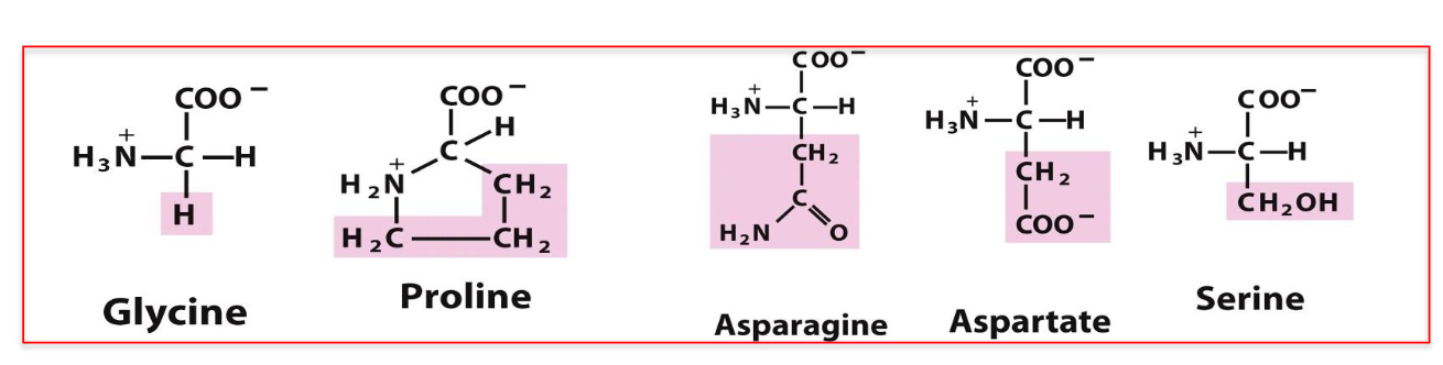

<ul><li><p><strong>Breakers:</strong> Gly, Pro, Asn, Asp, Ser (<strong>GPNDS</strong>).</p></li><li><p>Can interfere with regular secondary structure formation.</p></li><li><p><strong>Professor's rule:</strong> Two breakers within four amino acids can interrupt secondary structure.</p></li><li><p>Breaker clusters promote turns and flexible loops.</p></li><li><p>These regions allow the polypeptide backbone to change direction, sometimes by approximately 180°.</p></li></ul><p></p>
40
New cards

How can you predict secondary structure from an amino acid sequence?

  • Identify secondary structure breakers (GPNDS).

  • Look for clusters containing at least two breakers within four amino acids.

  • Use these clusters to divide the sequence into segments.

  • Examine the amino acid preferences within each segment.

  • Majority α-helix formers: Predict an α-helix.

  • Majority β-sheet formers: Predict a β-sheet.
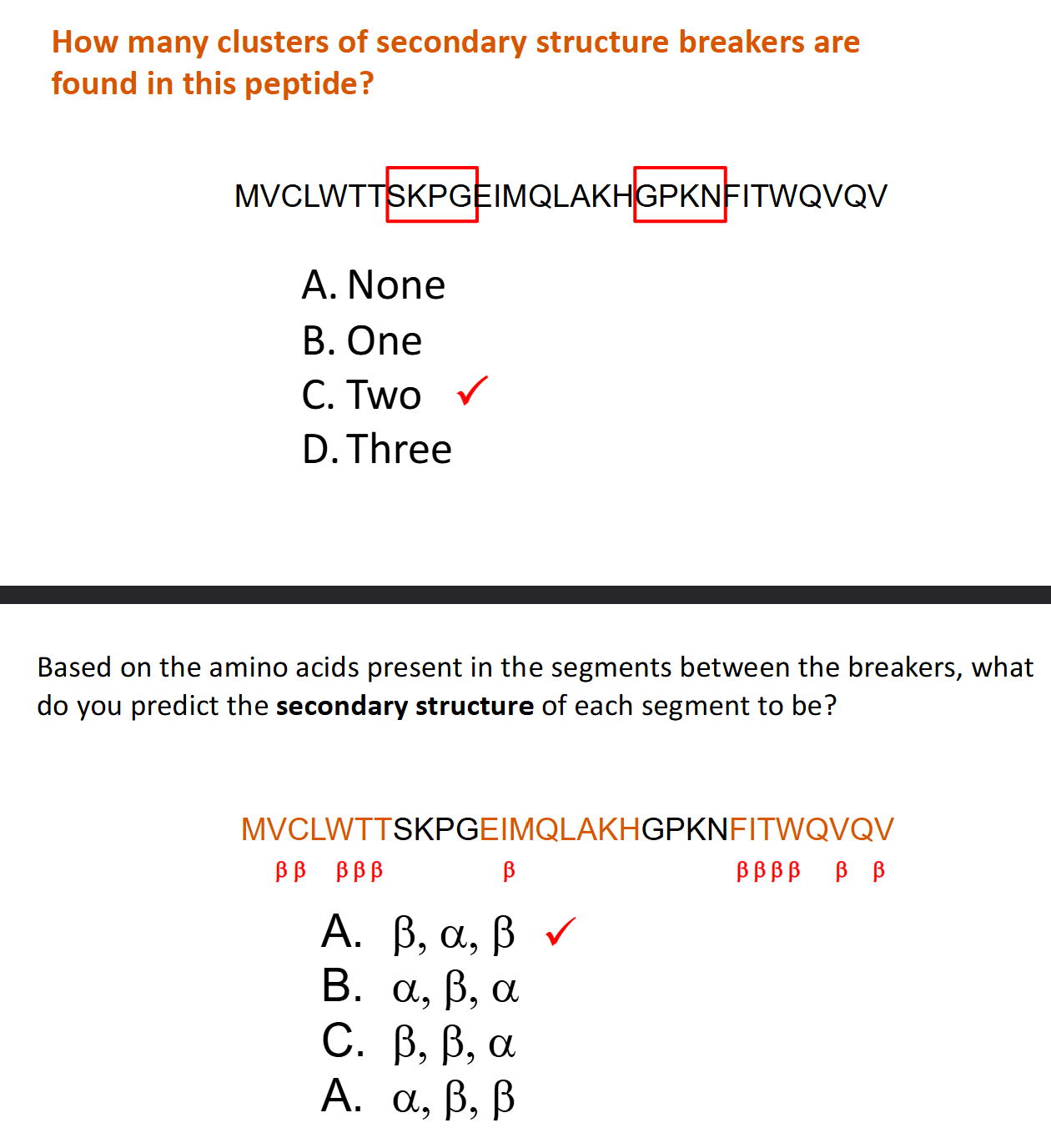

<ul><li><p>Identify secondary structure breakers (<strong>GPNDS</strong>).</p></li><li><p>Look for clusters containing at least two breakers within four amino acids.</p></li><li><p>Use these clusters to divide the sequence into segments.</p></li><li><p>Examine the amino acid preferences within each segment.</p></li><li><p><strong>Majority α-helix formers:</strong> Predict an α-helix.</p></li><li><p><strong>Majority β-sheet formers:</strong> Predict a β-sheet.</p></li></ul><p></p>
41
New cards

What is the difference between native and denatured proteins?

  • Native state: Protein is folded into its functional 3D tertiary structure.

  • Denaturation: Loss of normal protein folding.

  • Denatured proteins may become unstructured or aggregated.

  • Protein function is typically lost upon denaturation.

  • Causes: Heat, disruptive solvents, and harsh detergents (e.g., SDS).

  • Denaturation is often irreversible.

  • Denaturation is generally avoided during protein purification.


42
New cards

What is the simplest possible tertiary structure and three examples?

  • Tertiary structure is the overall pattern of folding of the whole polypeptide chain

  • The simplest possible tertiary structure is continuous secondary structure

  • α-keratin is totally α-helix

  • Fibroin (a β-keratin) is totally anti-parallel β-sheet

  • Collagen (tendon, bone and connective tissue) has a unique triple-helix structure

    • Collagen structure requires sequence with repeating units of three amino acids -Gly-Pro-X-

  • Secondary structure is rigid, so these are fibrous proteins


<ul><li><p><span>Tertiary structure is the overall pattern of folding of the whole polypeptide chain</span></p></li><li><p><span>The simplest possible tertiary structure is continuous secondary structure</span></p></li><li><p>α<span>-keratin is totally </span>α<span>-helix</span></p></li><li><p><span>Fibroin (a </span>β<span>-keratin) is totally anti-parallel </span>β<span>-sheet</span></p></li><li><p><span>Collagen (tendon, bone and connective tissue) has a unique triple-helix structure</span></p><ul><li><p><span>Collagen structure requires sequence with repeating units of three amino acids -Gly-Pro-X-</span></p></li></ul></li><li><p><span>Secondary structure is rigid, so these are fibrous proteins</span></p></li></ul><p></p>
43
New cards

How do secondary structure breakers contribute to globular protein formation?

  • Globular proteins require the polypeptide chain to fold back on itself.

  • Folding requires “breaks” in secondary structure, which is rigid

  • Breakers: Gly, Pro, Asn, Asp, Ser (GPNDS).

  • Clusters of 2–3 breakers within four amino acids can interrupt secondary structure.

  • These interruptions form flexible loops and turns.

  • Flexible regions allow the polypeptide to change direction and form compact tertiary structures.


<ul><li><p>Globular proteins require the polypeptide chain to fold back on itself.</p></li><li><p><span>Folding requires “breaks” in secondary structure, which is rigid</span></p></li><li><p><strong>Breakers:</strong> Gly, Pro, Asn, Asp, Ser (GPNDS).</p></li><li><p>Clusters of 2–3 breakers within four amino acids can interrupt secondary structure.</p></li><li><p>These interruptions form flexible loops and turns.</p></li><li><p>Flexible regions allow the polypeptide to change direction and form compact tertiary structures.</p></li></ul><p></p>
44
New cards

How does the hydrophobic effect drive protein folding?

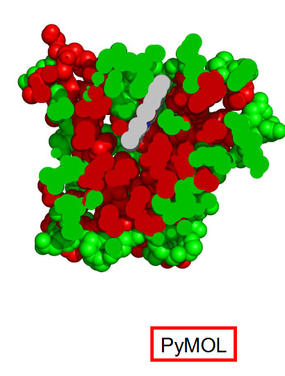
  • The hydrophobic effect is a major force driving protein folding.

  • Nonpolar amino acids: Cluster in the protein's interior to minimize contact with water.

  • Polar amino acids: Tend to remain on the exterior, interacting with water or ions.

  • Polar side chains can form hydrogen bonds with surrounding water.

  • This arrangement helps stabilize the folded structure of soluble globular proteins.

<ul><li><p>The hydrophobic effect is a major force driving protein folding.</p></li><li><p><strong>Nonpolar amino acids:</strong> Cluster in the protein's interior to minimize contact with water.</p></li><li><p><strong>Polar amino acids:</strong> Tend to remain on the exterior, interacting with water or ions.</p></li><li><p>Polar side chains can form hydrogen bonds with surrounding water.</p></li><li><p>This arrangement helps stabilize the folded structure of soluble globular proteins.</p></li></ul><p></p>
45
New cards

How do amino acid packing and van der Waals forces stabilize protein tertiary structure?

  • Amino acid side chains pack together like jigsaw puzzle pieces.

  • Good packing maximizes close atom-to-atom contacts.

  • Close contacts produce weak van der Waals (London dispersion) forces.

  • Each interaction contributes approximately 0.1–1 kJ/mol.

  • Hundreds of close contacts collectively help stabilize the protein.

  • Poor packing produces fewer favourable interactions.


<ul><li><p>Amino acid side chains pack together like jigsaw puzzle pieces.</p></li><li><p>Good packing maximizes close atom-to-atom contacts.</p></li><li><p>Close contacts produce weak <strong>van der Waals (London dispersion) forces</strong>.</p></li><li><p>Each interaction contributes approximately <strong>0.1–1 kJ/mol</strong>.</p></li><li><p>Hundreds of close contacts collectively help stabilize the protein.</p></li><li><p>Poor packing produces fewer favourable interactions.</p></li></ul><p></p>
46
New cards

Summary: How does the amino acid sequence determine a protein's tertiary structure?

  • Amino acid preferences influence α-helix and β-sheet formation.

  • Secondary structure breakers (GPNDS): Introduce flexible loops and turns.

  • Nonpolar amino acids: Tend to fold inward due to the hydrophobic effect.

  • Polar amino acids: Tend to remain exposed to water.

  • Side-chain packing: Pattern of large and small side chains is arranged so that secondary structure components (e.g. helices) pair up with best possible fit (Maximize Van der Wall Forces)

  • These factors collectively determine the protein's stable native structure.


47
New cards

Review: What are the three major tertiary structure patterns described in Lecture 8?

  • Mostly α-helical: Proteins containing primarily α-helical segments.

  • Mostly β-strand: Proteins containing primarily β-strands.

  • Mixed α/β: Proteins containing alternating α-helical and β-strand segments.

  • Amino acid sequence influences which tertiary structure pattern forms.


48
New cards

How do α-helix bundles form, and what is an example?

  • Proteins rich in α-helix-forming amino acids can form α-helix bundles.

  • Secondary structure breakers separate individual helices (set the limits of each helix)

  • Nonpolar amino acids every 3–4 residues create a hydrophobic stripe along each helix.

  • Nonpolar stripes face inward when helices pack together.

  • Example: Myoglobin is a bundle of 8 α-helix sections


<ul><li><p>Proteins rich in α-helix-forming amino acids can form α-helix bundles.</p></li><li><p>Secondary structure breakers separate individual helices (<span>set the limits of each helix)</span></p></li><li><p>Nonpolar amino acids every 3–4 residues create a hydrophobic stripe along each helix.</p></li><li><p>Nonpolar stripes face inward when helices pack together.</p></li><li><p><strong>Example:</strong> <span>Myoglobin is a bundle of 8 </span>α<span>-helix sections</span></p></li></ul><p></p>
49
New cards

How can antiparallel β-sheets have one polar face and one nonpolar face?

  • Antiparallel β-strands run in opposite N→C directions.

  • Their hydrogen bonds are relatively straight, making them more stable.

  • β-strand side chains alternate between opposite sides of the sheet.

  • Odd-numbered residues project toward one side; even-numbered residues project toward the other.

  • Alternating polar and nonpolar amino acids (NPNPNP) can create one polar face and one nonpolar face.


<ul><li><p>Antiparallel β-strands run in opposite N→C directions.</p></li><li><p>Their hydrogen bonds are relatively straight, making them more stable.</p></li><li><p>β-strand side chains alternate between opposite sides of the sheet.</p></li><li><p>Odd-numbered residues project toward one side; even-numbered residues project toward the other.</p></li><li><p>Alternating polar and nonpolar amino acids (NPNPNP) can create one polar face and one nonpolar face.</p></li></ul><p></p>
50
New cards

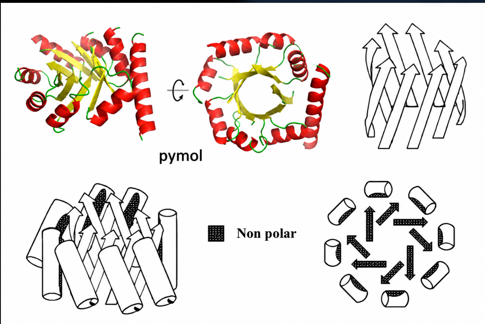
How do antiparallel β-barrels form?

  • β-Sheets with one polar face and one nonpolar face can curve to bury their nonpolar surface.

  • 3–5 β-strands (Smaller sheet): Form an open fold.

  • 6–8 β-strands (Larger sheet): Can wrap around to form an antiparallel β-barrel.

  • The nonpolar face points inward, while the polar face remains exposed to water.

  • Example: Green fluorescent protein (GFP).


<ul><li><p>β-Sheets with one polar face and one nonpolar face can curve to bury their nonpolar surface.</p></li><li><p><strong>3–5 β-strands (Smaller sheet):</strong> Form an open fold.</p></li><li><p><strong>6–8 β-strands (Larger sheet):</strong> Can wrap around to form an antiparallel β-barrel.</p></li><li><p>The nonpolar face points inward, while the polar face remains exposed to water.</p></li><li><p><strong>Example:</strong> Green fluorescent protein (GFP).</p></li></ul><p></p>
51
New cards

How do parallel β-sheets form, and what are their characteristics?

  • Alternating β–α–β–α sequences can form parallel β-sheets.

  • α-Helices connect β-strands running in the same N→C direction.

  • Connecting helices lie above or below the β-sheet.

  • Parallel β-sheets have angled hydrogen bonds and are less stable than antiparallel β-sheets.

  • They are generally buried in the protein's interior, away from water.

  • They typically contain mostly nonpolar amino acids.


<ul><li><p>Alternating β–α–β–α sequences can form parallel β-sheets.</p></li><li><p>α-Helices connect β-strands running in the same N→C direction.</p></li><li><p>Connecting helices lie above or below the β-sheet.</p></li><li><p>Parallel β-sheets have angled hydrogen bonds and are less stable than antiparallel β-sheets.</p></li><li><p>They are generally buried in the protein's interior, away from water.</p></li><li><p>They typically contain mostly nonpolar amino acids.</p></li></ul><p></p>
52
New cards

How does a parallel αβ-barrel form?

  • Forms from alternating α-helical and parallel β-strand segments.

  • Connecting α-helices lie on the same side of the β-sheet.

  • The β-sheet wraps around to form a central barrel.

  • α-Helices surround the outside of the barrel.

  • Example: Triose phosphate isomerase.


<ul><li><p>Forms from alternating α-helical and parallel β-strand segments.</p></li><li><p>Connecting α-helices lie on the same side of the β-sheet.</p></li><li><p>The β-sheet wraps around to form a central barrel.</p></li><li><p>α-Helices surround the outside of the barrel.</p></li><li><p><strong>Example:</strong> Triose phosphate isomerase.</p></li></ul><p></p>
53
New cards

How does a parallel αβ-sandwich form?

  • Forms from alternating α-helical and parallel β-strand segments.

  • Connecting α-helices lie on both sides of the β-sheet.

  • The nonpolar β-sheet forms the central layer between two layers of α-helices.

  • The β-sheet is often twisted to improve packing.

  • Example: Part of lactate dehydrogenase.


<ul><li><p>Forms from alternating α-helical and parallel β-strand segments.</p></li><li><p>Connecting α-helices lie on both sides of the β-sheet.</p></li><li><p>The nonpolar β-sheet forms the central layer between two layers of α-helices.</p></li><li><p>The β-sheet is often twisted to improve packing.</p></li><li><p><strong>Example:</strong> Part of lactate dehydrogenase.</p></li></ul><p></p>
54
New cards

What are protein domains, and why are larger proteins considered modular?

  • Domains are sections of a polypeptide chain that fold into distinct 3D structures.

  • Each of the structures outlined above forms from a chain of 10-20 kDa

  • Larger proteins fold up in 10-20 kDa sections; each section is called a domain

    • Ex. A protein of 50 kDa may have 3 or 4 domains

  • Larger proteins may contain multiple domains with different folding patterns.

  • This makes larger proteins modular.

  • Example: Lactate dehydrogenase contains two αβ-sandwich domains.


<ul><li><p>Domains are sections of a polypeptide chain that fold into distinct 3D structures.</p></li><li><p><span>Each of the structures outlined above forms from a chain of 10-20 kDa</span></p></li><li><p><span>Larger proteins fold up in 10-20 kDa sections; each section is called a domain</span></p><ul><li><p><span>Ex. A protein of 50 kDa may have 3 or 4 domains</span></p></li></ul></li><li><p>Larger proteins may contain multiple domains with different folding patterns.</p></li><li><p>This makes larger proteins modular.</p></li><li><p>Example: Lactate dehydrogenase contains two αβ-sandwich domains.</p></li></ul><p></p>
55
New cards

Review: The main forces that contribute to protein stability and function (2 really important ones!)

  • Covalent bonding links amino acids in a chain in a specific sequence

  • Non-covalent interactions dictate folding pattern and stability

  • Hydrophobic effect and van der Waals effect are the most important non-covalent interactions

  • Hydrophobic effect locates non-polar amino acids in core of folded protein, avoiding unfavourable interaction with H2O

    • Contributes ~50% of total energy stabilizing native state

    • –5 kJ/mol per CH, CH2 or CH3 moved out of contact with H2O

  • Van der Waals interaction – dipole-dipole interactions between atoms that are close, but not covalently bonded to each other – aka London dispersion forces

    • Contributes significantly if many atoms are in close contact


56
New cards

How do hydrogen bonds and ion pairs contribute to protein stability?

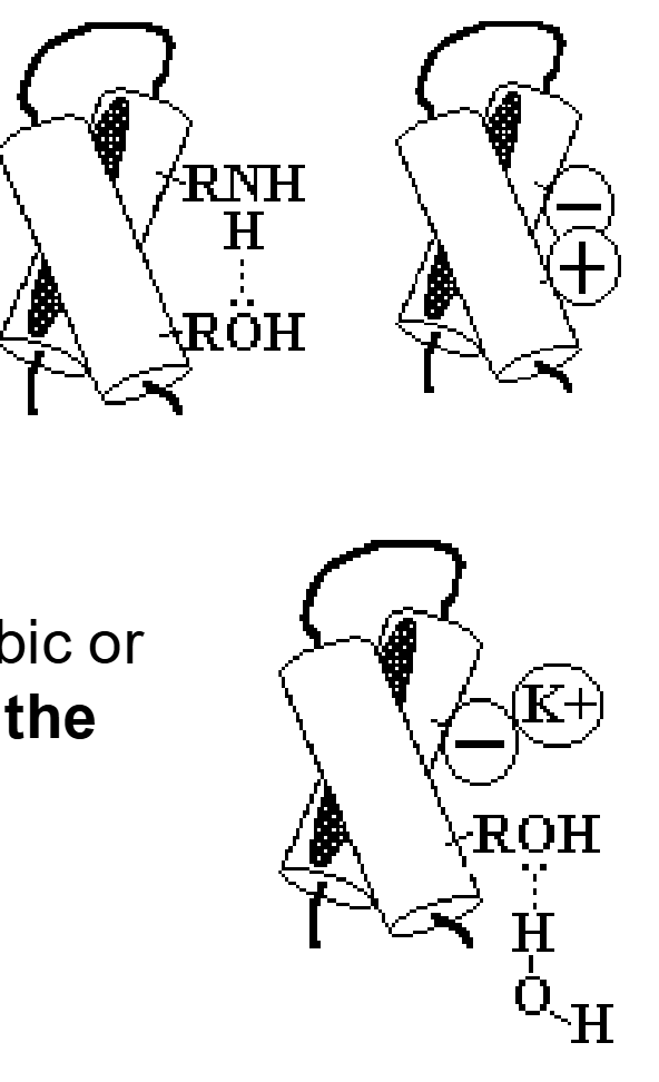
  • Hydrogen bonds form between appropriately aligned donors and acceptors.

  • Ion pairs (salt bridges) form through electrostatic attraction between oppositely charged side chains.

  • Both help stabilize the folded protein.

  • They generally contribute less than hydrophobic and van der Waals interactions because most polar groups face the protein's exterior.

    • Charged AAs pair with ions in external solution

    • H-bonding AAs bond to H2O in surroundings

<ul><li><p>Hydrogen bonds form between appropriately aligned donors and acceptors.</p></li><li><p>Ion pairs (salt bridges) form through electrostatic attraction between oppositely charged side chains.</p></li><li><p>Both help stabilize the folded protein.</p></li><li><p>They generally contribute less than hydrophobic and van der Waals interactions because most polar groups face the protein's exterior.</p><ul><li><p><span>Charged AAs pair with ions in external solution</span></p></li><li><p><span>H-bonding AAs bond to H2O in surroundings</span></p></li></ul></li></ul><p></p>
57
New cards

What are disulfide bonds, and how do they stabilize proteins?

  • Disulfide bonds form when pairs of Cys –SH groups react with O2, releasing H2O

  • Strong covalent S–S bonds formed by oxidation of two cysteine –SH groups.

  • Help hold different regions of a folded protein together.

  • Few proteins have disulfide bonds; mostly proteins designed to function outside cells since O2 needed

  • Example: Insulin.


<ul><li><p><span>Disulfide bonds form when pairs of Cys –SH groups react with O2, releasing H2O</span></p></li><li><p>Strong covalent S–S bonds formed by oxidation of two cysteine –SH groups.</p></li><li><p>Help hold different regions of a folded protein together.</p></li><li><p><span>Few proteins have disulfide bonds; mostly proteins designed to function outside cells since O2 needed</span></p></li><li><p>Example: Insulin.</p></li></ul><p></p>
58
New cards

What did Anfinsen's experiment demonstrate about protein folding?

  • The primary amino acid sequence contains the information required for a protein's secondary and tertiary structures.

  • Anfinsen denatured ribonuclease using urea and 2-mercaptoethanol.

  • When these chemicals were removed, ribonuclease regained its native structure and catalytic activity.

  • This process is called renaturation.


<ul><li><p>The primary amino acid sequence contains the information required for a protein's secondary and tertiary structures.</p></li><li><p>Anfinsen denatured ribonuclease using urea and 2-mercaptoethanol.</p></li><li><p>When these chemicals were removed, ribonuclease regained its native structure and catalytic activity.</p></li><li><p>This process is called renaturation.</p></li></ul><p></p>
59
New cards

What are the roles of urea and 2-mercaptoethanol in protein denaturation?

  • Urea: Weakens the hydrophobic effect, allowing proteins to unfold.

  • 2-Mercaptoethanol: Reduces disulfide bonds to cysteine –SH groups (converts disulfides back to the original unlinked Cys-SH groups).

  • Together, they disrupt interactions that stabilize the native protein structure.