The Cardiovascular System
 Call Kai
 Learn
Learn Practice Test
 Spaced Repetition
 Match
Match Flashcards
 Knowt Play
1/33

Earn XP
Description and Tags
My heart will stop
Name | Mastery | Learn | Test | Matching | Spaced | Call with Kai | Chat |
|---|
No analytics yet
Send a link to your students to track their progress
34 Terms

What is the cardiovascular system?
Includes the heart and the blood vessels
It brings nutrients to the cells and helps get rid of wastes
Blood is refreshed in the lung, kidneys, intestine and liver.
Lymphatic vessels help this system by collecting excess fluid surrounding tissues, and returning it to the cardiovascular system
Functions of the CVS
Generate blood pressure -Heart
Transport blood - Heat, Hormones, and electrolytes
Exchange of nutrients and wastes at the capillaries
Regulate blood flow as needed
Systolic pressure and diastolic pressure
Systolic pressure (top number)- Measure the pressure in your ateries when your heart beats and contracts
Dystolic pressure (bottom number)- Measures the pressures in your ateries when you heart rests between beats
What is the function of blood pressure?
To assist in the blood flow
The higher the blood pressure, the more blood flow you have
The average person had 5 L of blood, what happens when that blood is lost
Blood pressure decreases
What is the main pathway of blood in the body?
Heart → arteries → aterioles→ capillaries → venules → veins → back to the heart

Lymphatic Vessels
Branch throughout the body and help this system by collecting excess fluid, waste, and immune cells from surrounding tissues and returning it to the cardiovascular system

Arteries
Carry blood away from the heart, to the tissues, and their walls have three layers
Arteries branch out to form arterioles
Tunica Externa - Thick inner epithelium
Tunica Media - Thick smooth muscle layer
Tunica Interna - Outer connective tissue

Arterioles
Are small arteries that regulate blood pressure
They branch out
They regulate the blood pressure and control how much blood is supplied to any organ

Capillaries
Microscopic vessels between aterioles and venules and connect them together
Made of one layer of epithial tissue
Form capillarie beds of vessels where exchange with body cells occurs
Combined large surface area

Veins
Branches of venules come to form veins
Carry blood away from the heart
The lumin is much larger
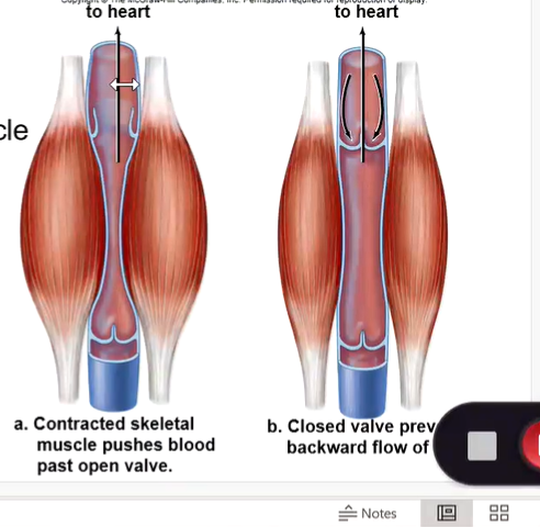
Veins that carry blood against gravity have valves to keep blood flowing towards the heart
Ensure the blood only flows one way
Most veins carry deoxygenated blood
Have a much thinner smooth muscle layer
Have valves
Valve Functions
Valves can open and close, and are present to ensure the blood only flows one way
Venules
Venules are small veins that receive blood from the capallaries

Anatomy of the Heart
Large, muscular organ consisting of mostly cardiac tissue and the myocardium
Surrounded by a sac called the pericardium
Consists of 2 sides, right and left, seperated by a septum
Consists of 4 chambers - 2 atria and 2 ventricles
2 sets of valves - semilunar valves and atrioventricular valves (AV)
Valves produce the lub, dub sounds of the heartbeat
Blood vessels gather and form a circuit called
Coronary circuit

Aorta
The main largest artery in the human body
Aortic root - The base attached to the left ventricle of the heart, housing the aortic valve and cornory artery openings.
Ascending arorta - The segment curving upward from the heart
Aortic Arch - The cane-like bend at the top that branches into vessels supplying the head, neck, and arms
Blood vessels that bring blood to the heart
Superior Vena Cava - Brings oxygen-poor blood from the the upper body, head, neck, arms, and chest to the heart
Inferior Vena Cava - Brings oxygen-poor blood from the lower body, trunk, and legs, to the heart
Pulmonary veins - Bring oxygen-rich blood from your lungs into your left atrium
Pulmonary Arteries
Brings blood from the heart, into your lungs
The only artery that carries deoxygenated blood
Pulmonary veins
Bring oxygenated blood from the lungs into the heart
Internal Anatomy of the Heart
Right Atrium - Receives deoxygenated blood, and then goes through the atrioventricular (tricuspid) valve into the right ventricle
Left Atrium - Recieves oxygenated blood, and then atrioventricular (bicuspid) valve and then into the left ventricle
Both recieve blood from the pulmonary veins
When the ventricles contract, that is when the valves close
Pulmonary Trunk
From the left ventricle goes into the pulmonary trunk, into the semilunar valves, from there the blood is distributed into through the pulmonary veins
How does blood flow through the heart?
Inferior and superior vena cava (1) dump blood into the right atrium (2)
Right ventricle (3)
2 pulmonary arteries (4) that leads to lungs (5) where blood becomes oxygenated
pulmonary veins (6) bring blood from the lungs to the left atrium (7)
Left ventricle (8) is large and muscular to pump blood into the aorta (9) and to the rest of the body (10)
Eventually blood will be pumped back to each vena cava (1)
Contraction and Relaxation
Together this makes a cardiac cycle
Can also be known as Systole, and diastole

Papillary muscles and chordae tendineae
Papillary muscles - Specialized cone shaped muscles inside the ventricles of the heart that anchor the antrioventricular valves to prevent blood from flowing backwards
Chordae tendineae - Commonly known as the heartstrings are strong chords of connective tissue located inside the ventricles
The left ventricle is much more —— than the right ventricle because it must pump blood throughout the entire body
muscular
Vein and Arteries
Veins - Have a thinner wall and a larger center to contain more blood
Arteries - Are more muscular than veins to withstand the pressure exerted on them and carry blood toward the heart

How does the heartbeat occur?
During systole the atria contract together followed by the ventricles contracting together.
This is followed by diastole, a rest phase when the chambers relax.
The cardiac cycle, or heartbeat, occur 70/times a minute on average.
Internal Control
The SA node in the right atrium initiates the heartbeat and causes the atrium to contract, artifical peacemakers
The impulse reaches the AV node, also in the right atrium, to send a signal down the AV bundle and Purkinje fibers that causes ventricular contraction.
These impulses travel between gap junctions at intercalated disks

How is heartbeat controlled?
External Control
Heartbeat is controlled by a cardiac center in the brains and hormones such as epinephrine and norepinephrine
The brain can only speed up the heart rate
Depolarization
The electrical activation of heart muscle cells that causes them to contract and pump blood
Repolarization
Repolarization is the biological process where a cell's membrane potential returns to a negative resting state after a depolarization event
What is blood pressure
It is the pressure against a blood vessel wall, usually measured in an artery of an arm
The highest pressure called the systolic pressure, is during blood ejection from the heart
The lowest pressure, the diastolic pressure, occurs when the ventricles relax.
Average blood pressure is recorded at about 120/80 mmHg
This is controlled by the aterioles
What is important about blood flow
Blood flow is under the highest blood pressure in the arteries but remember the thick, muscular walls.
Blood flow is slower in the capillaries which is important to allow time for exchange between the cells
Blood pressure is minimal in the veins and venules but blood flow increases
If blood pressure is so low in the veins, why does the blood flow increase?
They have help.
1. Skeletal muscle contraction
2. Breathing
3. Valves