Journal Club FIGURES (Ucar et al. 2021)
 Call Kai
 Learn
 Practice Test
 Spaced Repetition
 Match
 Flashcards
 Knowt Play
1/10

There's no tags or description
Looks like no tags are added yet.
11 Terms
What is the main purpose of Figure 1?
To determine whether pushing individual presynaptic boutons using fine glass pipettes change their glutamate-release responses
Stimulate the neuron → detect glutamate release → mechanically push the bouton → stimulate again → compare the responses

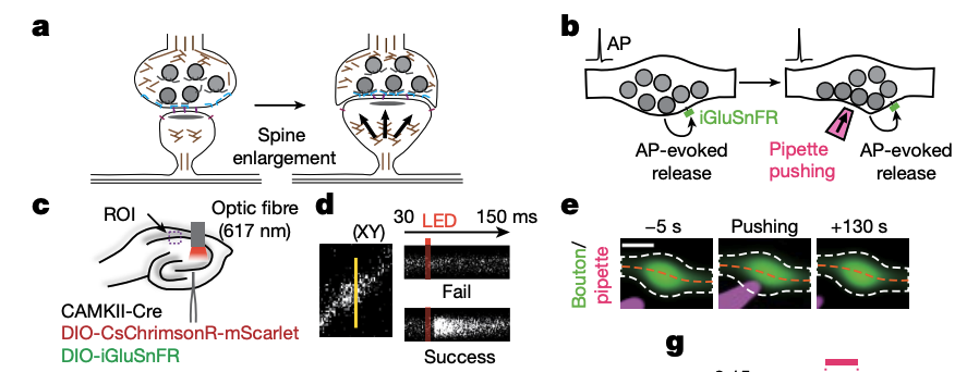
What do Figures 1A–C show?
A: The hypothesis can spine enlargement mechanically affect a bouton?
B: The experimental design comparing bouton's response to stimulation before and after mechanical pushing.
They wanted to ask a simpler question first (not beginning with spine enlargement): Is mechanical deformation alone sufficient to change presynaptic function?
C: The viral-expression and stimulation setup using CsChrimsonR and iGluSnFR.
CaMKII (CaMKIIα) = protein abundantly expressed in excitatory (glutamatergic) principal neuron
Cre Recombinase = An enzyme derived from P1 bacteriophage that recognizes specific DNA sequences called loxP sites.
Cre-Dependent Viral Constructs = A viral vector (AAV) carrying a gene of interest packaged in a Cre-dependent orientation (often called DIO, Double-Inverted Open Reading Frame).
In the virus, the target gene is initially flipped backwards or blocked by a "STOP" cassette so it cannot be expressed on its own.
When the virus infects a cell that also contains Cre recombinase (supplied by the CaMKII-Cre system), the Cre enzyme flips or excises the sequence into the correct forward orientation.
Consequently, the payload gene is expressed exclusively in CaMKII-positive excitatory neurons.

What do Figures 1D–G show?
D: iGluSnFR line scans distinguish detectable glutamate-release successes from failures.
success when the fluorescence signal exceeded three standard deviations above baseline within 20 milliseconds after stimulation.
E: Images show the bouton being physically pushed by the pipette.
Provides visual evidence that the pipette physically deforms the bouton and that the deformation is reversible.
F: Fluorescence profiles quantify the change in bouton width thru change in FWHM
Black: Before pushing.
Magenta: During pushing.
Cyan: After pushing.
G: Bouton width decreases during pushing and recovers after the pipette is withdrawn.
Shows that fine mechanical deformation is reversible, whereas the functional effects shown later last considerably longer
Interpretation: The manipulation physically deforms the bouton, but the deformation is reversible.
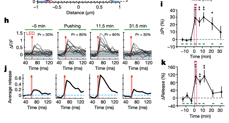
What do Figures 1H–K show?
H: Individual iGluSnFR traces show more frequent detectable responses after pushing.
the increase in release probability persisted long after (11.5 mins) the mechanical manipulation had ended.
I: Quantification shows increased release probability that persists for more than 20 minutes (combined multiple bouton data)
This result indicates that fine mechanical pushing can produce a long-lasting increase in the probability of evoked glutamate release.
J: Average fluorescence traces show increased glutamate-dependent responses.
Imagine two situations:
In the first, glutamate release occurs on 30% of trials.
In the second, glutamate release occurs on 80% of trials.
Even if the size of each successful event were unchanged, the second situation could produce a larger average response across all trials because successful responses occur more often.
K: Quantification shows the change in average response magnitude over time.
The manipulation is followed by an increase in the average glutamate-dependent fluorescence response, and this effect persists for several minutes.
Interpretation: Mechanical pushing enhances evoked glutamate release, and the effect lasts longer than the physical deformation.
What is the key conclusion of Figure 1?
Fine mechanical pushing increased evoked glutamate release by approximately two- to threefold, with effects lasting more than 20 minutes.
Why important? The persistent effect suggests that pushing triggers a longer-lasting functional change rather than simply causing a temporary change in bouton shape.
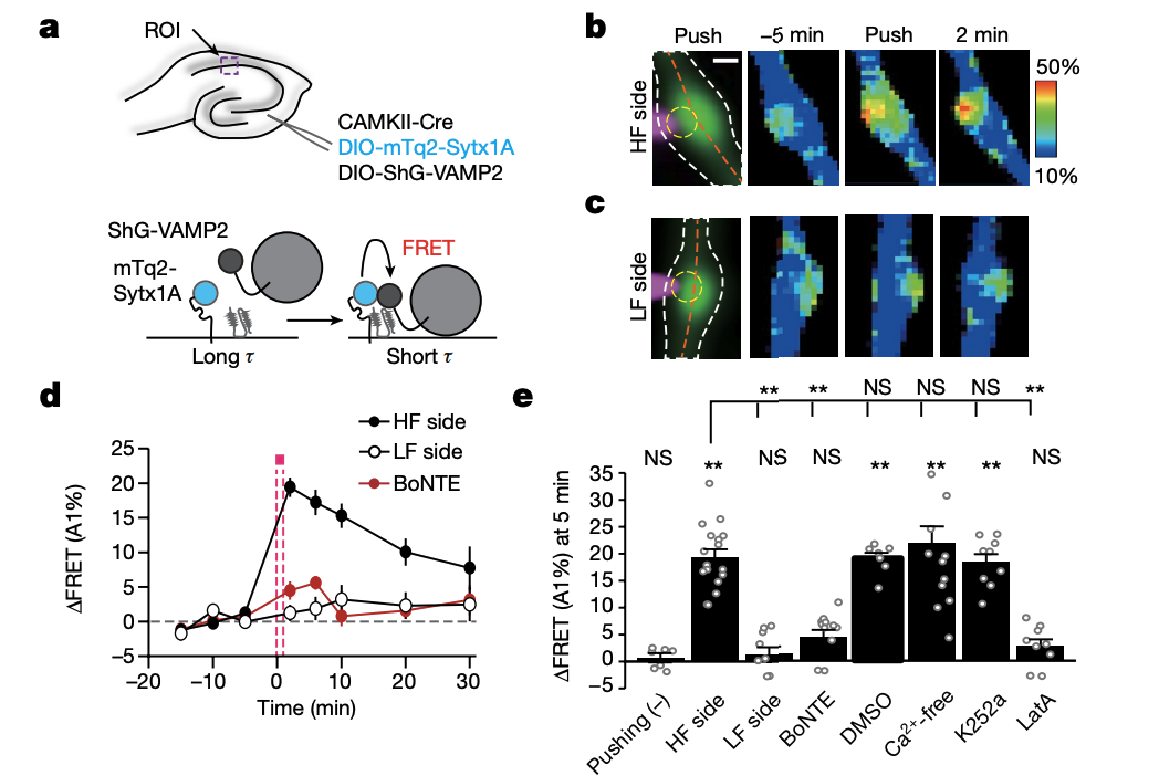
Figure 2A–E: How did researchers test whether mechanical pushing affects SNARE assembly?
A: Shows the viral-expression setup and FRET–FLIM probe design, using labelled syntaxin-1A and VAMP2 to examine SNARE-associated interactions.
B–C: FRET increased when the bouton was pushed from the higher-FRET side, but not from the lower-FRET side.
HF side = areas where energy transfer between syntaxin1a and vamp2 is high
D: Quantifies this response and shows that BoNTE, which disrupts SNARE function, prevents the normal FRET increase.
E: Compares pushing with controls and tests the effects of calcium removal, kinase inhibition (K252a), and actin-polymerization inhibition (LatA).
Meaning: Mechanical pushing can increase SNARE-associated FRET, but the response depends on where the force is applied and is sensitive to disruption of SNARE function.

Figure 2F–H: What factors contribute to the pushing-induced FRET response?
F: The FRET increase persists in calcium-free conditions.
G–H: Latrunculin A (LatA) blocks the response, whereas calcium removal and K252a do not.
The measured response does not require extracellular calcium or the kinase activity inhibited by K252a under these conditions, but it depends on actin polymerization. The effect also persists for over 20 minutes.

Figure 3A–E: Does sucrose treatment enhance glutamate release?
A: Shows the experimental setup using optogenetic stimulation and iGluSnFR to measure glutamate release.
B–C: Individual traces and quantification show increased release probability during and after application of 20 mM sucrose.
D–E: Average iGluSnFR traces and their quantification show increased glutamate-dependent response magnitude.
Meaning: A brief, modest hyperosmotic challenge enhances evoked glutamate release, with effects lasting after the sucrose application ends.
Why 20 mM? Under the tested conditions, it enhanced evoked release without itself triggering detectable miniature excitatory postsynaptic currents.

Figure 3F–I: Does sucrose also affect SNARE assembly?
F: FRET increases after 20 mM sucrose application.
G–H: LatA and BoNTE prevent the normal FRET response.
I: Quantification confirms that the increase occurs in the control condition but not with BoNTE or LatA.
Meaning: The sucrose-induced response involves SNARE-associated interactions and depends on functional SNARE machinery and actin polymerization.
Osmotic pressure reproduces important effects of mechanical pushing on both evoked glutamate release and SNARE-associated FRET.
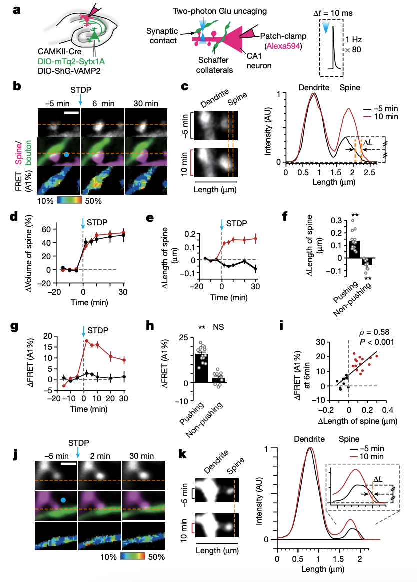
Figure 4A–F and J–K: How did researchers compare pushing and non-pushing spine–bouton pairs?
A: Shows the experimental setup and STDP protocol, which combines two-photon glutamate uncaging and a spike-timing-dependent plasticity (STDP) protocol to induce spine enlargement while monitoring SNARE-associated FRET in the bouton.
B–C: Images and measurements show spine elongation toward the bouton, alongside an increase in bouton FRET, in a pushing pair.
(B) Visually — When the spine elongated and pushed the bouton, the bouton's FRET signal increased.
(C) Quantifiably — The spine extended toward the bouton, supporting the idea that it physically deformed the presynaptic terminal.
D–F: Compare spine enlargement and length changes between pushing and non-pushing pairs.
(D = spine volume over time in pushing vs. nonpushing)
Pushing spines elongated, whereas non-pushing spines did not show the same elongation.
(F = spine length six minutes after STDP stimulation)
Pushing spines showed significant elongation, while non-pushing spines showed a significant decrease in length.
J–K: Show a non-pushing example and its spine-length measurements.
(J) Visually — Spine enlargement without the same physical pushing did not produce the strong FRET increase observed in panel B.
(K) Fluorescence profiles of the non-pushing spine before and after stimulation confirms it did not significantly elongate toward the bouton.
Meaning: The researchers compared pairs in which the enlarged spine physically pushed the bouton with pairs in which it did not, testing whether enlargement alone was sufficient.

Figure 4G–I: What happened when an enlarged spine pushed the bouton?
G–H: FRET increased in boutons pushed by elongating spines, but the increase was not statistically significant in the non-pushing group.
(G) FRET increased strongly when spines pushed boutons, but showed little change in the non-pushing group.
(H) The pushing group showed a significant FRET increase; the non-pushing group did not show a statistically significant increase relative to baseline.
I: shows relationship between spine length change and presynaptic FRET change.
Greater spine elongation was positively correlated with greater FRET increases.
Meaning: The results support the idea that physical pushing by an enlarging spine increases presynaptic SNARE-associated FRET.
The authors also report enhanced evoked glutamate release when spine enlargement pushes the bouton.
Postsynaptic structural change → mechanical force on the bouton → enhanced presynaptic response.