Cell Bio Lab #1 - Microscopy - Review
 Call Kai
 Learn
Learn Practice Test
 Spaced Repetition
 Match
Match Flashcards
 Knowt Play
1/34

There's no tags or description
Looks like no tags are added yet.
Name | Mastery | Learn | Test | Matching | Spaced | Call with Kai | Chat |
|---|
No analytics yet
Send a link to your students to track their progress
35 Terms
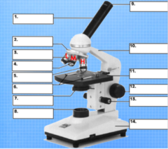
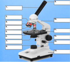
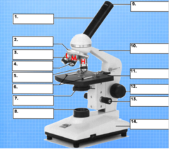
Body tube
Separates objective and eyepiece, allows alignment of optics, and brings the image to your eye
What is #1, and what is its function?

Revolving nosepiece
Holds objective lenses and is rotated to switch between them
What is #2, and what is its function?

Low objective
Likely to provide 4x magnification of the specimen. When combined with the eyepiece, it produces 40x magnification and is used to see larger structures
What is #3, and what is its function?

Medium objective
Likely to provide 10x magnification of the specimen. When combined with the eyepiece, it produces 100x magnification and is used to see specimen components closer than the low objective, but still zoomed out enough compared to the high objective
What is #4, and what is its function?

High objective
Likely to provide 40x magnification of the specimen. When combined with the eyepiece, it provides 400x magnification and provides more magnification than the medium objective, but not enough as an oil-immersion objective lens.
What is #5, and what is its function?

Stage clips
Holds the slide in place
What is #6, and what is its function?
Condenser
Focuses/concentrates light onto the specimen
What is #7, and what is its function?

Light source (lamp)
Illuminates the specimen and allows light to pass through it, enabling an image to be produced
What is #8, and what is its function?

Eyepiece
The lens the viewer looks through to see a specimen which also provides 10x magnification
What is #9, and what is its function?

Arm
Connects the body tube to the base of the microscope and acts as a structure to carry the scope by
What is #10, and what is its function?
Stage
The flat platform that supports the slide
What is #11, and what is its function?

Fine adjustment
Brings specimen into sharp focus under low power and used for all focusing with high power objectives
What is #12, and what is its function?
Coarse adjustment
Moves the stage up and down to bring the specimen into focus
What is #13, and what is its function?

Base
Bottom of the microscope used for support
What is #14, and what is its function?

Bright field microscope (lab - light compound microscope)
This type of microscope is (1) the most widely used, (2) it forms its image when light is transmitted through the specimen, (3) the specimen produces an image that is darker than the surrounding illuminated field, and (4) it can be used with live, unstained and preserved, stain specimens
Phase contrast microscope
This type of microscope is (1) used for live specimens, (2) the specimen is contrasted against a gray background, and (3) it is excellent for internal cell details
Differential-interference microscope
This type of microscope (1) allows for detailed view of live, unstained specimens, (2) it includes two prisms that add contrasting colors to the image, and (3) the image is colorful and three-dimensional
Fluorescence microscope
This type of microscope (1) includes a UV radiation source and a filter that protects the viewer's eyes, (2) it is used with dyes that show fluorescence under UV rays, (3) it forms a colored image against a black field, and (4) it is used in diagnosing infections caused by specific bacteria, protozoa, and viruses using fluorescent antibodies
Confocal microscope
This type of microscope (1) allows for viewing cells art higher magnifications using a laser beam of light to scan various depths in the specimen, and (2) it is most often used on fluorescently stained specimens.
Electrons
This energy source used in microscopy (1) travels in wavelike patterns 1000x shorter than visible light waves, (2) it increases resolving power tremendously, (3) allows for magnification between 5000x and 1000000x for biological specimens, and (4) allows scientists to view the finest structure of cells
Transmission electron microscope (TEM) (up to 100,000x)
This type of microscope (1) is often used to view structures of cells and viruses, (2) it transmits electrons through the specimen, (3) the specimen must be very thin and stained to increase image contrast, and (4) the dark areas of this microscope's image represents thicker or denser parts.
Scanning electron microscope (SEM) (up to 650,000x)
This type of microscope (1) creates an extremely detailed 3D view of all kinds of objects, (2) its electrons bombard the surface of a whole metal-coated specimen, (3) electrons deflected form the surface are picked up by detectors(s), (4) the electron pattern is displayed as an image on a television screen, (5) contours of specimens resolved with this are very revealing and surprising, and (6) the images produced from this may be computer enhanced to give them color.
Actin
This is a structural component of the cells involved in forming the contractile filaments of muscle cells and is a critical feature responsible for movement in other cells
Breast epithelial (MCF-10A) and breast cancer (MDA-MB-231) cells
What types of cells did we examine in this experiment?
Excitation
This is an increased state of energy compared to a molecules ground state which causes it to absorb a certain wavelength of light
Emission
This occurs when an excited molecule returns to its ground state and releases a wavelength of light that can be detected
Fluorescence
This is the emission of light by a substance that has absorbed light or other electromagnetic radiation
Fluorophore
This type of molecule can absorb and emit photons with a range of wavelengths (often in the excitation and emission spectra). These become excited, absorbing a certain wavelength of light, and when the molecule goes from its excited state to its ground state, this emits a different wavelength of light.
Photobleaching
This is when a fluorophore loses its fluorescence due to damage induced by light (often is white light)
Fluorophores have specific excitation or emission spectra which determine the intensity and production of fluorescence
Excitation is similar to the specific wavelengths the molecules can absorb, and emission refers to the specific wavelengths of light that are released. The spectra between the two corresponds with certain wavelengths of light
What do you understand by excitation or emission spectra in fluorescence microscope?
1. A phase contrast microscope uses different phases that light passes through unlike basic microscopy
2. Phase contrast microscopy uses a condenser annulus, a -90° phase shift ring, and a gray filter ring. These components are not present in basic light microscopes.
3. Phase contrast is used for live specimens, for internal cell details, and if it is against a gray background - basic microscopy can have dead, live, stained specimens and views them against a bright background
How is phase contrast microscopy different from basic microscopy?
DAPI stains DNA and can pass through cell membranes. DAPI is a fluorescent stain that binds to DNA and makes it appear blue under UV light, creating an image depicting the stained DNA under the microscope
What is the use of DAPI in fluorescent microscopy?
Paraformaldehyde has the purpose of fixing cells to the plate
Why was paraformaldehyde used in the protocol?
Triton-X-100 is a detergent for permeabilizing the cells so that they are able to accept the dye. This allows dye to enter the cells, stain them, and be visible under UV light
Why was triton-X-100 used in the protocol?
PBS - phosphate buffer saline
What buffer solution was used in the experiment?