1.2 and 1.3 - Cytology
 Call Kai
 Learn
Learn Practice Test
 Spaced Repetition
 Match
Match Flashcards
 Knowt Play
1/70

There's no tags or description
Looks like no tags are added yet.
Name | Mastery | Learn | Test | Matching | Spaced | Call with Kai | Chat |
|---|
No analytics yet
Send a link to your students to track their progress
71 Terms
TEM
What type of microscopy is this?

SEM
What type of microscopy is this?
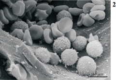
Light microscopy
What type of microscopy is this?

E-face
face of the plasma membrane that faces the extracellular matrix
P-face
face of the plasma membrane that faces the cytoplasm
Passive transport
doesn't require energy
Active transport
movement against the concentration gradient that requires the input of energy
vesicular transport
transportation of material into or out of the cell membrane by bound vesicles
Exocytosis
release of material via vesicular transport. Vesicles fuse with the plasma membrane and release cellular products
Exocytosis
What is this an example of?

Regulated secretion
vesicles congregate near plasma membrane awaiting a signal to release into the extracellular space
mast cells
Example of a regulated secretion
Constitutive secretion
continuous secretion that doesn't need a signal leading to a lack of accumulation around the membrane
Plasma cells
Example of constitutive secretion
Endocytosis
uptake of material via vesicular transport
Plasma membrane
During endocytosis, what folds to form the vesicle containing the inbound material
Pinocytosis, phagocytosis, and receptor mediated endocytosis
3 types of endocytosis
pinocytosis
cellular drinking found in all cells. drinks the water and small dissolved solutes using pinocytotic vesicles
giving water to lysosomes and maintaining cell size
2 important functions of pinocytosis
pinocytosis
What is this an example of - endothelial cells lining capillaries and sucking fluid from the lumen to be used by the cell

Phagocytosis
cellular eating by engulfing large particles, cell debris, and bacteria
pseudopodia
extensions of a cell that is phagocytizing that engulf material
phagosomes
The vesicles created by pseudopodia after engulfing material
give to lysosome or present to immune cell
2 things that a phagocytotic cell can do with the material it collects
Phagocytosis of a macrophage
What is this a picture of

Receptor mediated endocytosis
when a ligand/molecule binds to its certain receptor on the cell surface and is then brought into the cell
Heterochromatin
A

nuclear pores
B
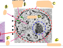
Nuclear envelope
C

chromatin
D - all the stuff in the middle (general)

nucleolus
E

Euchromatin
F

Euchromatin
uncoiled DNA undergoing active transcription that is electron lucent
Heterochromatin
condensed DNA that is inactive and is electron dense
1
Which cell is more active

Ribosomes
translate mRNA and synthesizes proteins
cytoplasmic proteins
What type of proteins do free ribosomes make
secretory proteins
what type of proteins to ER-bound proteins make
rER
interconnected, flattened membranous sacs for protein synthesis
ribosomes
What can be found on the outside surface of the rER
cisterna
the inner lumen of the rER
cisterna
where do the rER ribsosomes deposit the proteins
rER
What is this a picture of

making secretory proteins
what could be the function of this cell

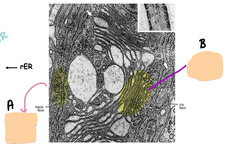
Golgi apparatus
the UPS of the cell responsible for receiving and shipping proteins synthesized by the rER
cis face
the face of the golgi that faces the rER
trans face
the face of the golgi that faces the membrane
Golgi apparatus
B

rER
A
Lysosomes
Little packages of whatever enzyme the cell may need that can process and degrade its contents
True
T/F - lysosomes will fuse with autophagosomes, endocytotic vesicles, and occasionally phagosomes
mitochondria
site of ATP synthesis used for chemical conversion
Smooth and folded
What are the 2 membrane types of the mitochondria
cristae
Inner layer of MOST mitochondria that is shelf-like
vesicular mitochondria
rare mitochondria that has tubular folds instead of cristae
steroid hormone producing cells
In what type of cells can vesicular mitochondria be found
vesicular mitochondria
Identify this photo
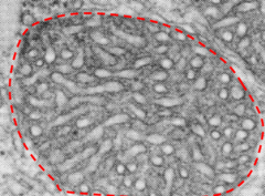
Mitochondria
Identify this photo

smooth ER
interconnected membrane-closed tubules that synthesize steroid hormones and lipids and is present in ALL cells
rER
top arrow

smooth ER
bottom arrow

cytoskeleton
upholds the cell shape, functions in motility, and interacts with the ECM
Actin
the smallest filament that helps anchor cells to eachother and the ECM. Can also help with movement
Actin
Identify this picture

intermediate filaments
Filament that functions in cell-cell and cell-ECM adhesion and is very strong. Can be made up of a variety of materials (ex. keratin, neurofibrils)
intermediate filaments
identify this yellow arrow

microtubules
the largest cytoskeletal component that provides rigidity, aids in intracellular transport, and aids in movement of the cell, cilia, and flagella
a lumen
When looking at actin and microtubules, what makes it a microtubule
microtubules
Identify this photo

Inclusions
Additional molecules or substances found within the cytosol that are not membrane bound
Glycogen, pigment, and lipids
3 examples of inclusions that may be found in the cytosol