Eye
 Call Kai
 Learn
 Practice Test
 Spaced Repetition
 Match
 Flashcards
 Knowt Play
1/205

There's no tags or description
Looks like no tags are added yet.
206 Terms
What components make up the fibrous outer coat?
Sclera
Cornea
What structures make up the uvea?
Iris
Ciliary body
Choroid
What is the neurosensory retina?
The retina that lines the vitreous chamber and sits on the retinal pigment epithelium (RPE)
What structures bound the anterior chamber?
Cornea anteriorly
Trabecular meshwork laterally
Iris posteriorly
Where is the posterior chamber located?
Between the iris and lens
What is the normal pathway of aqueous humor?
Pars plicata of the ciliary body → posterior chamber → around lens → through pupil → anterior chamber → trabecular meshwork → Schlemm canal
What happens when aqueous humor outflow is impaired?
Intraocular pressure (IOP) increases, potentially producing glaucoma
Why does an increase in orbital contents cause proptosis?
The orbit is relatively closed:
medially
laterally
and posteriorly
so increased orbital contents push the eye forward
What are some consequences of proptosis?
Eyelids may not completely cover the eye
Tear-film distributed unevenly
Chronic corneal exposure
Predisposes to:
Corneal ulceration
Infection
Axial vs Positional proptosis
What is axial proptosis?
Displacement of the eye straight forward
Usually from a mass within the muscle cone
Most common:
Optic nerve glioma
Optic nerve meningioma
What is positional proptosis?
Displacement of the eye in a direction OTHER than straight forward
Ex. Lacrimal gland neoplasm or inflammation displaces the eye inferiorly and medially
What causes proptosis in Graves disease?
Accumulation of extracellular matrix proteins within the rectus muscles
Accompanied by variable degrees of fibrosis
How does Graves disease (thyroid ophthalmopathy) produce proptosis?
Enlarged extraocular muscles increase orbital contents and push the eye forward
How can sinus infections spread to the orbit?
The orbital floor forms the roof of the maxillary sinus
The medial orbital wall separates the orbit from the ethmoid sinuses
What types of uncontrolled sinus infection can spread to the orbit?
Acute bacterial infection
Fungal infection
What type of tumors predominate among primary orbital neoplasms?
Vascular tumors
Capillary hemangioma
Lymphangioma
Cavernous hemangioma
What is the typical age group for capillary hemangioma of the orbit?
Infants and young children
(Unencapsulated)
What is the typical demographic for cavernous hemangioma?
Adults
How does a cavernous hemangioma differ from capillary hemangioma?
Cavernous hemangiomas are typically encapsulated and occur in adults
What is a key feature of lymphangioma?
It is unencapsulated
What are the basic components of the eyelid?
Skin externally
Mucosa/conjunctiva on the surface facing the eye
What is the function of structures within the eyelid?
They contribute to formation of the tear film
What is a chalazion?
A chronic inflammatory granulomatous lesion caused by obstruction of structures at the eyelid margin
Result in lipid extravasation into surrounding tissue
Extravasated lipids provoke a granulomatous inflammatory response
What is the most common eyelid malignancy?
Basal cell carcinoma
Favors:
Lower eyelid
Medial canthus
What is the characteristic behavior of eyelid BCC?
It is locally very invasive
What are the common presentations of sebaceous carcinoma of the eyelid?
May form a mass that mimics chalazion
May diffusely thicken the eyelid
Where does sebaceous carcinoma initially spread?
To the parotid and submandibular lymph nodes
How can sebaceous carcinoma spread locally?
It can spread through the
Conjunctival epithelium
Epidermis
into the lacrimal drainage system and nasopharynx
What eyelid/conjunctival tumor is associated with AIDS?
Kaposi sarcoma
What are the three histologically distinct zones of the conjunctiva?
Palpebral conjunctiva
Bulbar conjunctiva
Conjunctival fornix
What is the palpebral conjunctiva?
The conjunctiva lining the inner surface of the eyelid
What is the bulbar conjunctiva?
The conjunctiva covering the anterior sclera up to the limbus
What is distinctive about the conjunctival fornix cell types?
It contains pseudostratified columnar epithelium rich in goblet cells
Why is the conjunctival fornix important in viral conjunctivitis?
It contains numerous lymphoid cells involved in the immune response
VERY prominent
What type of primary lymphoma most commonly develops in the conjunctival fornix?
Marginal zone B-cell lymphoma
What systemic disease can produce granulomas in the conjunctival fornix?
Sarcoidosis
How can ocular sarcoidosis be diagnosed in the conjunctival fornix?
Conjunctival biopsy can demonstrate granulomas
What is a pterygium?
A submucosal elevation of fibrovascular connective tissue
Caused by chronic actinic/sun damage that grows onto the cornea
What is a pinguecula?
A small yellowish submucosal elevation of the conjunctiva
Associated with chronic sun exposure that does NOT invade the cornea
Benign
What is the major distinction between pterygium and pinguecula?
Pterygium → grows onto cornea
Pinguecula → does NOT invade cornea

What can occur if a pinguecula produces an uneven tear film?
Focal dehydration can produce a dellen
A saucer-like corneal depression

Why should excised pterygia be submitted for pathology?
Precursors to:
Squamous cell carcinoma (SCC)
Melanoma
(can occasionally be present)
Typically NOT a threat to vision
What is the usual resolve of bacterial and viral conjunctivitis?
Most cases resolve without sequelae
Which organism can cause significant conjunctival scarring?
Chlamydia trachomatis
Where do squamous and melanocytic conjunctival neoplasms tend to arise?
At the limbus
What is the precursor lesion to conjunctival squamous cell carcinoma?
Conjunctival intraepithelial neoplasia (CIN)
Analogous to CIN of the cervix — Cervical intraepithelial neoplasia
What is the common presentation of conjunctival nevi?
Common neoplasm
Seldom invade the cornea

What is characteristic of compound conjunctival nevi?
They characteristically contain subepithelial cysts lined by surface epithelium
What precursor lesion can give rise to conjunctival melanoma?
Arises through an intraepithelial phase called primary acquired melanosis with atypia

How should pigmented conjunctival lesions be regarded clinically?
They warrant evaluation for:
Melanoma, or
A melanoma precursor
Where can conjunctival melanoma metastasize?
Parotid lymph nodes
Submandibular lymph nodes
Approximately 25% are fatal
What is the sclera primarily composed of?
Collagen, with relatively few blood vessels and fibroblasts
Wounds and surgical incisions heal POORLY
What conditions can produce blue sclera?
Scleritis
Staphyloma
Osteogenesis imperfecta
Congenital melanosis oculi
What are common causes of scleritis?
Autoimmune disease
Infection
Injury
Idiopathic

What is staphyloma?
Scleral thinning/ectasia associated with very high intraocular pressure
Lined by uveal tissue

Why does osteogenesis imperfecta produce blue sclera?
Defective type I collagen produces a thin sclera through which underlying choroidal veins can be seen

What is congenital melanosis oculi?
A heavily pigmented congenital nevus of the underlying uvea
What forms the major refractive surface of the eye?
The cornea and its overlying tear film
NOT the lens
What causes myopia (nearsightedness)?
The eye is too long for its refractive power
The most common refractive error
What causes hyperopia (farsightedness)?
The eye is too short
What does LASIK accomplish?
It surgically reshapes the cornea to alter its refractive properties
What does “LASIK” stand for?
Laser-assisted in situ keratomileusis
What are the five major layers of the cornea?
From anterior to posterior:
Epithelium — Rests on BM
Bowman layer
Stroma
Descemet membrane
Endothelium
What is the Bowman layer?
An acellular layer beneath the epithelial basement membrane
Acts as a barrier between:
Malignant epithelial cells
Stroma
Where does calcium deposit in calcific band keratopathy?
In the Bowman layer
What is a histologic hallmark of keratoconus involving Bowman layer?
Breaks in the Bowman layer
Why is the corneal stroma transparent?
It lacks blood vessels and lymphatics
Its collagen fibers are precisely organized
What can cause vascularization of the corneal stroma?
Chronic edema
Inflammation
Scarring
What is Descemet membrane?
The basement membrane of the corneal endothelium
What deposits in Descemet membrane in Wilson disease?
Copper → Produces the Kayser-Fleischer ring

What is the characteristic location of granulomatous inflammation in chronic herpes simplex keratitis?
The Descemet membrane
What happens in Fuchs endothelial dystrophy?
Primary loss of endothelial cells causes corneal/stromal edema
What infections commonly produce corneal ulceration?
Various pathogens, particularly:
Herpes simplex virus (HSV)
Herpes zoster
What is the histologic hallmark of chronic herpes simplex keratitis?
Granulomatous inflammation of Descemet membrane
How can chronic proptosis predispose to corneal ulceration?
Incomplete eyelid coverage causes chronic corneal exposure
Promotes ulceration and infection
(Overall chronic corneal exposure predisposes to ulceration/infection)
How do corneal degenerations and dystrophies differ in laterality?
Degenerations — May be unilateral or bilateral
Dystrophies — Typically bilateral
How do corneal degenerations and dystrophies differ in inheritance?
Degenerations — Generally nonhereditary
Dystrophies — Hereditary
How do corneal degenerations and dystrophies differ in the layers affected?
Degenerations — Varies by disorders
Dystrophies — May affect selective layers or be distributed through multiple layers
What are some examples of corneal degenerations?
Band keratopathies
Keratoconus
What are some examples of corneal dystrophies?
Fuchs endothelial dystrophy
Stromal dystrophies
What are the two forms of band keratopathies?
Calcific band keratopathy
Actinic band keratopathy
What is calcific band keratopathy?
Calcium deposition in the Bowman layer
What causes actinic band keratopathy?
Chronic exposure to high levels of UV light
What happens to the superficial corneal collagen in actinic band keratopathy?
It develops solar elastosis
In the sun-exposed interpalpebral fissures
Why can actinic band keratopathy be mistaken for pinguecula?
Sun-damaged collagen may develop a yellow appearance
“Oil-droplet keratopathy”
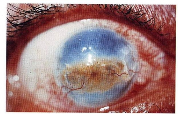
What is keratoconus?
Progressive thinning and ectasia of the cornea
WITHOUT inflammation or vascularization
What shape does the cornea take in keratoconus?
A conical shape
Rather than spherical shape

What visual problem does keratoconus cause?
Astigmatism
Often difficult to correct with spectacles
What other disorders are associated with keratoconus?
Down syndrome
Marfan syndrome
Atopic disorders
What are the major histologic hallmarks of keratoconus?
Corneal thinning
Breaks in the Bowman layer
What is the primary defect in Fuchs endothelial dystrophy?
Loss of corneal endothelial cells, resulting:
Stromal edema
Thickening
What is one of the principal indications for corneal transplantation in the US?
Fuchs endothelial dystrophy
What are the manifestation results from endothelial cell loss in Fuchs dystrophy?
Stromal edema
Bullous keratopathy
Why does Fuchs dystrophy impair vision?
Stromal edema makes the cornea less transparent
Chronic corneal edema can promote vascularization
What is stromal dystrophies?
Stromal deposits that generate discrete opacities
May compromise vision
What can deposits in stromal dystrophies lead to?
Deposits may cause painful epithelial erosions
Scarring of the Bowman layer may further impair vision
Where is aqueous humor produced?
The pars plicata of the ciliary body
Where does aqueous humor drain?
Through the trabecular meshwork at the anterior chamber angle
What is the pathway of how the aqueous humor enters?
Enters the posterior chamber → Bathes the lens → Circulates through the pupil into the anterior chamber
What can impaired aqueous humor outflow cause?
Can raise intraocular pressure