(A&P 1) Lab 9: Spinal Cord, Spinal Nerves and Reflex Physiology
 Call Kai
 Learn
 Practice Test
 Spaced Repetition
 Match
 Flashcards
 Knowt Play
1/29

There's no tags or description
Looks like no tags are added yet.
30 Terms
Where does the spinal cord arise from, what canal does it pass through, and where does it end?
It arises from the brainstem at the foramen magnum of the skull, passes through the vertebral canal, and extends as far as the inferior margin of the first lumbar vertebra (L1)

How many pairs of spinal nerves are there, and where do they exit the vertebral column?
There are 31 pairs of spinal nerves:
8 cervical (C1–C8), 12 thoracic (T1–T12), 5 lumbar (L1–L5), 5 sacral (S1–S5), and 1 coccygeal (Co).
The first pair passes between the skull and vertebra C1, while the rest pass through the intervertebral foramina.
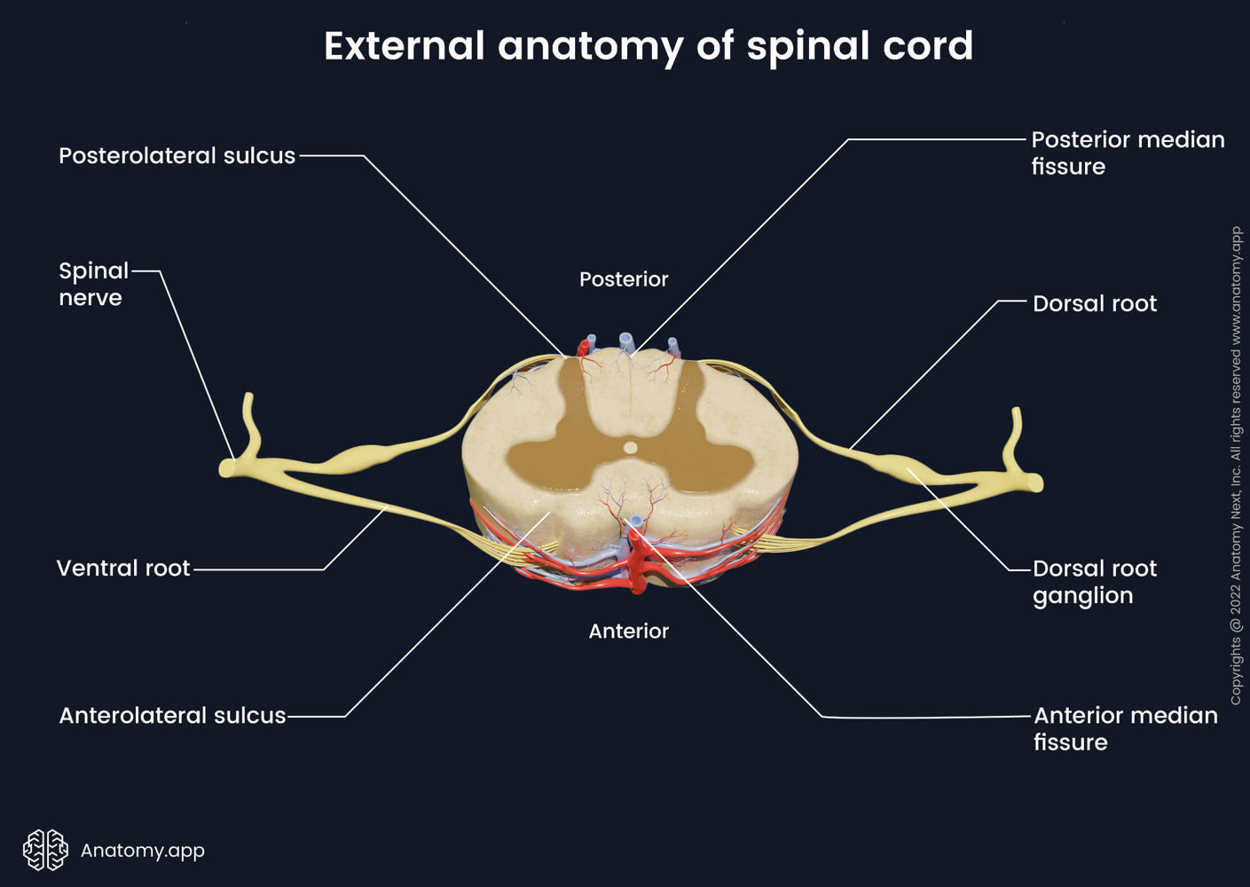
What are the two longitudinal grooves on the anterior and posterior sides of the spinal cord?
The anterior median fissure (a deeper groove on the ventral side) and the posterior median sulcus (a shallower groove on the dorsal side).

What are the four main anatomical regions of the spinal cord?
Cervical, thoracic, lumbar, and sacral regions.

What are the two thickened enlargements of the spinal cord, what vertebral levels do they span, and what do they innervate?
• Cervical enlargement (C4–T1): Gives rise to nerves of the upper limbs.
• Lumbar enlargement (T11–L1): Issues nerves to the pelvic region and lower

Define the medullary cone (conus medullaris), cauda equina, and filum terminale.
• Medullary cone (conus medullaris): The tapered inferior end of the spinal cord proper.
• Cauda equina: The bundle of dorsal and ventral nerve roots occupying the vertebral canal from L2 to S5 that innervates the pelvic organs and lower limbs.
• Filum terminale (terminal filum): A strand of pia mater extending beyond the medullary cone that fuses with the dura mater at S2 (within the lumbar cistern) to form the coccygeal ligament.

Describe the three spinal meninges and their associated spaces from superficial to deep.
• Epidural space: between dural sheath and vertebral bones; occupied by blood vessels, adipose tissue (fat), and loose connective tissue.
• Dura mater: Tough, collagenous membrane that forms the dural sheath.
• Subdural Space: Narrow space beneath the dura mater.
• Arachnoid mater: Simple squamous epithelium (arachnoid membrane).
• Subarachnoid space: Gap between the arachnoid membrane and pia mater filled with cerebrospinal fluid (CSF).
• Pia mater: Delicate, transparent membrane that closely follows the contours of the spinal cord.

Contrast the composition and function of gray matter and white matter in the spinal cord.
Gray matter: Contains little myelin and is made up of neuron somas, dendrites, and proximal axons.
neural integration (synaptic contact)
White matter: Contains an abundance of myelin; composed of bundles of axons (tracts)
carry signals from one part of the CNS to another.
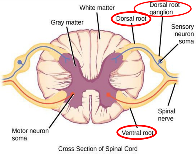
What is the functional difference between the posterior (dorsal) root, dorsal root ganglion, and anterior (ventral) root/horn?
• Posterior (dorsal) root: Carries afferent sensory nerve fibers into the spinal cord.
• Dorsal (posterior) root ganglion: Swelling on the dorsal root containing the somas (cell bodies) of sensory neurons.
• Anterior (ventral) horn & root: contains the large somas of somatic motor neurons, whose axons exit via the anterior (ventral) root to carry efferent motor signals.
Differentiate between ascending tracts and descending tracts.
Ascending tracts carry sensory (afferent) information up the spinal cord to the brain.
Descending tracts conduct motor (efferent) impulses down the spinal cord from the brain.

Define decussation, contralateral, and ipsilateral in relation to spinal tracts.
• Decussation: The crossing over of nerve fibers from one side of the central nervous system to the other.
• Contralateral: When the origin and destination of a tract are on opposite sides of the body (due to decussation).
• Ipsilateral: When the origin and destination of a tract are on the same side of the body (no decussation).

Describe the three connective tissue wrappings of a peripheral nerve from deep (innermost) to superficial (outermost).
• Endoneurium: Thin sleeve of loose connective tissue (over a basal lamina) surrounding each individual nerve fiber (axon).
• Perineurium: Sheath wrapping a bundle of nerve fibers called a fascicle.
• Epineurium: Outer fibrous coat wrapping several fascicles together to compose the nerve as a whole.

Why are most spinal nerves classified as "mixed nerves"?
Because they consist of both sensory (afferent) fibers and motor (efferent) fibers, conducting signals in two directions.

What branches does a spinal nerve split into after exiting the intervertebral foramen?
It splits into a posterior (dorsal) ramus, an anterior (ventral) ramus, a meningeal branch, and communicating rami

What virus causes chickenpox and shingles, where does it remain dormant, and how does it cause a shingles outbreak?
Pathogen: Varicella-zoster virus.
Dormancy: After childhood chickenpox, the virus hides for life in the posterior (dorsal) root ganglia.
Reactivation: If immune system weakens in adulthood, the virus travels via fast axonal transport to cause shingles

What is Shingles?
A painful trail of skin discoloration and fluid-filled vesicles along the nerve's dermatome.

Name the five major nerve plexuses and their general functions.
Cervical plexus (neck)
Brachial plexus (near shoulder)
Lumbar plexus (lower back)
Sacral plexus (pelvis)
Coccygeal plexus.
They carry somatosensory signals (touch, heat, cold, stretch, pressure, pain), proprioception (body position/movement for balance), and motor commands
Define a reflex and list its four main properties.
A reflex is a quick, involuntary, stereotyped reaction of glands or muscles to stimulation.
1. Requires stimulation: Not spontaneous; it is a response to a sensory stimulus.
2. Quick: Involves few or no interneurons and minimum synaptic delay.
3. Involuntary: Occurs without intent, often without awareness, and is difficult to suppress.
4. Stereotyped: Occurs in essentially the same predictable way every time.
Differentiate between an inborn (intrinsic) reflex and a learned (acquired) reflex.
• Inborn (intrinsic) reflex: Unlearned and involuntary motor response built into our neural anatomy (regulated by the brainstem and spinal cord).
• Learned (acquired) reflex: Results from practice or repetition (driving a car).
List the five components of a Reflex Arc in order from stimulus to response.
1. Receptor: Site of stimulus action.
2. Sensory Neuron: Transmits afferent impulses to the CNS (via the dorsal root).
3. Integration Center: Synapse(s) within the CNS gray matter—can be a monosynaptic reflex (direct synapse between sensory and motor neuron) or polysynaptic reflex (involves one or more interneurons).
4. Motor Neuron: Conducts efferent impulses from the integration center to the effector (via the ventral root).
5. Effector: Muscle fiber or gland that responds to the impulse.
Contrast Somatic Reflexes, Autonomic Reflexes, and Spinal Reflexes
• Somatic reflexes: Activate skeletal muscle.
• Autonomic reflexes: Activate visceral effectors (smooth muscle, cardiac muscle, or glands).
• Spinal reflexes: Somatic reflexes mediated directly by the spinal cord (clinically assessed via stretch, flexor, and superficial reflexes).

Define Spinal Shock
Occurs when the spinal cord is transected, immediately depressing all functions controlled by the cord below the injury.

What are muscle spindles?
Muscle spindles are stretch receptors in skeletal muscles that monitor muscle length using intrafusal (gamma) and extrafusal (alpha) fibers.

How does the Stretch Reflex work?
When a muscle is suddenly stretched (tapping the patellar tendon in the patellar/knee-jerk reflex), it reflexively contracts, increases tone, and feels stiffer to help maintain equilibrium and posture.

What are Golgi tendon organs?
Tendon organs (Golgi tendon organs) are proprioceptors located in a tendon near its junction with a muscle that measure tension.

How does the Tendon Reflex work?
The tendon reflex responds to excessive tension on a tendon by moderating (inhibiting) muscle contraction before the force tears the tendon.
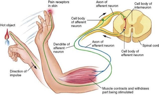
Contrast the Flexor (Withdrawal) Reflex and the Crossed Extension Reflex.
• Flexor (withdrawal) reflex: The quick contraction of flexor muscles resulting in the withdrawal of a limb from an injurious/noxious stimulus (e.g., touching a flame or stepping on glass); an ipsilateral reflex.
• Crossed extension reflex: The simultaneous contraction of extensor muscles in the limb opposite from the one that is withdrawn (contralateral), extending that limb to support body weight and maintain balance.

How is the Plantar Reflex tested?
Elicited by drawing a blunt object downward lateral aspect of the plantar surface of the foot, tests the integrity of the spinal cord from L4–S2 and the corticospinal tracts
What is the difference between a normal response and Babinski's sign? (in Plantar Flexion)
Normal response: Downward flexion (curling) of the toes.
Abnormal response (Babinski's sign): Dorsiflexion (upward extension) of the great toe and lateral fanning of the smaller toes

How is the Abdominal Reflex tested, what spinal levels does it assess, and what is the expected response?
Elicited by stroking the skin of the lateral abdomen above the umbilicus; checks the integrity of the spinal cord and ventral rami from T8–T12 and the corticospinal tracts.
Normal response is the contraction of abdominal muscles causing the umbilicus (navel) to move toward the stimulated site.