2: Epithelial & Connective Tissue, Integumentary System
 Call Kai
 Learn
Learn Practice Test
 Spaced Repetition
 Match
Match Flashcards
 Knowt Play
1/75

There's no tags or description
Looks like no tags are added yet.
Name | Mastery | Learn | Test | Matching | Spaced | Call with Kai | Chat |
|---|
No analytics yet
Send a link to your students to track their progress
76 Terms
epithelium (epithelia)
covering + lining
covers exposed body surfaces + protects from external environment
ex. epidermis
lines internal body cavities, hollow organs, organ passageways for absorption/secretion
ex. GI + respiratory tract lining
glandular
collections of ____ cells that secrete substances into ducts or bloodstream
ex. sweat glands, sebaceous glands, salivary glands, pancreatic glands
covering/lining epithelium
type of epithelium
has adjacent cells attached via cell junctions
has 3 cell surfaces
lateral - side surface
apical - free surface
basal - bottom surface
has basement membrane, separating from connective tissue
basal lamina (facing upwards)
reticular lamina (facing downwards)
avascular - relies on diffusion from underlying loose connective tissue
cell junctions
composed of specialized transmembrane proteins; 3 types:
tight
gap
anchoring
epithelial membrane
epithelial layer + underlying connective tissue layer
consists of:
epithelial layer
basement membrane
connective tissue layer
types:
mucous
serous
cutaneous (skin)
basement membrane
barrier separating epithelial layer from underlying connective tissue layer
top layer facing epithelium = basal lamina
bottom layer facing connective tissue = reticular lamina
layer to which epithelial cells can attach + anchor
tight junctions
bind cells together to form leak-proof sheets
happens closer to apical surface
found on lateral surface of cells

gap junctions
allow communication b/w adjacent cells via open pore

anchoring junctions
prevent cells from being pulled apart + anchors cells to underlying membrane
functions via 3 different proteins
desmosome
adherens
hemidesmosome
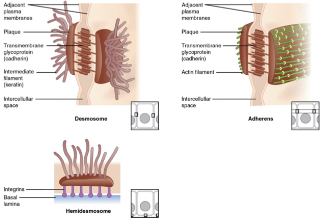
desmosome
forms anchoring junctions
transmembrane proteins attach like velcro to e/o, connecting cells
pass through PM
proteins further anchored to intermediate filaments inside cells

adherens
forms anchoring junctions
come all around cell + interact w/ e/o to keep cells together

hemidesmosome
forms anchoring junctions
looks like half of a desmosome
found on basal surface of cells
anchors cells to basement membrane
Velcro-like pieces reach down into basement membrane
protein itself is anchored to cell

simple epithelium
1 layer of epithelial cells
designed for exchange of materials
all cells anchored to basement membrane
classified as:
squamous
cuboidal
columnar (ciliated or non-ciliated)
pseudo stratified columnar (usually ciliated)
pseudostratified epithelium
1 layer of epithelial cells but looks like multiple
designed for exchange of materials + protective barrier
nuclei are scattered + cells are bunched together
stratified epithelium
multiple layers of epithelial cells
designed for protective barrier
only deepest layer anchors to basement membrane
classified as:
squamous
cuboidal (uncommon)
columnar (uncommon)
transitional
squamous epithelium
flat-shaped epithelial cells
designed for rapid exchange of materials
cuboidal epithelium
cube-shaped epithelial cells
designed for production, secretion, absorption of materials
columnar epithelium
column-shaped epithelial cells
designed for production, secretion, absorption of materials
simple squamous
single layer of flat cells
all cells in contact w/ basement membrane
ex. in lung alveoli → permits diffusion of gases b/w alveoli + pulmonary capillaries
simple cuboidal
single layer of cube-shaped cells
nuclei centrally located
most of digestive system lined w/ this
ex. pancreatic duct lining → secrete H2O + bicarbonate
simple columnar
single layer of rectangular-shaped cells
can be non-ciliated (small intestine epithelium) or ciliated (oviduct epithelium)
nuclei located near basal surface of cell
often features goblet cells
goblet cells
modified columnar cells that produce mucus
mucus forms layer on apical surface
lubricates, moistens, protects epithelium
pseudostratified columnar
single layer of cells that appear stratified
nuclei located at different levels
all cells attached to basement membrane BUT not all reach apical surface
largely found in respiratory tract
ex. lining trachea → prepares air for gas exchange in lungs
stratified squamous
layers closes to apical surface = squamous
deeper cell layers = cuboidal or columnar
can be keratinized or non-keratinized
keratinized = outside of body = protection
non-keratinized = inside of body (still has keratin, just not as much)
transitional epithelium
type of stratified epithelium
apical layer changes shape depending on organ state
stretched = squamous (flat)
relaxed = cuboidal (square)
ex. epithelium lining urinary bladder, ureter
gland
single cell or group of epithelial cells that:
secrete substances into ducts or onto surface = exocrine (outside/indirect)
secrete substances directly into bloodstream = endocrine (inside/direct)
exocrine pancreas
aka pancreatic acini
secrete digestive enzymes into ductal system → empties to 2nd part of duodenum
endocrine pancreas
aka islet of Langerhans
collection of cells that secrete hormones that regulate blood glucose homeostasis
hormones deposited into interstitium + enter blood vessels directly
main cells:
alpha = secrete GLUCAGON → ⬆ blood glucose
beta = secrete INSULIN → ⬇ blood glucose
connective tissue
functions in structural support + packaging
abundant + widely distributed throughout body
greater space b/w cells compared to epithelial tissue
components:
several cell types → subset responsible for maintaining ECM
varying amounts ECM → determines quality/consistency
protein fibers
ground substance
does not occur on free surface
vascular + innervated except for cartilage
connective tissue classification
connective tissues
connective tissue proper → packaging, repair, protection
loose
dense
fluid connective tissue → transportation
blood
lymph
supporting connective tissue → structure, framework
cartilage
bone
connective tissue cells
type caries according to connective tissue type
fibroblasts
macrophages
plasma cells
mast cells
adipocytes
fibroblasts
cell found in connective tissue
numerous - in many connective tissue types
synthesize + secrete:
proteins → assembled into long fibres (ex. collagen)
ECM ground substance components (ex. proteoglycans)
adipocytes
cell found in connective tissue
triglycerides stored in large fat droplet
nucleus + organelles pushed to side
extracellular matrix (ECM)
material b/w cells in connective tissues
composition varies b/w different tissues
ex. bone tissue → calcium phosphate (hydroxyapatite)
ex. tendon/ligament → tightly packed collagen fibres
composed of:
protein fibers
collagen
elastic
reticular
ground substance
H2O + large organic molecules
hydroxyapatite (mineral deposition) in bone
collagen fibers
found in ECM of connective tissue
most abundant protein in body (~25%)
type I and II most common
synthesized by fibroblasts
form long fibres → arranged in bundles
provide tensile strength + flexibility
scurvy
condition due to vitamin C deficiency
vitamin C required for collagen synthesis
deficiency affects areas w/ high collagen turnover
causes teeth loss → periodontal ligament holds teeth in place
causes poor wound healing → collagen needed to form scars
elastic fibers
found in ECM of connective tissue
thin, wavy fibres consisting of protein elastin + glycoprotein fibrillar
provides elasticity
can be stretched + returns to normal when relaxed
reticular fibers
found in ECM of connective tissue
thin fibres (type III collagen) that form interwoven, branched network
synthesized by reticular cells (fibroblast)
forms framework (storma) → strengthens, supports soft organs, ex. liver, spleen
ground substance
non-fibrous material b/w cells + protein fibres in connective tissue
contains
water
proteoglycans
complexes of polysaccharides (ex. glycosaminoglycans - GAG) + proteins
have (-) charge → attracts cations, ie. Na+
Na+ pulls H2O w/ it
proteoglycans trap + hold H2O in matrix
forms gel-like, viscous substance
provides resistance to compressive forces
connective tissue proper
classified based on [protein fibres] relative to ground substance
loose
dense
loose connective tissue
type of connective tissue proper
loosely packed = ⬇ protein fibers + ⬆ ground substance
functions in protection, support, insulation, energy storage
types:
areolar
adipose
reticular
dense connective tissue
type of connective tissue proper
densely packed = ⬆ protein fibers + ⬇ ground substance
functions in structure, support, withstanding tensile strength
types:
regular
irregular
elastic
areolar connective tissue
type of loose connective tissue proper
widely distributed throughout body
provides strength + support
has all 3 protein fibres
collagen
elastic
reticular
viscous ground substance
ex. connective tissue underlying epithelium
lamina propria of mucous membranes
superficial dermis of skin
adipose tissue
type of loose connective tissue proper
found within areolar connective tissue
comprised of adipocytes
highly vascularized
needs vascular network to get energy molecules out from stored triglycerides
ex. surrounding organs → protection, thermal insulation, energy storage
reticular connective tissue
type of loose connective tissue proper
contains network of interwoven reticular fibres
ex. in lymph nodes → filters lymph + provides supporting framework
dense regular connective tissue
type of dense connective tissue proper
bundles of collagen fibres arranged in parallel
flattened fibroblasts
sparse ground substance
able to withstand tensile/pulling stress in one direction
ex. tendons, ligaments
dense irregular connective tissue
type of dense connective tissue proper
sheets of closely packed, irregularly arranged collagen fiber bundles
able to within stand pulling forces in many directions
ex. fibrous sheets surrounding organs, fibrous joint capsule, deep dermis of skin
elastic connective tissue
type of dense connective tissue proper
densely packed elastic fibres (appear wavy)
allows stretching + recoil back to original shape
ex. aortic walls → stretching when heart contracts + recoil when heart relaxes
membrane
flat sheets of tissue covering body surface/structure or lining body cavity/hollow organ
2 types:
epithelial
connective tissue
connective tissue membrane
consists of connective tissue layer
no epithelial layer, ex. synovial membrane
mucous membrane (mucosa)
type of epithelial membrane
lines hollow passageways in body that are directly open to external environment
ex. respiratory tract, GI tract
epithelial layer:
type of epithelium varies w/ location
basement membrane
connective tissue layer
lamina propria = areolar connective tissue layer
respiratory mucosa
type of epithelial mucous membrane
epithelial layer
pseudostratified ciliated columnar
cells anchored to basement membrane
goblet cells secrete mucous
precent apical surface from drying
traps fine particulates in incoming air
connective tissue layer
lamina propria = areolar connective tissue
has blood vessels, lymphatics, nerves
support/anchor for epithelial layer
protection for underlying structures
serous membranes (serosa)
type of epithelial membrane
lines internal body cavities + covers visceral organs within body cavities
epithelial layer
simple squamous epithelium = mesothelium
secretion of serous fluid into cavity → ⬇ friction between/w organs, prevents adhesion
basement membrane
connective tissue layer
areolar connective tissue
two layers w/ cavity in b/w:
parietal
visceral
parietal layer
type of serous membrane (epithelial membrane)
outer layer
adheres to body cavity wall
visceral layer
type of serous membrane (epithelial membrane)
inner layer
adheres to organ surface
layers of skin
epidermis
dermis
hypodermis
epidermis
superficial epithelial layer + basement membrane
avascular keratinized stratified squamous epithelium
multiple layers of flattened cells w/ lots of keratin = protective layer
thick + thin skin
thick → has extra layer = stratum corner
cell types:
keratinocytes
melanocytes
Langerhans cells
Merkel cells
keratinocytes
most abundant cells in epidermis (90%)
produces:
keratin
tough, fibrous protein that fills cell
lipid-rich lamellar granules
contains water repellent/sealant released into extracellular environment
protects → abrasion, heat, injury, microorganism entry
prevent excessive water loss
fungal infections
affects hair, nails + top layer of skin
fungi survive by feeding on keratin in these areas
make enzymes that digest keratin
melanocytes
comprises ~8% of epidermal cells
found in deepest layer of epidermis = stratum basal
produces melanin
black-brown/red-yellow pigment
contributes to skin colour
absorbs UV light + prevents DNA damage in skin cells
have cytoplasmic extensions → deliver melanin to keratinocytes
melanosomes cluster above nucleus toward skin surface → protect DNA
UV light = ⬆ melanocyte enzymatic activity
melanocytes nevus (mole)
benign tumour of melanocytes
melanoma
superficial spreading → treated by excision
can progress to vertical growth phase into dermis → metastatic potential
grows past basement membrane into dermis = cancerous
ABCDE mnemonic
asymmetry
border irregularities
colour variation
diameter
evolution
langerhans cells (dendritic cells)
type of macrophage
produced in bone marrow → migrates to epidermis (stratum spinosum)
engulfs antigens that invade epidermis
travel via lymphatic vessels to lymph nodes
present antigens to lymphocytes = immune response
merkel cells
least numerous epidermal cell
in stratum basale → deepest epidermis layer
cells contact specialized flattened sensory nerve endings → detect light touch sensations
epidermis cell layers
4 (thin) or 5 (thick) layers w/ keratinocytes in various stages of development:
corneum
lucidum (only in thick)
granulosum
spinosum
basale
stratum basale
deepest epidermal layer w/ only single row of cells
in contact w/ basement membrane
contains:
Merkel cells
melanocytes
basal (stem) cells
divide via mitosis
1 daughter cell pushed up to replace cells shed at apical surface
other daughter cell remains as stem cell
dysplasia of basal cells → basal cell carcinoma
psoriasis
autoimmune condition that stimulates rapid basal cell division = thickening of skin (plaques)
cells shed prematurely → flaky, silver scales at surface
appears red due to underlying inflammation
chronic + recurring
stratum spinosum
8-10 layers of keratinocytes produced by stem cell vision in basal layer
dysplasia of keratinocytes → squamous cell carcinoma
packed closely + anchored together by desmosomes
keratinocytes producing keratin
contains:
melanocyte processes
Langerhans cells
stratum granulosum
3-5 layers of flattened keratinocytes
keratin synthesis complete → cells release lipid-rich granules
cells start to die = apoptosis
organelles degenerate + flatten
stratum lucidum
3-5 layers of flattened, dead keratinocytes
cells filled w/ large amounts of keratin
only in thick skin, eg. fingertips, palms, soles
stratum corneum
15 (face) to 100 (soles) layers of dead keratinocytes
exposed layer → facing external environment
cells continuously shed + replaced by underlying layer
takes 21-30 days for cell to move from stratum basal to top layer
callus
thickening of stratum corneum
form in response to repeated friction (protective)
atopic dermatitis (eczema)
chronic inflammatory skin condition characterized by dry skin, redness, pruritis (itching)
weakness in skin barrier → underlying tissues susceptible to external irritants/allergens
dermis
connective tissue layer in cutaneous membrane
2 layers:
papillary layer
reticular layer
contains:
blood vessels
sensory receptors (nerve endings)
nociceptors = pain/itch
thermoreceptors = temperature
mechanoreceptors = pressure/touch, vibration, stretch
sweat (sudoriferous) + sebaceous (oil) glands
hair follicles
papillary layer
layer of dermis → superficial 1/5
areolar connective tissue
forms fingerlike structures = dermal papillae
extend upwards into epidermis → ⬆ SA for attachment to epidermis for diffusion from capillary loops
contains:
capillary loops → nourish epidermis
Meissner corpuscles → light touch encapsulated receptors
reticular layer
layer of dermis → deep 4/5
dense irregular connective tissue
⬆ collagen + elastic fibers = support, strength, elasticity
contains:
vascular network → supplies capillary loops in papillary region
sensory receptors
Ruffini corpuscles = stretch
Pacinian corpuscles = pressure/vibration
sweat (sudoriferous) + sebaceous (oil) glands
hair follicles
hypodermis
subcutaneous layer (below skin) - NOT part of integument
areolar connective tissue w/ adipose tissue deposits
anchors skin to deep tissues