Exam 2 (Ch.8, 7, 2, 9)
 Call Kai
 Learn
 Practice Test
 Spaced Repetition
 Match
 Flashcards
 Knowt Play
1/87

There's no tags or description
Looks like no tags are added yet.
Name | Mastery | Learn | Test | Matching | Spaced | Call with Kai | Chat |
|---|
No analytics yet
Send a link to your students to track their progress
88 Terms
What is membrane trafficking?
The movement of proteins, lipids, and other materials between different compartments of a cell
Cells have diff organelles that perform specific functions
Proteins & lipids must reach correct organelle to function
Cell uses vesicles to transport many of these materials
What is the Endomembrane System?
A group of membranes and organelles that work together to:
Produce, modify, and transport lipids & proteins
Secrete substances outside the cell (exocytosis)
Bring substances into the cell (endocytosis)
Important organelles
Endoplasmic reticulum (ER)
Golgi
Endosomes
Lysosomes

Endoplasmic reticulum (ER) function
Protein & lipid production
Golgi apparatus function
Protein modification and sorting
Endosomes function
Sort internalized materials
Lysosomes function
Digest and recycle materials
What are vesicles?
A small membrane-bound sac that transports material (protein, lipids, etc) between organelles/compartments
Form by budding from one membrane
Deliver materials by fusing w/ another membrane
How does vesicle transport work?
1) A protein is synthesized in ER
2) Portion of the ER membrane buds outward
3) Vesicle containing the protein separates from ER
4) Vesicle travels to Golgi
5) Vesicle fuses w/ Golgi membrane
6) Protein is delivered to Golgi

Soluble proteins vs. Membrane proteins
Soluble proteins: Located inside vesicle & released into recipient organelle’s interior after fusion
Membrane proteins: Embedded in vesicle membrane and transferred into recipient organelle’s membrane when fusion occurs
Why do vesicles fuse/go through Golgi?
To deliver proteins that either need to function in Golgi or need further modifications before reaching their final destination
1) Golgi req proteins to perform its function
some proteins transported from ER are meant to stay in Golgi
2) Proteins could also need further modifications that take place in Golgi
Proteins synthesized in rough ER may not be fully processed or ready to function, so vesicle transports these proteins from ER to Golgi to perform additional modifications
Directions of vesicle transport/movement
Anterograde: Moves material forward through secretory pathway
ER → Golgi
Golgi → plasma membrane
Retrograde: Moves material backward or returns/recycles material
Golgi → ER
Plasma membrane → endosome
Allows proteins that belong in ER but accidentally travel to Golgi to return to ER
Why do we need both directions?
Proteins must move forward to reach their destinations, but certain proteins & membrane components need to be returned so organelles maintain their proper functions
How do scientists study membrane trafficking?
Pulse-chase autoradiography: determines where newly synthesized proteins travel inside the cell
Conclusion: proteins move through secretory pathway from ER to Golgi & then toward plasma membrane
GFP fusion proteins: allows scientists to directly observe protein movement w/out radioactive labeling
Conclusion: ER → Golgi
Cell-free systems
Conclusion: Proteins synthesized by ER-bound ribosomes can enter ER lumen
Mutant screens: scientists mutate genes to determine which proteins are required for membrane trafficking
If a cell produces many vesicles but they cannot fuse w/ their destination, the vesicles accumulate → tells scientists that the mutated protein normally helps vesicles fuse
What is secretion/exocytosis?
Process of releasing large molecules, such as proteins, from inside the cell to outside the cell
Occurs through secretory pathway, which transports proteins through diff organelles before they are released
Main secretory pathway:
Rough ER → Golgi → secretory vesicle → plasma membrane → outside cell
First stop: Endoplasmic Reticulum (ER)
Endoplasmic Reticulum (ER)
The 1st organelle involved in secretory pathway
3 main functions: Synthesizes proteins, begins glycosylation, & performs quality control
1) Protein/lipid synthesis
Rough ER: synthesizes proteins that will be secreted, inserted into membranes, or transported within endomembrane system
proteins synthesized in RER can undergo modifications before being transported to Golgi
Smooth ER: synthesizes lipids
2) Protein glycosylation: modification of proteins by adding several oligosaccharides → proteins become glycoproteins
Helps proteins fold properly
Important for protein quality control
Helps proteins function correctly
3) Protein quality control
ER checks whether newly synthesized proteins are folded correctly
Correctly folded proteins → can leave ER and travel to Golgi
Incorrectly folded proteins → retains in ER for further folding attempts
Proteins that can’t fold correctly may be sent for degradation
What is the secretory pathway?
Transports proteins from rough ER through Golgi to their final destinations aka transports proteins through endomembrane system
some proteins are secreted outside the cell, while others become part of the plasma membrane or are delivered by lysosomes
The process by which proteins are synthesized in rough ER, transported to Golgi for modification and sorting, and then delivered to their final destination
Main pathway:
ER → Transport vesicle → Golgi → Transport vesicle → Final destination/plasma membrane
Proteins may be delivered to:
plasma membrane (*major pathway) → membrane proteins
extracellular environment
lysosomes → digestive enzymes
other endomembrane compartments

Rough ER
Has ribosomes attached to its surface
Responsible for synthesizing proteins destined for secretion, membranes, and endomembrane system
Located near nuclear envelope
Smooth ER
Doesn’t have ribosomes attached
Synthesizes lipids & steroid hormones
Participates in detoxification
Stores Ca2+ ions
Free ribosomes
Located in cytoplasm
Produce proteins that may function in cytoplasm or be imported into organelles such as the nucleus & mitochondria
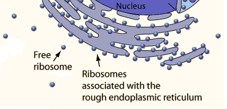
ER-bound ribosomes
Attached to rough ER during translation
Produce proteins that enter endomembrane system, be secreted from the cell, or become membrane proteins
Go to these destinations:
Endomembrane system
ER
Golgi
Vesicles
Plant vacuoles
Secretion
Become integral membrane proteins (TM proteins)

How does a soluble protein know to go to ER?
They contain an ER signal sequence (6-15 hydrophobic amino acids at protein N-term)
acts like zip code directing protein to ER
Signal sequence is bound by Signal Recognition Particle (SRP)
Signal Recognition Particle (SRP)
A molecule that recognizes the ER signal sequence on a growing protein
Steps
1) Translation begins on a free ribosome
2) ER signal sequence emerges from ribosome
3) SRP-GTP recognizes & binds the signal sequence
4) SRP temporarily pauses translation
Pauses translation so ribosome can be directed to ER before protein continues being synthesized
5) SRP brings the ribosome to the ER membrane
6) SRP binds to an SRP receptor
7) The ribosome becomes positioned near a translocon
Translocon: a protein channel in ER membrane
8)GTP hydrolysis helps release SRP which enters translocon (SRP-GTP → SRP GDP)
9) Translation resumes, & the growing protein enters the ER
What is a translocon?
A protein channel in ER membrane
Allows growing proteins to enter ER lumen
Can open to allow the protein to pass through
Can also release hydrophobic transmembrane segments into lipid bilayer
Cotranslation translocation
Process where a protein enters ER while it’s being synthesized (protein doesn’t have to be completely synthesized before entering ER)
Translation & translocation occur simultaneously
Translation: ribosome makes a protein by joining amino acids together
Translocation: growing protein moves into ER through protein channel called translocon
Purpose: Allows a growing, mostly unfolded protein to enter ER through translocon → makes transport easier b/c unfolded proteins can pass through the narrow channel
THEN once inside ER, chaperones help protein fold into its functional 3D shape
Steps:
1) Ribosome begins translation in cytosol
2) SRP recognizes signal sequence & pauses translation
3) SRP directs ribosome to SRP receptor on rough ER
4) Ribosome attaches to translocon, & SRP is released
5) Translation resumes → growing protein enters ER through translocon (translation & translocation occurring simultaneously)
6) BiP & other chaperones help protein fold correctly
7) Signal peptide is often removed by signal peptidase
8)Once translation finishes, a soluble protein can be released into ER lumen

Start-transfer sequence vs. Stop-transfer sequence
Start-transfer sequence: A hydrophobic sequence that initiates movement of a protein through the translocon
Stop-transfer sequence: A hydrophobic sequence that stops movement of the protein into ER lumen & becomes embedded in the membrane
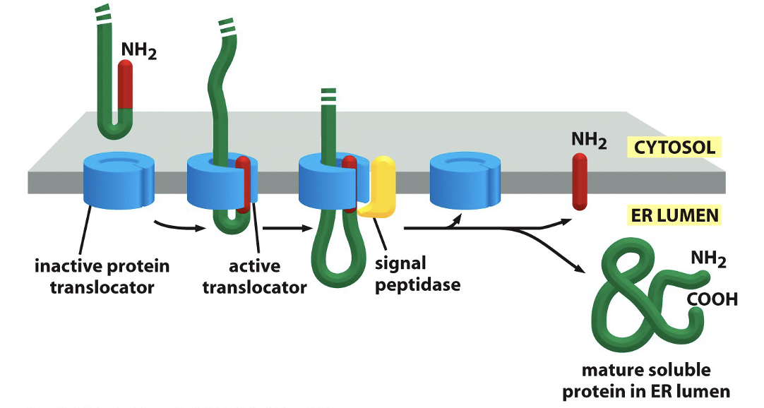
Soluble protein translocation into ER
After proteins are synthesized by ribosomes, they need to enter ER lumen or become embedded in ER membrane
This protein enters ER lumen completely
Steps
1) Protein synthesis begins on ribosome in cytoplasm
2) The protein contains a hydrophobic signal peptide that acts as a start-transfer sequence
It directs the protein to the ER & initiates movement through translocon
3) The growing protein passes through translocon into ER lumen
4) Signal peptidase cleaves the signal peptide during translocatioin
5) The mature protein is released into ER lumen & folds
Single-Pass Transmembrane Protein: Method 1
Purpose: Produce a protein that crosses the ER membrane once
Requires both a start-transfer & a stop-transfer sequence
Steps:
1) Protein synthesis begins in cytoplasm
2) Protein contains an N-terminal signal peptide that acts as a start-transfer sequence
Initiates movement through translocon into ER lumen
3) A second hydrophobic sequence called the stop-transfer sequence reaches the translocon
Stops further movement of the protein into ER lumen
4) The stop-transfer sequence is released sideways from translocon into lipid bilayer
It becomes the transmembrane segment that anchors the protein
5) Signal peptidase cleaves the original signal peptide
6) Translation finishes, leaving the protein embedded in ER membrane

Single-Pass Transmembrane Protein: Method 1 Final Result
N-terminus → ER lumen
C-terminus → Cytoplasm
Stop-transfer sequence → Embedded in ER membrane

Single-Pass Transmembrane Protein: Method 2
Purpose: Produce a single-pass protein using an internal ER recognition sequence
Uses an internal start-transfer sequence that isn’t removed
Steps:
1) Protein synthesis begins in cytoplasm
2) The protein contains an internal hydrophobic signal sequence instead of an N-terminal signal peptide
3) SRP recognizes the internal signal sequence & directs the ribosome to ER
4) The internal signal sequence inserts into translocon and acts as both a start-transfer sequence & a membrane anchor
N-terminal portion remains in cytoplasm
C-terminal portion enters ER lumen
5) Internal signal sequence remains in embedded in ER membrane and is not cleaved

Single-Pass Transmembrane Protein: Method 2 Final Result
N-terminus → Cytoplasm
C-terminus → ER lumen
Internal signal sequence → Embedded in ER membrane

Single-Pass Transmembrane Protein: Method 3
Purpose: Produce a single-pass protein w/ the opposite orientation from Method 2
Uses internal signal sequences like Method 2, but the protein inserts in opposite orientation
Steps:
1) Protein synthesis begins in cytoplasm
2) The protein contains an internal hydrophobic ER recognition sequence
3) Internal sequence directs insertion into translocon
4) Internal sequence inserts in the opposite orientation compared w/ Method 2
N-terminal portion enters ER lumen
C-terminal portion remains in cytoplasm
5) Internal sequence remains embedded in ER membrane and is not cleaved
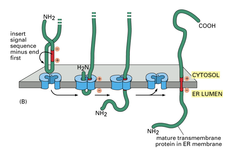
Single-Pass Transmembrane Protein: Method 3 Final Result
N-terminus → ER lumen
C-terminus → Cytoplasm
Internal signal sequence → Embedded in ER membrane

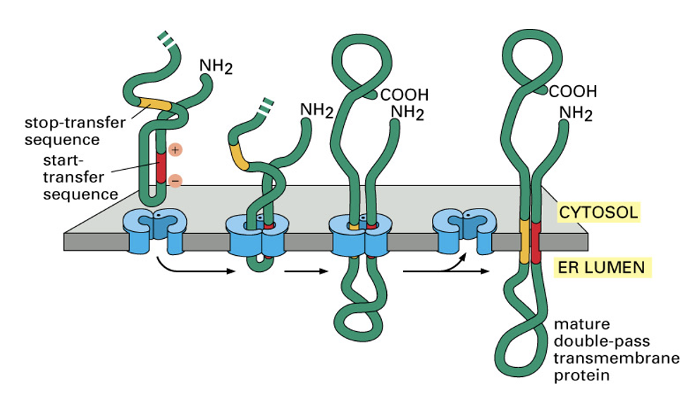
Double-Pass & Multipass Transmembrane Proteins
Double-pass proteins: Cross ER membrane twice
Multipass proteins: Cross ER membrane multiple times
These proteins contain multiple hydrophobic sequence that directs their insertion into membrane
Steps of multipass transmembrane proteins
1) A start-transfer sequence initiates movement of protein into ER
2) A stop-transfer sequence stops movement into ER lumen
3) Another start-transfer sequence can initiate translocation again to enter ER lumen again
4) Another stop-transfer sequence can stop translocation and stop proteins from entering again
5) Repeated start-transfer and stop-transfer sequences creates multiple membrane-spanning regions
Final result: protein crosses ER membrane multiple times
Rough ER…
Makes transmembrane proteins
Determines their orientation in membrane
Prepares them for their functions at plasma membrane
How does rough ER make transmembrane proteins?
TM proteins = proteins that span across phospholipid bilayer
synthesized by ribosomes attached to rough ER
contain 1 or more hydrophobic transmembrane segments that become embedded in membrane
some become receptors on plasma membrane
Steps:
1) A ribosome begins making a protein, which is directed to rough ER
2) The growing protein enters translocon in ER membrane
3) When hydrophobic transmembrane segment reaches translocon, it can move sideways into phospholipid bilayer
4) The hydrophobic segment stays embedded in membrane, anchoring the protein
5) Depending on its amino acid sequence, diff parts of protein face cytoplasm or ER lumen
What determines orientation of TM proteins?
The distribution of charged amino acids surrounding hydrophobic TM segment
More positively charged amino acids side of TM segment remain facing cytoplasm
Opposite side tends to face ER lumen (Note: negatively charged amino acids don’t always have to face ER lumen)
Two ends:
a) N terminus, NH2 (beginning of protein)
b) C-terminus, COOH (end of protein)
When a protein travels from ER to plasma membrane, its orientation is preserved b/c its orientation is established during synthesis in ER
Part facing ER lumen (space inside ER) will eventually face extracellular environment
Part facing cytoplasm (an area inside cell) will remain facing cytoplasm
Example of Proteins Having Diff Orientations: LDL Receptor
LDL: a particle that transports cholesterol & other lipids through blood
LDL receptor: a transmembrane protein that binds LDL & helps bring it into cell through receptor-mediated endocytosis
LDL receptor orientation:
N-terminus has negatively charged aa close by: faces ER lumen during synthesis & eventually faces outside cell
Contains region that binds LDL
C-terminus has positively charged aa close by: faces cytoplasm
Contains signals needed to recruit machinery for endocytosis
TM domain: anchors receptor in membrane
LDL receptor needs its N-terminus outside to bind LDL & its C-terminus inside to help bring LDL into cell
Example of Proteins Having Diff Orientations: Transferrin receptor
Transferrin (TF): A protein that carries iron in blood
Transferrin receptor: a transmembrane protein that binds iron-loaded transferrin & helps bring it into cell through receptor-mediated endocytosis
Transferrin receptor orientation:
N-terminus has positively charged aa close by: faces cytoplasm
Contains signal needed for endocytosis
C-terminus has negatively charged aa close by: faces ER lumen during synthesis & eventually faces outside the cell
Binds transferrin, allowing receptor to capture iron-loaded transferrin
TM domain: anchors receptor in membrane
Transferrin receptor has opposite N/C orientation from LDL receptor, but both perform receptor-mediated endocytosis
How are multiple TM segments inserted?
They cross the membrane several times
The 1st TM segment helps establish orientation
Subsequent TM segments alternate their directions across the membrane
What is protein glycosylation?
The addition of carbohydrate chains (oligosaccharides) to proteins → glycoproteins
Why is it important?
Helps protein fold properly
Supports cell-to-cell interactions
Contributes to protein function
2 types:
a) N-linked: oligosaccharide attached to asparagine (Asn)
Begins in ER, modified further in Golgi
b) O-linked: oligosaccharide attached to serine (Ser) or threonine (Thr)
Occurs in Golgi, modified in Golgi

How does N-linked glycosylation happen in rough ER?
1) Glycosyl transferase (enzyme) helps assemble the branched oligosaccharide containing 3 glucose molecules at its tip → adds sugars
2) Branched oligosaccharide (sugar chain) containing 3 glucose molecules at its tip is attached to asparagine (Asn)
This occurs co-transitionally (while translation is still happening)
Transfer occurs on ER lumen side
3) The sugar chain is modified in ER
Glucose molecules are removed from sugar chain by enzymes
These modifications help the protein fold correctly
4) Protein is transported from ER to Golgi in transport vesicle
In Golgi, additional sugars may be added or removed to further modify the protein
5) Glycosylated protein travels to final destination
Can become a plasma membrane TM protein or a secreted protein
Protein quality control in ER
ER checks whether newly synthesized proteins have folded correctly
Important proteins:
BiP: chaperone that helps proteins fold
Calnexin: chaperone involved in glycoprotein folding
Calreticulin: another ER chaperone
Glucosidases: removes glucose residues from glycoproteins
Steps:
1) A newly synthesized protein enters ER
2) Chaperones help the protein fold
3) ER checks whether the protein is folded correctly
4) If correctly folded, protein can leave ER
5) If incorrectly folded: protein may undergo another folding cycle
6) If protein cannot fold correctly, it may be sent to cytoplasmic proteasome for degradation
What happens when misfolded proteins accumulate in ER?
Response: Unfolded Protein Response (UPR) → a cellular response triggered by accumulation of misfolded proteins in ER
UPR sensors: proteins located in ER membrane that detect problems w/ protein folding
Under normal conditions:
BiP charperone not busy and is available b/c few misfolded proteins in ER
BiP binds UPR sensors & keeps them inactive → UPR is OFF
When misfolded proteins accumulate:
BiP is recruited to help these proteins fold & is busy
BiP dissociates from UPR sensors which makes them become active
UPR is ON and initiate signaling pathways that help cell respond to ER stress
2 responses:
1) Phosphorylate translation factor elF2α to stop translation/production of more proteins
2) Part of cytoplasm is cut, released, & acts as transcription factor that needs to go to nucleus to make more charperone proteins to reinforce proper protein folding
What is the Golgi apparatus?
Responsible for:
Modifying proteins
Processing proteins
Sorting proteins (secretion, PM, lysosomes)
Packaging proteins for transport
Regions of Golgi:
Cis-Golgi Network
Cis-Golgi
Medial-Golgi
Trans-Golgi
Trans-Golgi network
Regions of Golgi: cis-Golgi network
Receives vesicles from ER
Closest to nucleus/ER
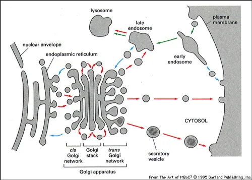
Regions of Golgi: trans-Golgi network
Sorts proteins/vesicles to different destinations
Farthest from nucleus/ER
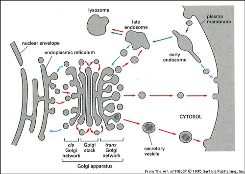
Regions of Golgi: cis, medial, trans-Golgi
Protein processing
cis: early protein processing
medial: further modification
trans: later modification

Protein processing in Golgi: Proteolysis
The cleavage of a protein to make shorter functional proteins
Why? → Some proteins become functional only after being cleaved into smaller pieces
Ex. POMC
One gene produces one transcript
The transcript produces one polypeptide
The polypeptide can be processed into multiple peptide hormones

Protein processing in Golgi: Glycosylation
Adding or modifying sugar chains attached to proteins
Important for:
Protein folding & stability
Cell recognition & cell-cell interactions
Protein trafficking & function
2 types:
N-linked: begins at rough ER, modified in Golgi
O-linked: Begins in Golgi, modified in Golgi
Why are Golgi regions important?
Different regions of Golgi contain different enzymes that carry out different modifications → Golgi functions are regionalized
Proteins progress through these regions:
1) Cis-Golgi: early modifications, including removal of mannose residues
2) Medial Golgi: further mannose removal & addition of other sugars
3) Trans-Golgi: later modifications, including addition of galactose & sialic acid in many glycoproteins
A Glycosylation Pattern: Enzyme α-Mannosidase I
Located in cis-Golgi
Function: Removes mannose residues

A Glycosylation Pattern: Enzyme GlcNAc-transferase I
Located in medial-Golgi
Function: Adds N-acetylglucosamine (GlcNAc)

A Glycosylation Pattern: Enzyme α-Mannosidase II
Located in medial-Golgi
Function: Removes additional mannose residues
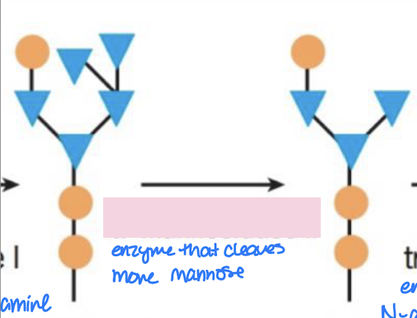
A Glycolsylation Pattern: Enzyme GlcNAc-transferase II
Located in medial-Golgi
Function: Adds another N-acetylglucosamine
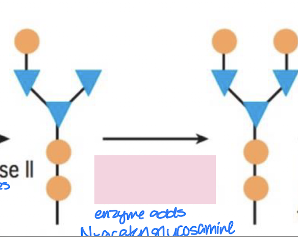
A Glycosylation Pattern: Enzyme Fucosyltransferase
Located in medial-Golgi/trans-Golgi
Function: Adds frucose

A Glycosylation Pattern: Enzyme Galactosyltransferase
Located in trans-Golgi
Function: Adds galactose

A Glycosylation Pattern: Enzyme Sialyltransferase
Located in trans-Golgi
Function: Adds sialic acid

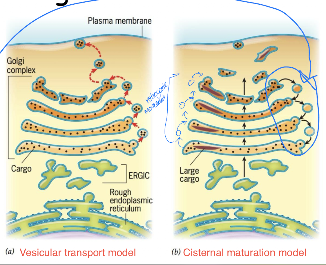
How do proteins move through Golgi?
2 Models:
a) Vesicular transport model
Vesicles carry cargo from one Golgi cisterna to the next
Golgi enzymes remain in their compartments
b) Cisternal maturation model
Cargo remains inside a cisterna as it matures
Golgi enzymes move backward through retrograde vesicles
Note: current evidence supports cisternal maturation model
What is exocytosis?
The release of substances outside cell through vesicle fusion w/ plasma membrane
2 Types:
a) Constitutive secretion
Not regulated b/c cells know its molecules to throw away (excreted products, damaged parts) → doesn’t require a specific release signal
Occurs continuously
b) Regulated secretion
Is regulated → requires a signal
i.e. secretion of neurotransmitters
What is synaptic vesicle exocytosis?
An example of regulated secretion
Steps
1) Synaptic vesicles contain neurotransmitters
2) Vesicles dock near plasma membrane since they need to fuse w/ it and waits for signal to fuse
3) An action potential arrives
4) Voltage-gated Ca2+ channels open
5) Ca2+ enters the cell
6) The signal triggers vesicle fusion
7) Neurotransmitters are released

What are lysosomes?
Digestive organelles
Contain acid hydrolases
Have acidic interior (~ pH 4.6)
Break down old organelles & other materials
Participate in autophagy
Help certain immune cells digest bacteria
Is a secretory pathway destination
How do lysosomal enzymes reach lysosomes?
They follow secretory pathway:
Synthesized in ER → Modified in Golgi → Endosomes → Lysosomes
Why? B/c lysosomal enzymes are proteins that enter endomembrane system, so they must be synthesized, processed, sorted, & delivered to lysosomes
Lysosomes are part of antegrade movement & endocytosis movement
Lysosomal enzymes are translated/synthesized in ER-bound ribosomes
Where are proteins going through endomembrane trafficking system synthesized?
In the rough ER
How are proteins transported through the secretory pathway?
The cell needs several types of proteins to go from Rough ER → Golgi → Transport vesicle → Final destination
Coat proteins: Help membrane bend to form a vesicle & select which cargo goes inside
Small GTPases: Regulate vesicle formation & help coat proteins attach/targetting
Motor proteins: Move vesicles along cytoskeleton toward their destinations
Rab proteins: Help vesicles recognize & target specific compartments
are small GTPases
SNAREs and SNAPs: Help regulate vesicle docking & fusion → brings vesicle & target membranes together so they can fuse
What are the main functions of coat proteins?
2 major jobs:
a) Promote membrane curvature
More coat proteins = more curvature → creates vesicle
b) Select cargo and specifies movement to where
Types of Coat Proteins: COPII
Form around vesicles that go ER → Golgi
Transport type: Anterograde
Helps proteins leave ER
2 Functions:
a) Promote membrane curvature to help form vesicles
b) Select cargo that needs to leave ER
Types of cargo it can transport:
Soluble proteins: proteins located inside ER lumen that need to travel to Golgi
Membrane proteins: proteins embedded in ER membrane that need to be transported
Cargo receptors: Membrane proteins that bind certain soluble cargo molecules & help package them into COPII vesicles
Its coat assembly is regulated by a small GTPase called Sar1

How does COPII coat assembly work?
Sar1: small GTPase that regulates COPII assembly
Sar1-GDP = Inactive
Sar1-GTP = Active
Steps:
1) Sar1 is initially inactive, bound to GDP
Sar1-GDP = primarily soluble in cytoplasm
Not yet recruiting COPII coat proteins
2) GEF activates Sar1
GEF in ER membrane promotes release of GDP
GTP binds to Sar1 → Sar1 becomes active
3) Sar1-GTP becomes embedded in ER membrane
GTP binding causes conformational change in Sar1 which exposes an ampihpathic helix that’s inserts into ER membrane
Active Sar1 can now recruit COPII coat proteins
4) Sar1-GTP recruits Sec23 and Sec24
Sec23/24 form inner COPII coat
Sec24 helps select cargo proteins
Cargo receptors help capture soluble cargo
5) Sec13 and Sec31 are recruited
Sec13/Sec31 form outer COPII coat
Outer coat helps stabilize & shape curved membrane
Developing vesicle bud becomes more pronounced
6) COPII vesicle buds from ER
Vesicle separates from ER membrane and carries selected cargo toward Golgi
Sar1 eventually hydrolyzes GTP to GDP, promoting coat disassembly
Uncoated carrier can then undergo targeting & fusion

Types of Coat Proteins: COPI
Form around vesicles that go: Golgi → ER
a) trans-Golgi → cis-Golgi
b) cis-Golgi → ER
Transport type: Retrograde
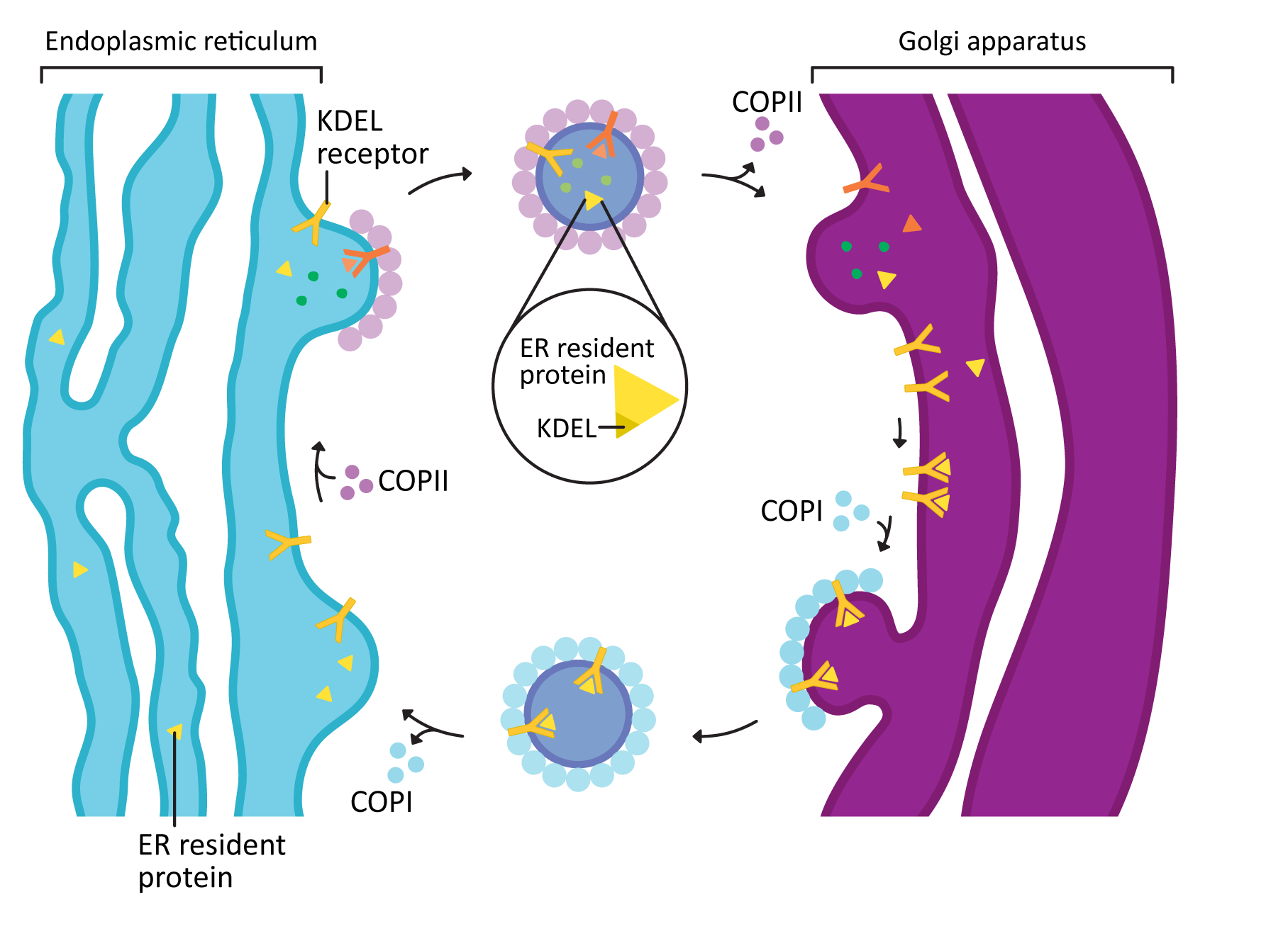
Helps return proteins to ER
Why does the cell need this coat protein?
Some ER resident proteins (that contain KDEL) accidentally enter COPII vesicles
These proteins reach Golgi even though they belong in ER
Cell recognizes them using retrieval signals
KDEL: indicates that protein needs to go back to rough ER
K= lysine
D= Aspartic acid
E= Glutamic Acid
L = Leucine
COPI-mediated transport helps return them to ER

How does COPI return proteins to ER?
An ER-resident protein containing KDEL accidentally reaches Golgi
A KDEL receptor in Golgi recognizes & binds COPI coat
COPI coat machinery is recruited to membrane
The membrane bends & forms a COPI-coated vesicle
The vesicle transports the protein back toward the ER
The vesicle fuses w/ ER membrane, & the protein is released into ER lumen
Types of Coat Proteins: Clathrin
Helps forms vesicles during endocytosis & during sorting of certain proteins toward lysosomal pathway
Transport routes:
trans-Golgi network → Endosome → Lysosome
Plasma membrane → Endosome → Lydosome (sometimes)
Structure:
Forms a 3-legged structure called triskelion
each triskelion contains 3 heavy chains & 3 light chains
many triskelions assemble into a coat that helps curve the membrane
Important for endocytosis & cargo transport toward endosomes

What are lysosomal enzymes?
Organelles that break down & recycle materials inside the cell
Contain digestive enzymes called acid hydrolases which break down protein, lipids, carbohydrates, and other molecules
Are synthesized on rough ER and transported through Golgi
For these enzymes to function in lysosomes, they must be delivered into correct destination
How does the Golgi know which proteins should go to lysosomes instead of being secreted?
Certain lysosomal enzymes in the Golgi are tagged w/ Mannose-6-phosphate
Lysosomal enzymes are glycoproteins, meaning they contain attached carbohydrate chains
Mannose = a sugar within those carbohydrate chains
Golgi adds a phosphate-containing tag to mannose, producing mannose-6-phosphate which identifies the enzyme as cargo destined for the lysosome
How does the lysosomal enzyme get packaged into a vesicle?
1) Mannose 6-phosphate (M6P) receptor binds M6P tag on lysosomal enzyme in trans-Golgi network
2) Arf1 (a small GTPase) binds GTP & becomes active
Active Arf1 associates w/ Golgi membrane & recruits GGA adaptor proteins
3) GGA interacts w/ Arf1-GTP & the cytoplasmic tail of M6P receptor and helps recruit clathrin
4) Clathrin proteins assemble around budding membrane → helps form a transport vesicle containing the lysosomal enzyme & its receptor
5) The vesicle separates from Golgi membrane and travels toward an endosome
Its clathrin coat is removed before vesicle fuses w/ its target memrbane
6) Lysosomal enzymes separate from M6P receptors in endosome
7) M6P receptors recycle back to trans-Golgi network to bind to other M6P-tagged lysosomal enzymes
8)Lysosomal enzymes are delivered to lysosomes
What is an effector for Arf1?
An adaptor protein that recruits the clathrin coat proteins
How do vesicles move?
Vesicles use motor proteins that move along cytoskeletal tracks
Motor protein type changes depending on direction of movement:
Toward microtubule (+) end → carried by Kinesin
Toward microtubule (-) end → carried by Dynein
Vesicle tethering, docking, and fusion
Tethering
Brings a vesicle near its target membrane
Tethers can capture vesicles from approx 50-200 nm away
Docking
Positions a vesicle very close to target membrane
Prepares vesicle for fusion
v-SNAREs (located on vesicle membrane) interact w/ t-SNAREs (located on target membrane)
Their interaction helps position & hold vesicle against target membrane
Fusion
SNARE proteins form a tight complex through their alpha-helical regions
This pulls the two membranes close enough to promote fusion
The vesicle releases its contents into the target compartment

SNARE proteins
Help w/ vesicle docking & membrane fusion
v-SNARE and t-SNARE interact to form a SNARE complex
v-SNARE: located on vesicle membrane
ex. Synaptobrevin
t-SNARE: located in target membrane
ex. SNAP-25 and Syntaxin
SNARE complex helps dock the vesicle & pulls the two membranes together to promote fusion
Bacterial neurotoxins that target SNAREs
Clostridium botulinum and Clostridium tetani = proteases that cleave SNARE proteins
This prevents synaptic vesicle fusion and disrupts neurotransmitter release
What is protein folding?
Proteins are made of chains of amino acids called polypeptides
A polypeptide must fold into a specific 3D shape to function properly
Correctly folded functional shape = native conformation
Protein foldings is influenced by interactions between amino acid side chains (R groups)
Why is protein folding complicated? + Folding Pathways
Scientists don’t completely understand every detail of how proteins fold
Diff proteins can follow diff folding pathways
Secondary structures (α-helices and β-sheets) can form at diff stages of folding
Protein collapse: Unfolded protein becomes more compact as hydrophobic aa side chains tend to move toward inside, away from water
Two possible folding pathways:
Pathway A: Secondary structure forms FIRST
Unfolded protein → Secondary structure forms (α-helices and β-sheets forms) → Protein collapses/becomes compact → Native protein
Pathway B: Protein collapses FIRST
Unfolded protein → Protein collapses/becomes compact → Secondary structure forms (α-helices and β-sheets form) → Native protein

Why do most proteins need help folding?
Main problem: Hydrophobic regions can aggregate which prevents proteins from reaching their correct native conformation
Aggregation: proteins sticking together, often b/c exposed hydrophobic regions interact
How?
Hydrophobic amino acids have non-polar side chains & tend to avoid water
During folding, these hydrophobic regions may be temporarily exposed
Exposed hydrophobic regions can interact with:
Hydrophobic regions of same protein, potentially causing incorrect folding
Hydrophobic regions of other proteins, causing proteins to stick together
What are molecular chaperones?
Proteins that help other proteins fold properly
They bind to & temporarily shield exposed hydrophobic regions of unfolded or partially folded proteins
This prevents unwanted aggregation & allows proper folding to occur
How do they work?
1) A polypeptide is synthesized by a ribosome
2) Hydrophobic regions of the polypeptide become exposed
3) Molecular chaperones bind to these hydrophobic regions
4) This prevents the polypeptide from aggregating w/ other proteins
5) Chaperones release the polypeptide, allowing it to continue folding into its native conformation
Note: Chaperones don’t act as exact molds that determine final shape of proteins, but, instead, help proteins fold by preventing inappropriate interactions
Hsp70 Family of Chaperones
Stands for Heat Shock Protein 70
Is a family of molecular chaperones
Can function co-transitionally (while polypeptide is still being synthesized)
How do they work?
1) A ribosome begins producing a polypeptide
2) Hsp70 binds to exposed hydrophobic regions of the growing polypeptide
This prevents aggregation while translation continues
3) The polypeptide is released & can fold into its native structure

Hsp70 and High Temps
High temps can cause proteins to unfold or become denatured
Denaturation: a protein loses its normal 3D structure
2 Responses to High Temp
a) Promotes misfolding of proteins
b) Promotes transcription from chaperone encoding genes → make more chaperones that will try to fold misfolded proteins properly
This creates process
1) High temp promotes protein misfolding
2) Heat shock promotes transcription of chaperone-encoding genes
3) More chaperone proteins are synthesized
4) These chaperones help protect proteins from aggregation & assist their refolding
What are Chaperonins?
Another type of specialized molecular chaperone
Made up of multiple protein subunits
They form barrel-shaped chambers in which proteins can fold
These chambers help isolate unfolded proteins from other proteins that could cause aggregation
2 examples:
TRiC: a barrel-shaped chaperonin complex
GroEL: a bacterial chaperonin belonging to Hsp60 family
GroEL
Folding chamber
A chaperonin found in bacteria
Belongs to Hsp60 family
Contains 14 polypeptide subunits
These 14 subunits form 2 stacked rings, each containing 7 subunits
The rings form chambers where proteins can fold
GroES
A separate protein complex that acts as a cap for GroEL → cap that closes the chamber
Binds to one end of GroEL complex
Helps enclose the unfolded protein inside the folding chamber
How does GroEL and GroES Help Proteins Fold
1) GroEL binds an unfolded protein
When GroES is not attached, the binding surface inside GroEL is hydrophobic
The hydrophobic surface binds exposed hydrophobic regions of an unfolded protein
This temporarily holds the protein inside the chamber
Why? → Hydrophobic regions of the unfolded protein interact w/ hydrophobic binding surfaces of GroEL
2) ATP and GroES bind to GroEL
ATP binds to GroEL
GroES attaches to GroEL and forms a cap
The unfolded protein becomes enclosed inside chamber
3) GroEL chamber changes shape
When ATP & GroES bind:
GroEL undergoes a conformational change
Chamber enlarges
Chamber’s inner surface becomes hydrophilic
Unfolded protein is released from its interactions w/ chamber wall
4) Protein folding occurs
The protein folds inside enclosed chamber
GroES keeps protein separated from other proteins in cell
ATP is hydrolyzed to ADP during the folding cycle
5) GroES detaches & the protein is released
GroEL/GroES complex undergoes additional ATP-dependent changes
GroES detaches
Protein is released from chamber
Protein may be correctly folded or may still be misfolded
If protein is not correctly folded, it may undergo another folding cycle

Why is correct protein folding important?
Proteins must fold correctly to perform their normal cellular functions
If proteins misfold they …
May lose their normal function
May expose hydrophobic regions that should be normally buried
May aggregate w/ other proteins
Protein misfolding can contribute to disease
Common features of protein folding diseases
Conversion of α-helices into β-sheets → indicates that hydrophobic regions aren’t folded properly & interacts w/ other hydrophobic regions which forms aggregates
