Chapter 9; Anatomy + Physiology
 Call Kai
 Learn
 Practice Test
 Spaced Repetition
 Match
 Flashcards
 Knowt Play
1/69

There's no tags or description
Looks like no tags are added yet.
70 Terms
what 3 prefixes are used to commonly denote muscle
myo-, mys-, and sacro-
what is the cytoplasm in a muscle cell referred to
sarcoplasm
what is the cell membrane of muscle cell referred to
sacrolemma
which muscle tissue types consist of elongated cells (muscle fibers)
skeletal and smooth
(cardiac cells are not muscle fibers)
3 key words used to describe SKELETAL muscle tissue
skeletal, voluntary, striated
SVS; Stop Vans Slowly
3 key words used to describe CARDIAC muscle tissue
cardiac, involuntary, striated
CIS; Cars in Slime
3 keys words used to describe SMOOTH muscle tissue
visceral, involuntary, nonstriated
VIN; Vans in Need
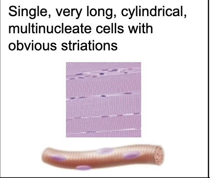
which muscle cells are long, cylindrical, multinucleate, with obvious striations
skeletal muscle cells

which muscle cells are branching chains of cell, uni/binucleate, and have striations
cardiac muscle cells

which muscle cells are single, spindle-shaped, uninucleate and have no nucleus
smooth muscle cells
where is smooth muscle usually found (1 main spot)
hollow visceral organs (stomach/bladder)
also in intrinsic eye muscles + large arteries
what are the 4 functional characteristics shared by all muscle cells
contractility
excitibility
extensibility
elasticity
CEEE
muscle contractility definition
ability of muscle to shorten forcibly
difference between muscle extensibility and elasticity
extensibility; ability to be stretched/extended
elasticity; ability to recoil and resume resting length
4 main FUNCTIONS of muscle tissue in the body
produce movement
maintain body posture/position
stabilize joints
generate heat during contraction
layer of connective tissue sheath that surrounds the entire skeletal muscle organ
epimysium (dense irregular connective tissue)

what structural unit of skeletal muscle is surrounded by the perimysium?
fasicle (group of muscle fibers wrapped in perimysium
discrete bundle of muscle fibers wrapped in fibrous connective tissue)
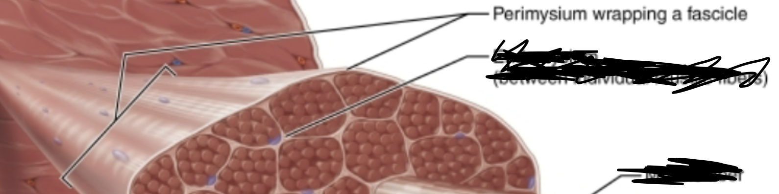
what connective tissue sheath surrounds individual skeletal MUSCLE FIBERS
endomysium
fine areolar connective tissue
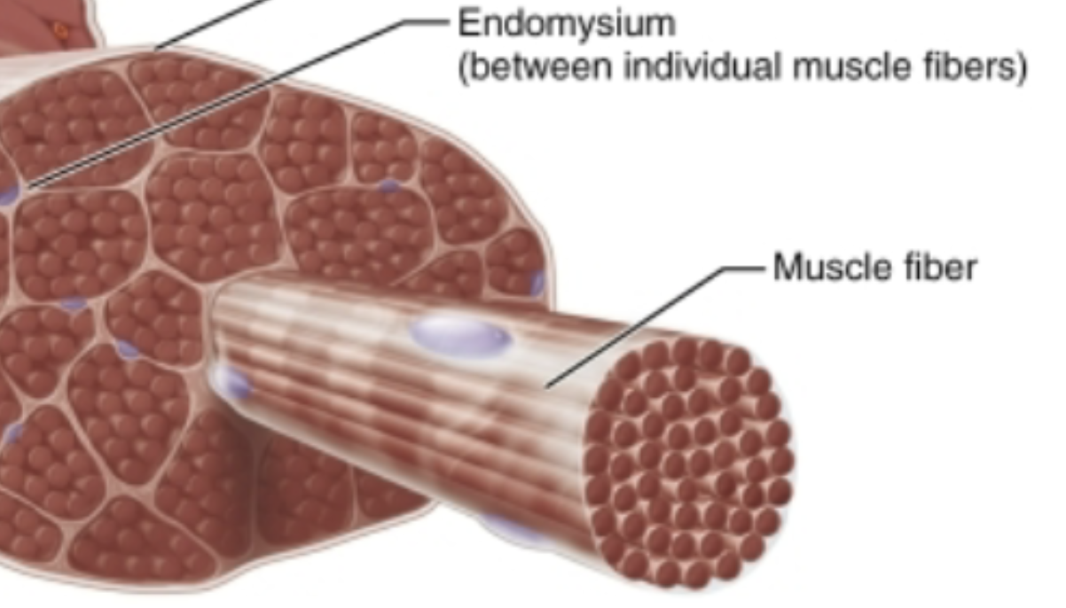
what sheath wraps a fascicle and what sheath wraps the individual skeletal muscle fibers
fascicle; perimysium
muscle fibers; endomysium
direct (fleshy) attatchments vs. indirect attatchments
direct attachments; connection is formed by the epimysium fusing directly to the periosteum of the bone
indirect attatchments; connection is formed by using tendons or sheetlike aponeurosis
in indirection attachments, what terms are used to describe the ropelike and sheetlike structures
ropelike; tendons
sheetlike; aponeurosis
order of structural levels of a skeletal muscle organ from largest to smallest (5 levels)
fascicle
muscle fiber
myofibril
sarcomere
myofilaments
what 2 specialized cellular structures exist in the sarcoplasm of skeletal muscle fibers to store carbohydrates and oxygen?
glycosomes; store carbohydrates
myoglobin; store oxygen
what proportion of total skeletal muscle cell volume is occupied by myofibrils
80%
what creates the visible striations along the length of skeletal myofibrils
repeating series of dark bands (A bands) and light bands (I bands) along the myofibrils

in myofibril striations, what is the lighter zone in the middle of the dark A band called
H zone
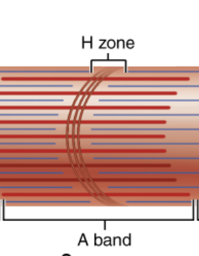
what protein LINE bisects the H zone vertically in the center of the sarcomere
M line
made of myomesin

what is the coin shaped protein sheet that anchors thin thilaments in the midline of a light I band
Z disc

what is the smallest functional contractile unit unit of a skeletal fiber
sarcomere
definition; region of myofibril between 2 Z discs

what are the band components of 1 sarcomere
(what kinds of bands)
1 complete A band
½ of a I band at each end
location of actin (thin) vs. myosin (thick) filaments in a sarcomere
actin (thin); extend across the I band partway into the A band, anchor to the Z discs
myosin (thick): extend the full length of A band and connect to M line
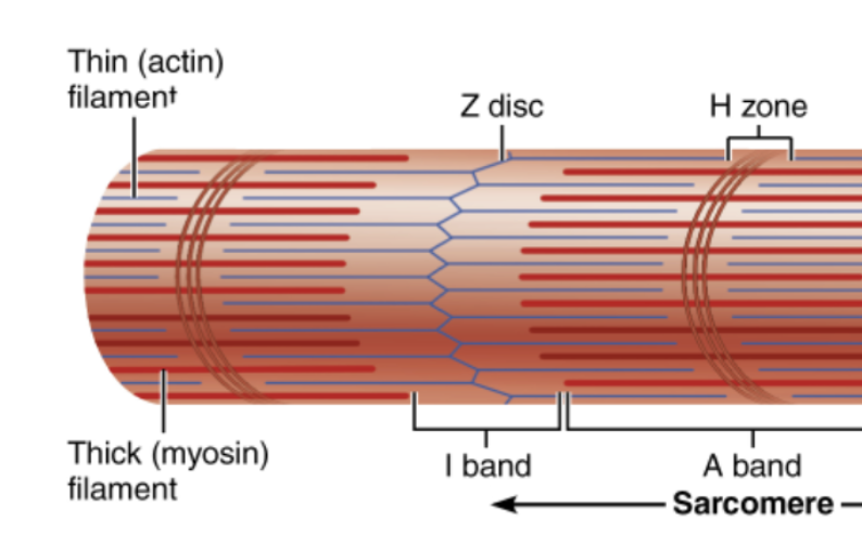
how are thin and thick filaments arranged
hexagonal arrangement
1 thick filament per 2 thin filaments
what polypeptide chains form the tail and globular heads of a myosin molecule
2 heavy polypeptide chains form the myosin tail
4 light polypeptide chains form the 2 globular myosin head
what 2 essential binding sites are on the globular heads of myosin
actin-binding sites
ATP-binding sites
in thick filaments, how are myosin heads arranged along the length of the thick filament
myosin molecules are bundled with their tails intertwined and heads sticking out

how does globular G actin subunits and long fibrous F actin relate in thin filament structure
G actin; link together into long strands of F actin
F actin; 2 F actin strands twist together in helix to form the filaments

what roles do tropomyosin and troponin play in regulating muscle contraction?
tropomyosin; blocks myosin binding sites on actin in relaxed muscle
troponin; binds Ca2+ to move tropomyosin away from active sites
exposes active sites of the myosin and actin for cross bridge formation (contraction)
structural role of elastic filaments made from the protein titin (3 things)
holds filaments in place
alows recoil after stretching
resists excessive stretching
what structural protein links thin actin filaments directly to integral proteins of the sarcolemma
dystrophin
what structural proteins assist in maintaining myofibril alightment + sarcomere stability
(other than titin and dystrophin)
nebulin, myomesin, C proteins
what organelle network surrounds each myofibril and regulates intracellular calcium levels
sarcoplasmic reticulum (SR)
elaborate smooth endoplasmic reticulum that restores + stores Ca2+
what enlarged SR structures form perpendicular cross channels at the A-I band junctions
terminal cisterns
what is a T tubule + what is its main function in skeletal muscle
what it is; deep invagination (intrusion) of the sarcolemma (skeletal muscle plasma membrane) into the cell interior
function; conducts electrical impulses deep into the muscle fiber to reach the myofibril
what 3 membrane structures create a triad
one T tubule with terminal cistern on either side

how do the proteins in the T tubules interact with the sarcoplasmic reticulum to release calcium?
when electrical impulses pass by T tubules, it changes shape
the change in shape causes SR proteins to change shape aswell and release Ca2+ into the sarcolemma
during contraction, how do thin and thick filaments move
during contraction, thin filaments slide past thick filaments
actin and myosin overlap even more
what happens when the nervous system stimulates a muscle fiber
begins the sliding filament model
(excitation contraction coupling)
what structural changes occur to the I bands, H zones, Z discs, and A bands as the sarcomere fully contracts
I bands; shorten
H zones; disappear
Z discs; move closer to eachother 9sarcomere shortens)
A bands; move closer together (maintains constant length
what does the H zone look like in a fully relaxed muscle vs. a contracted one
relaxed; visible in the center of the A band
contracted; thi filaments overlap and the H zone disappears
how is action potential (AP) moved from the neuron (the structure who begins movement) to the muscle cells
neurotransmitter ACh acetylcholine
2 exampels of ion channels
chemically gated; open in response to chemical messengers (ligands)
voltage gated; opened in response to changing membrane potential
what kind of neurons stimulate skeletal muscle fibers to contract
somatic motor neurons
what are axons?
long threadlike extensions of the nervous system that connect to skeletal muscle

what is the name of the region where axons connect to the skeletal muscle fiber
neuromuscular junction

name of the gel-filled space between the axon terminal (end of an axon) from the muscle fiber’s sarcolemma
synaptic cleft

what is the name of the vesicle inside the axon terminal that hold ACh
synaptic vesicle
(active transport)
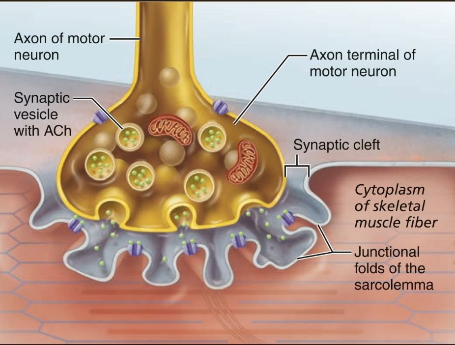
on the sarcolemma, where are the chemically gated ACh recepters on the neuromuscle junction
junction folds

what are the 3 main parts of the neuromuscle junction
axon terminal from the somatic motor neuron
synaptic cleft
junction folds of sarcolemma
what enzyme breaks down acetylcholine and stops its activity in the synaptic cleft
acetylcholinesterase
when ACh binds to its receptor, what is let into the skeletal muscle sarcolemma?
large amounts of Na+ ions
ACh receptors open their gates which lets the Na+ ions in
why does action potential traveling down the T tubules trigger Ca+ to be released
when T tubules sense an electrical charge, they change shape
when T tubules change shape, it causes the sarcoplasmic reticulum to change shape and release Ca+
what are the three main electrical phases that generate an action potential across the sarcolemma?
generation of end plate potential (change in electronegativity)
depolarization (AP generation + propagation b/c Na+ channels)
repolarization (resotration of resting membrane potential via K+ channels)
what step in the 3 phases that generate action potential in the sarcolemma is the following;
ACh binding to ACh receptors which open its chemically gated ion channels which lets Na+ diffuse inside resulting in the interior of the sarcolemma being more positive
generation of end plate potential

what ion flux is responsible for depolarization
influx of Na+ into the sarcoplasm
a result from the change in membrane potential reaching a threshold which opens voltage-gated channels to open allowing even more Na+ inside

what channel event causes the repolarization of the sarcolemma back to resting membrane voltage
the closing of the voltage-gated Na+ channels and the opening of the K+ channels
allows rapid release of K+ out of the cell

what is the peak voltage the sarcolemma reachings during the depolarization
+30mV
rises from -90mV
what is the refractory period during muscle fiber polarization
period during repolarization where the muscle fiber cannot be stimulated again until repolarization is complete
what is the ACTIVE TRANSPORT mechanism that restores original resting ionic concentration gradients (Na+ and K+) after repolarization
Na+ and K+ pump
the surplus Na+ that came in is pumped out, the K+ that was pumped out comes back inside
definition of the excitation-concentration (E-C) coupling
sequence of events by which transmission of an action potential along the sarcolemma (axon, ACh interactions, T tubules, etc) causes sliding of myofilaments (cross bridge)
complete when myosin heads bind to active sites on actin
basically how a skeletal muscle contracts
when neural AP stimulation ceases, what happens to Ca+ concentration and muscle state?
Ca+; Ca+ channels close and is pumped back into the sarcoplasmic reticulum
muscle state; without Ca+ tropomyosin moves back to blocking the myosin binding sites on actin and muscles become relaxed