Functional Anatomy 1 - Lecture 5 - Lungs & Lymph
 Call Kai
 Learn
Learn Practice Test
 Spaced Repetition
 Match
Match Flashcards
 Knowt Play
1/56

There's no tags or description
Looks like no tags are added yet.
Name | Mastery | Learn | Test | Matching | Spaced | Call with Kai | Chat |
|---|
No analytics yet
Send a link to your students to track their progress
57 Terms
carbon dioxide
drives respiration
mediastinum
central compartment of thoracic cavity
occupied by viscera between pulmonary cavities

conduction zone
the goal is to get gas to the areas that diffuse the gas
also humidifies and warms the air
includes
nose, pharynx, larynx
trachea
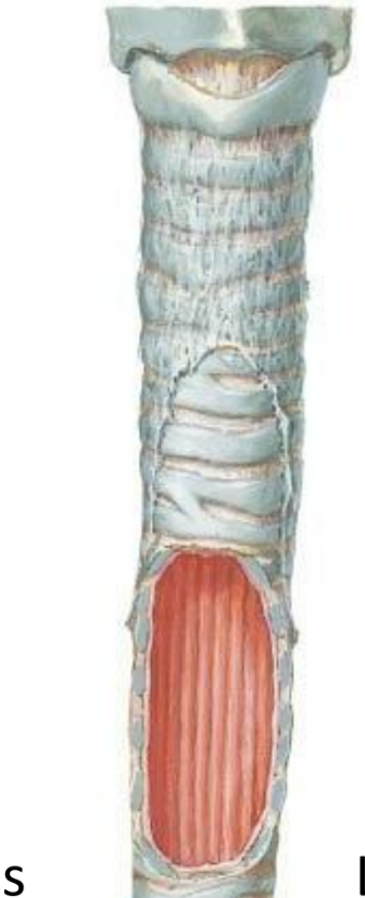
bronchioles
smooth muscle
can change diameter
bronchoconstriction (parasympathetic)
bronchodilation (sympathetic)
alveolar ducts
alveolar sacs
gas exchange

transitional zone
separates conduction system from the respiratory portion
includes:
terminal bronchioles
respiratory bronchioles
respiratory zone
site of gas exchange
includes
alveolar ducts
alveolar sacs
deoxygenated blood comes from heart via pulmonary artery
oxygenated blood back to heart via pulmonary vein

alveoli
thinly lined walls that allow for easy gas diffusion into the capillary bed
all of these are interdependent of each other
an increase in volume of one alveolar sac increases the volume in the adjacent sac
this is done via cross ventilation from the pores of Kohn and Lambert’s canals
blood vessel branching

lungs
light, soft, spongy
conical in shape, apex, base, costal surface, medial surface, hilum
right lung
three lobes: superior, middle, inferior
oblique and horizontal fissure
left lung
two lobes, superior and inferior
lingula
cardiac notch for apex of heart
oblique fissure
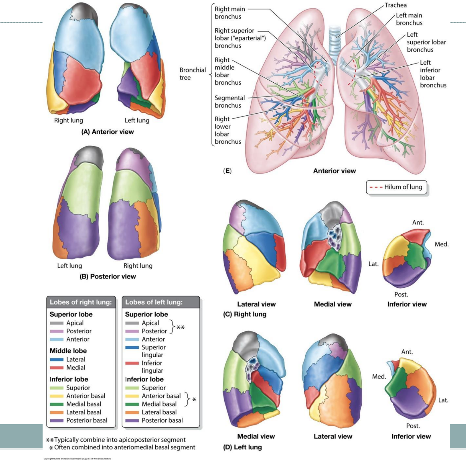
bronchopulmonary segments
root of the lungs
group of structures from the lungs to the mediastinum
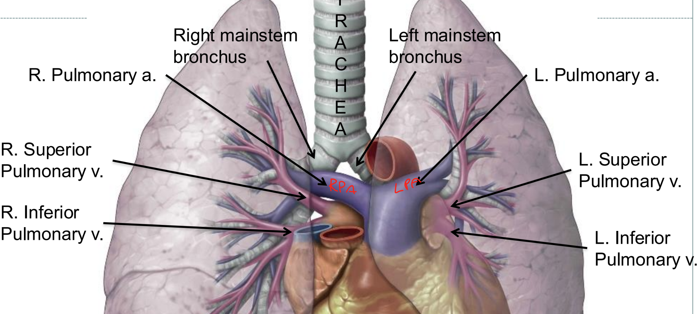
hilum
the region at which vessels, nerves, etc. enter or emerge from a part/organ

bony anatomy of chest wall
sternum
manubrium
body
xiphoid process
ribs
costochondral
costosternal
costovertebral

respiratory muscles: ventilation
these create a rhythmic pump to displace the chest wall
the displacement creates a negative pressure inside of the lungs by pulling the elastic lungs “open”
they also help the circulatory system by creating the thoracic pump to move venous blood back toward the heart
inhalation
diaphragm depresses
exhalation
diaphragm returns/elevates
diaphragm
primary inspiratory muscle
innervated by the phrenic nerves (C3,4,5 help us breathe and stay alive)
extremely fatigue resistant!
high amounts of mitochondria
dense/large capillary beds
capillary beds have a large ability to vasodilate to maximize blood flow
accessory inspiratory muscles
external intercostals
sternocleidomastoid
scalenes (ant, mid, post)
upper trapezius
pectoralis (major, minor)
serratus (ant, post)
latissimus dorsi
quadratus lumborum
diaphragm innervation
phrenic nerves (C3,4,5)
diaphragm: sternal portion
starts at xiphoid process
diaphragm: costal portion
comes from the transverse abdominal, last 6 ribs and costal cartilage
diaphragm: lumbar portion
attached to the bodies of the lumbar vertebrae to form the left and right crura
diaphragm attachments
xiphoid process
costal margin of thoracic wall
ribs 11 and 12
L1-L3 bodies
converge to a central tendon (pericardium attaches here)
diaphragm action
contraction increases thoracic volume (inspiration)
aortic aperture of diaphragm
aorta, thoracic duct
posterior to diaphragm
T12
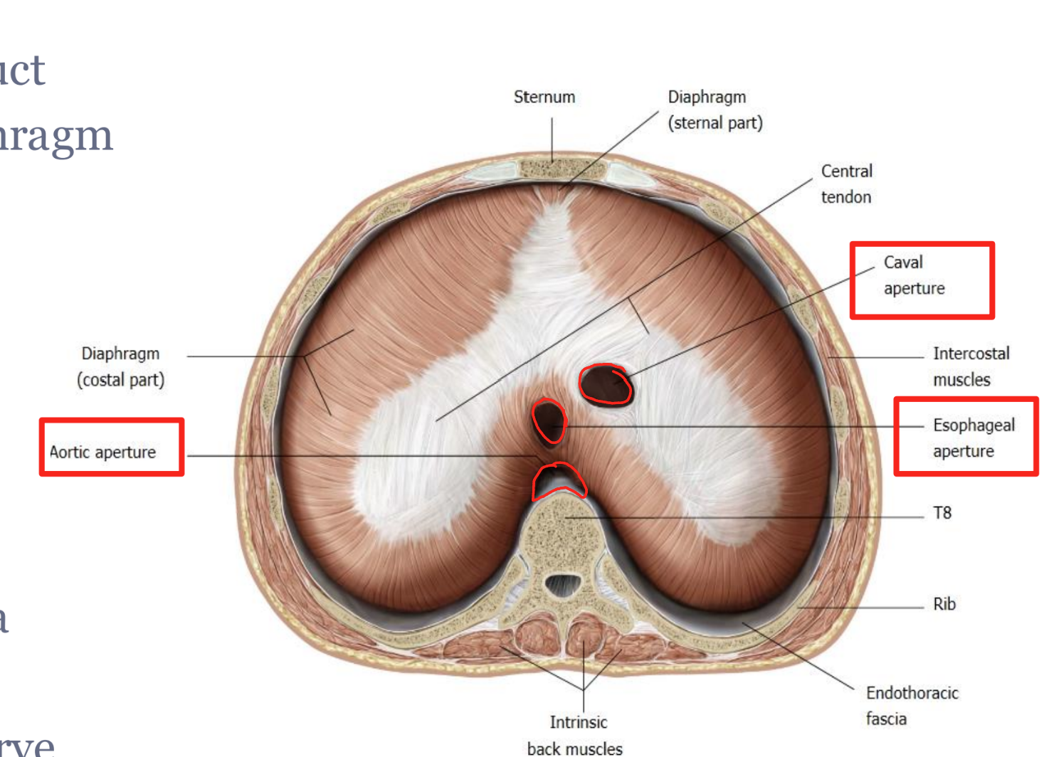
esophageal aperture of diaphragm
esophagus
vagus nerve
T10

caval aperture of diaphragm
inferior vena cava
T8
right phrenic nerve

contracts, down
the diaphragm _____ as the crural and rib attachments pull the central diaphragm _____
this creates 2 distinct different movements:
lateral costal expansion (bucket handle)
anterior/posterior upper chest wall movement (pump handle)

none!
primary muscle of exhalation
accessory muscles
used to create even higher negative pressures and to overcome increased pulmonary and/or chest wall compliance
this is needed with activity when greater ventilation is needed (minute ventilation) due to exercise or pathology
exhalation
passive; abdominal and internal intercostal muscles are accessory muscles used for this
elastic recoil
alveolar liquid/air interface
elastin & collagen fibers
pleura
secretes serous fluid
surround each lung
visceral (inside) and parietal (outside) parts are continuous
pleural cavity
fluid to prevent rubbing of surfaces during in/exhalation
cervical, costal, diaphragmatic, mediastinal
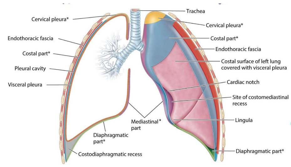
costodiaphragmatic recess

tracheobronchial tree innervation
parasympathetic via CN 10 (vagus)
efferent function
bronchoconstriction
afferent function
responsible for cough reflex
sympathetic
T1-T5 (pre-ganglionic) and paravertebral ganglia (post-ganglionic) to cardiopulmonary splanchnic nerves
adrenergic response (adrenal gland, epinephrine/norepinephrine)
bronchodilation
not sensitive
visceral pleura is ___ ________ to pain
innervation of the parietal pleura
costal: intercostal nerve
mediastinal: phrenic
diaphragmatic phrenic
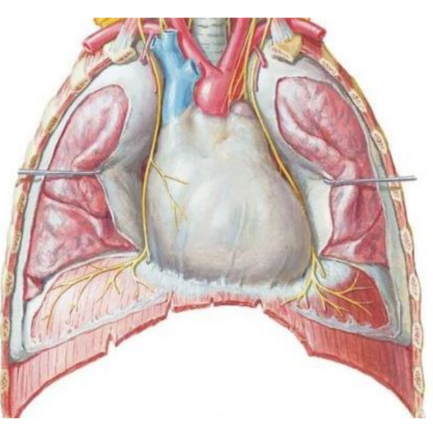
innervation
sympathetic
sympathetic trunk (T1-5) via cardiopulmonary splanchnic nerves
bronchodilation
parasympathetics
vagus
bronchoconstriction

Blood Supply
blood delivered to lung tissue via bronchial arteries
from aortic arch (left) or posterior intercostal artery (right)
venous blood drained via bronchial veins
into azygous system

pulmonary embolism
obstruction of pulmonary artery
acute respiratory distress
obstructive disorders of the lung
narrow airway-difficulty exhaling air, remains in lungs, shortness of breath
chronic obstructive pulmonary disease, asthma, cystic fibrosis
restrictive disorders of the lung
stiffness of lungs/chest, weak muscles - difficulty filling lungs with air, shortness of breath
pulmonary fibrosis, obesity, ALS
Innervation and Blood Supply of diaphragm

crura of the diaphragm
musculotendinous bundles
arise from anterior bodies L1-L3
L and R converge to form the median arcuate ligament
thoracic aorta
posterior intercostal vasculature
internal thoracic, musculophrenic
anterior intercostal vasculature
lymphatic system
drainage of surplus tissue fluid
removal of cellular debris and infection
maintains blood volume
lymph, drains into lymph vessels and is emptied back into blood stream
1. intracellular = in cells
2. intravascular = in veins
3. everywhere else
increased
______ prominence of lymph nodes could mean underlying infection
right lymphatic duct
right thorax
right upper limb
right head
thoracic lymphatic duct
drains lymph from the rest of the body
lymphatic system
plays a major role in body defense mechanism
lymph contains lymphocytes and macrophages
lymph capillaries are found in tissues closely related to arterioles and venules
drain to larger lymphatic vessels that ultimately empty into subclavian veins
filtering out pathogens
lymph nodes are found in strategic locations and process lymph passing through it by
spleen
has rich blood supply, process incoming blood, engulf bacteria, viruses, worn out RBCs
thymus
near the heart shrinks with age as it is most important in infancy
trains lymphocytes to be effective in immune system as they mature in thymus and become capable of attacking specific pathogens
tonsils
back of mouth, throat
protect the upper GI and respiratory systems from bacteria from air and food