lecture 24 - bone tissue pt.2
 Call Kai
 Learn
 Practice Test
 Spaced Repetition
 Match
 Flashcards
 Knowt Play
1/60

Earn XP
Description and Tags
DO NOT SHUFFLE
61 Terms
way to classify bone tissue
organization of collagen fibres within bone matrix
types of bone tissue
woven bone
lamellar bone
woven bone
type of bone tissue
randomly oriented collagen fibres
type that is made when skeleton is first created as an embryo
type that is first made when repairing a broken bone
lamellar bone
type of bone tissue
mature bone
organized in sheets/layers (lamellae)
type of bone seen in all bones of body
2 types
lamellae
sheets/layers of bone matrix that lamellar bones are organized into
2 types of lamellar bone
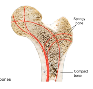
spongy/cancellous/trabecular bone
compact/cortical bone

spongy/cancellous/trabecular bone
type of lamellar bone
less bone matrix, more space
porous appearance
protected by compact bone

where is spongy/cancellous/trabecular bone found?
found on inside of bone
fills spaces of irregular bones or ends of long bones
forms a thin layer where there are cavities inside bones

cancellous bone makes up __% of our skeleton in terms of mass
20

compact/cortical bone
type of lamellar bone
dense, less space
surrounds spongy/cancellous/trabecular bone

where is compact/cortical bone found?
outside of all bones
majority of shafts of long bones
compact/cortical bone makes up __% of our skeleton in terms of mass
80

spongy/cancellous/trabecular bone structure
bone matrix organized into trabeculae
spaces in b/w trabeculae filled w/ bone marrow (typically red bone marrow, can have yellow bone marrow) + blood vessels
in b/w sheets of bone matrix are the osteocytes
nutrients diffuse through canaliculi to reach osteocytes

trabeculae
connecting rod/plate-like structures that the bone matrix of spongy/cancellous/trabecular bone is organized into
oriented along lines of stress (where the most force is applied on the bone)
osteocytes within the innermost layers of lamellae
has osteoclasts + osteoblasts on surface (many more osteoblasts)
has osteochondral progenitor cells

compact/cortical bone structure
has very organized sheets of lamellar bone (organized in 3 ways)
outside is periosteum
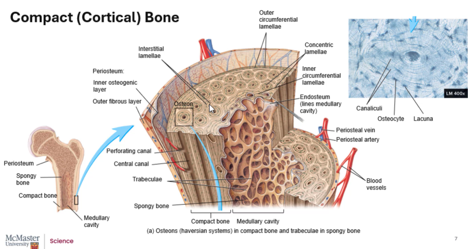
periosteum
2 layers
outer fibrous layer: dense fibrous CT (containing fibroblasts)
inner osteogenic layer: has osteoblasts + osteoclasts + osteochondral progenitor cells
surrounds the outside of all bones

the periosteum + perichondrium are very similar in structure + shape, its just the ___ types found in the inner layer that differ: in cartilage, they’re __________, and in bone, they’re ________ and _________
cell, chondrocytes, osteoblasts, osteoclasts

medullary cavity
space within shaft of bone
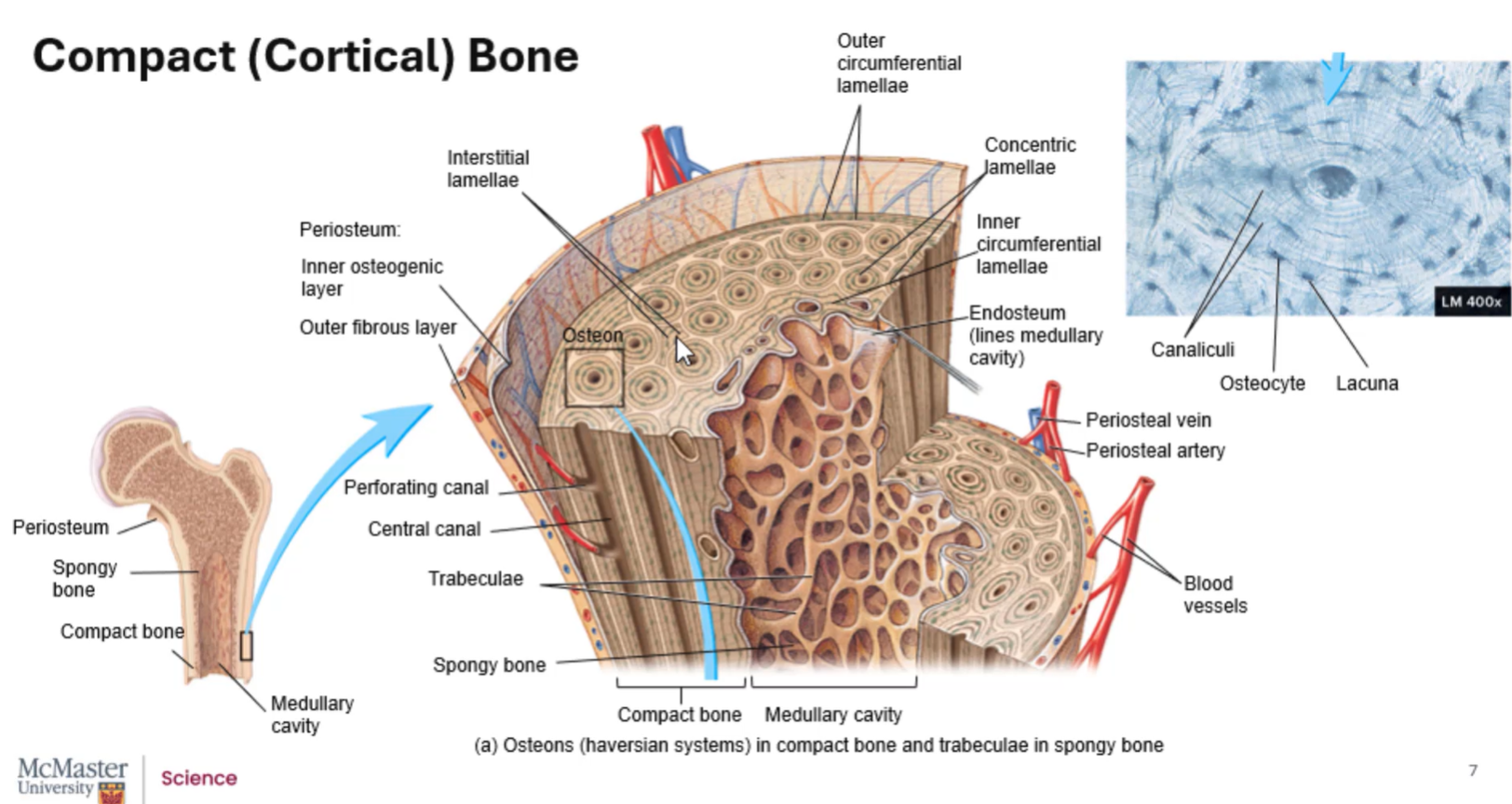
3 ways that lamellar bone is organized within compact/cortical bone
concentric lamellae
circumferential lamellae (has inner and outer)
interstitial lamellae
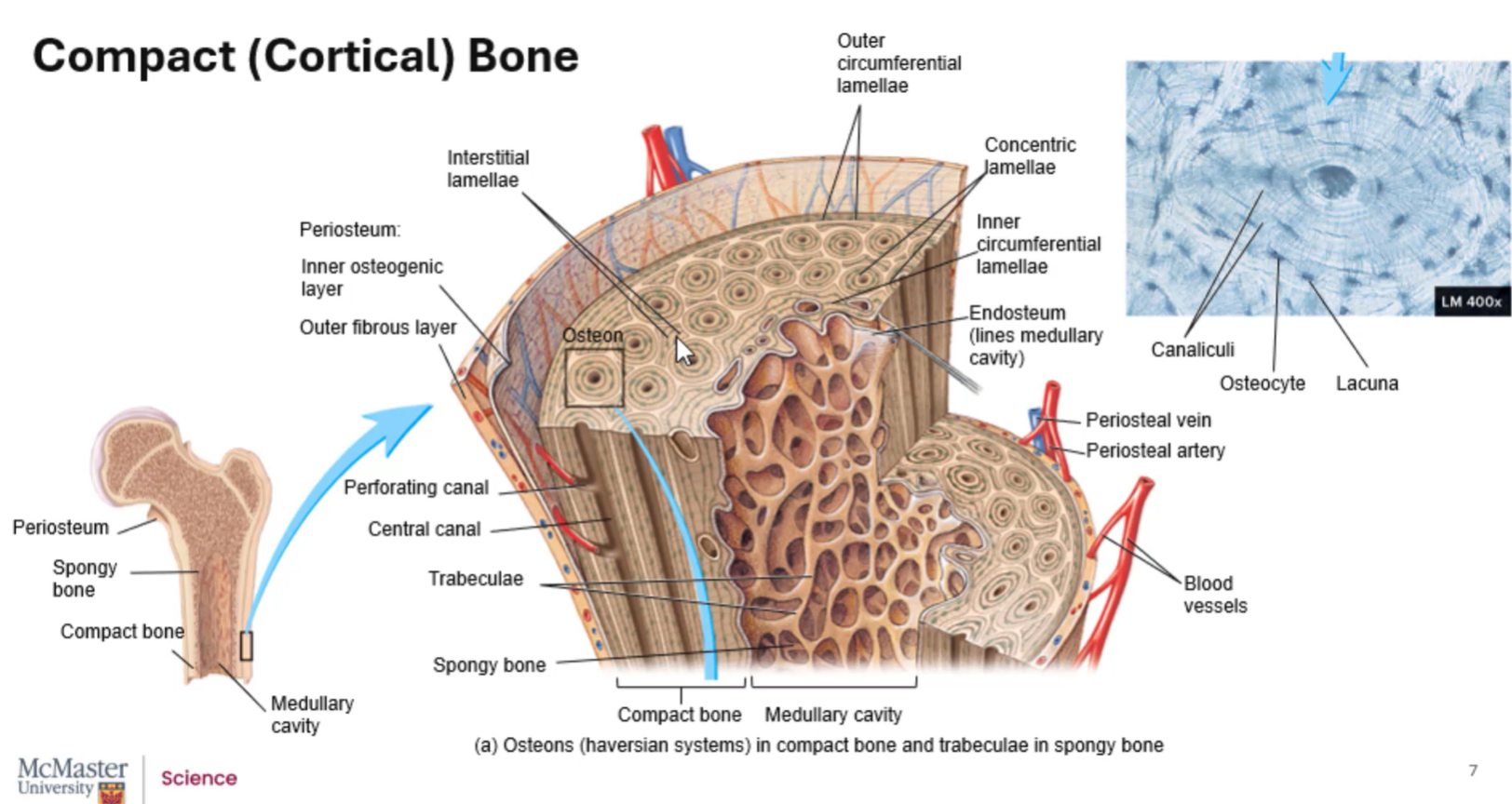
endosteum
layer surrounding the medullary cavity
has osteoblasts + osteoclasts + osteochondral progenitor cells

the ____ layer of the periosteum is where blood supply to the bone is found
outer

periosteal vein/artery
blood vessels associated with the periosteum

perforating/Volkmann’s canals
canals that run perpendicular to the length of the bone
where blood vessels enter into bone

how do blood vessels travel through bone?
enter through Volkmann’s/perforating canals
travel to organized units of bone → run parallel to bone in the center of those organized units via central/Haversian canals
continue to run through perforating canals to reach other central canals
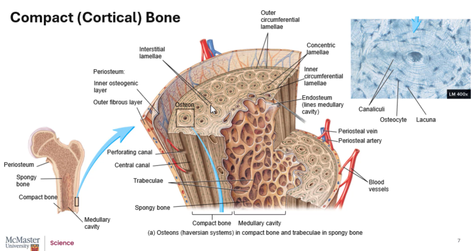
central/Haversian canals
form parallel tunnels within the organized units of bone

concentric lamellae
lamellae that form concentric circles around blood vessels found in a central canal
has osteocyte cells sandwiched b/w lamellae w/ canaliculi forming pathways b/w the layers
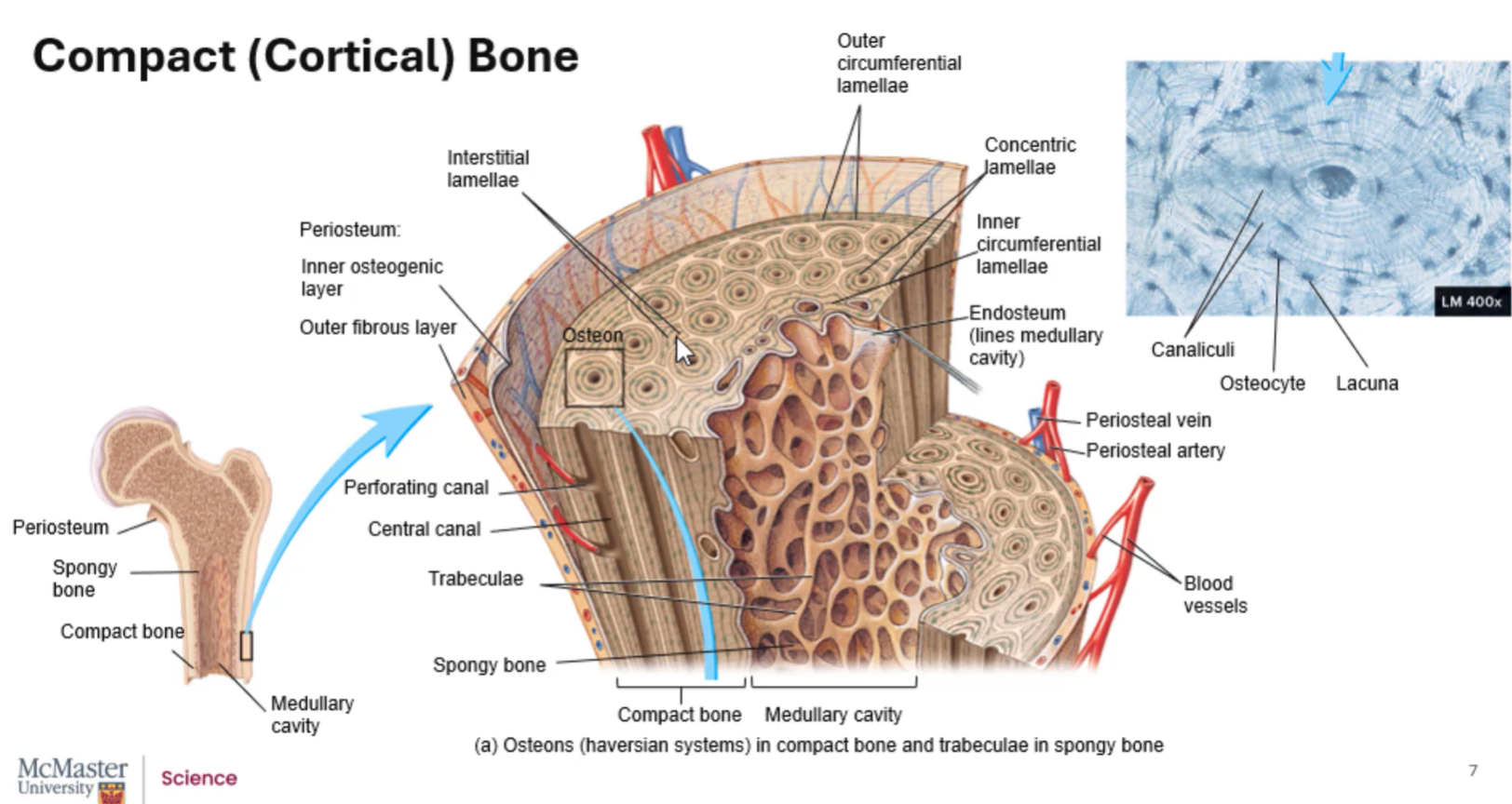
osteon
organization of bone into concentric lamellae around a central canal
in compact bone
many of them

how are nutrients/gases delivered in compact/cortical bone?
blood vessels are in the central canals, so nutrients/gases move outward toward osteocytes, then osteoblasts and osteoclasts

outer circumferential lamellae
bundle osteons tgth within compact bone
runs around outside of entire bone
a few layers thick
just underneath periosteum

inner circumferential lamellae
surround the medullary cavity (superficial to the endosteum)

interstitial lamellae
short pieces of lamellar bone
leftover remnants of osteons that have been broken down by osteoclasts
fill spaces b/w osteons

an osteon is also referred to as a ______ system
Haversian
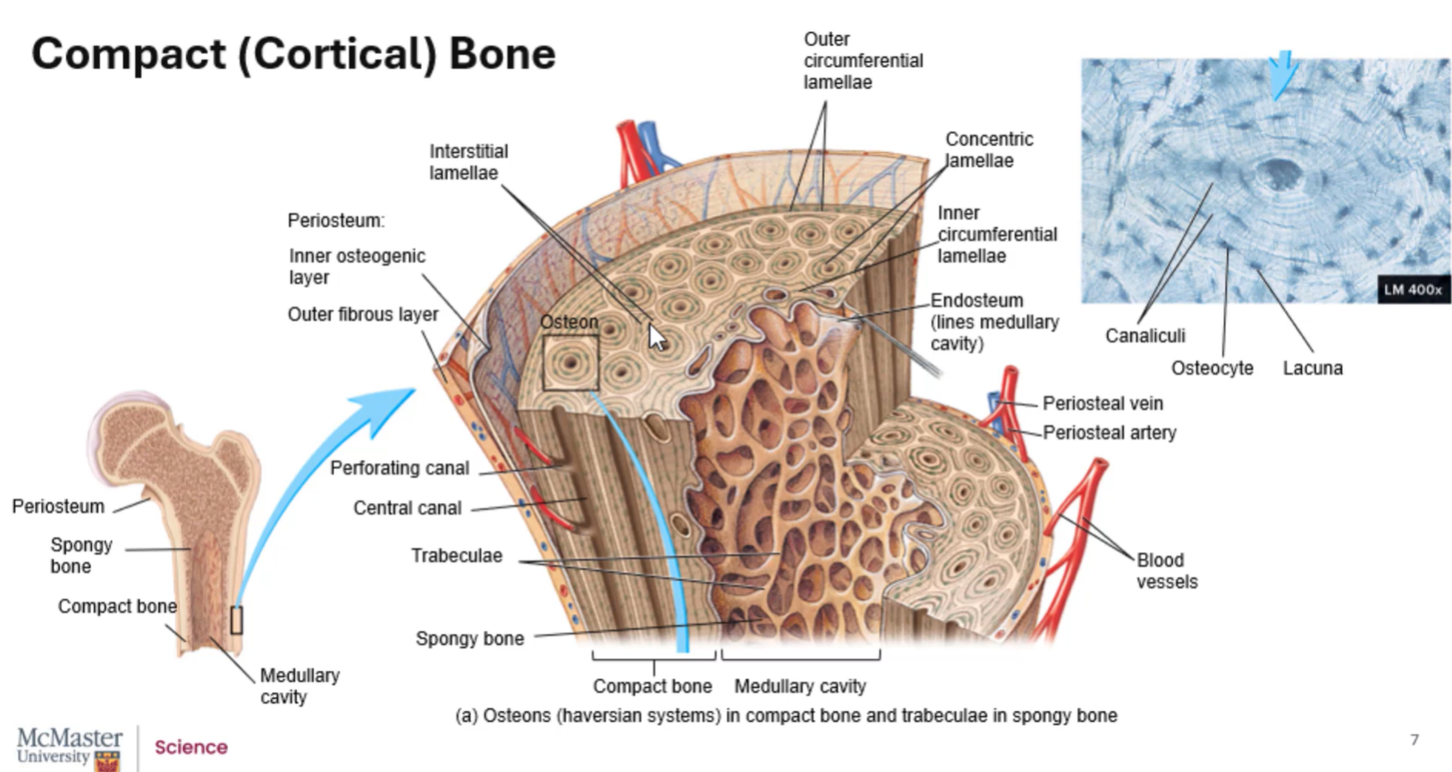
long bones
bones that are longer in length than in width

long bone structure
→→

epiphysis
region that is closest to the end of the bone
pointed towards the trunk of the body: proximal epiphysis
pointed away/farther from the trunk of the body: distal epiphysis

diaphysis
shaft
mostly made of compact bone
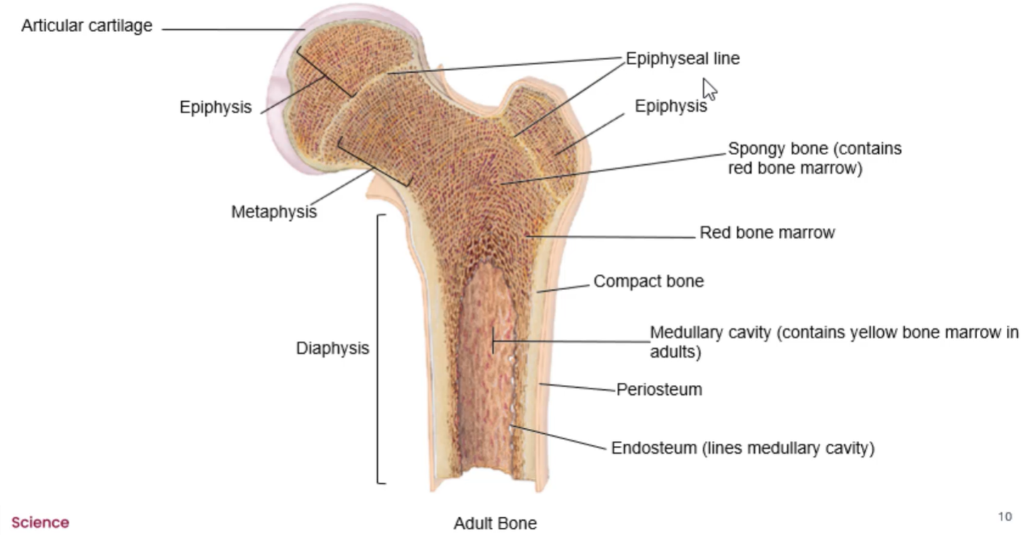
metaphysis
connects the diaphysis and epiphysis
located in regions where bone growth occurs
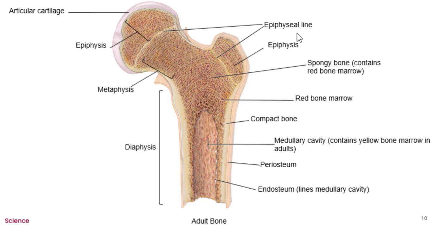
articular cartilage
surrounds outside of epiphysis
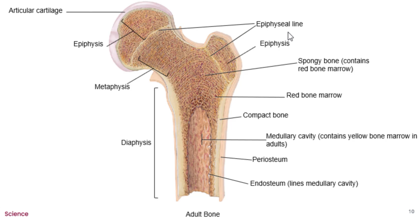
ends of long bones
primarily spongy bone (contains red bone marrow)

epiphyseal/growth plate
b/w the epiphysis and diaphysis
layer of hyaline cartilage that allows bone to grow throughout childhood; calcifies into epiphyseal line when full height is reached
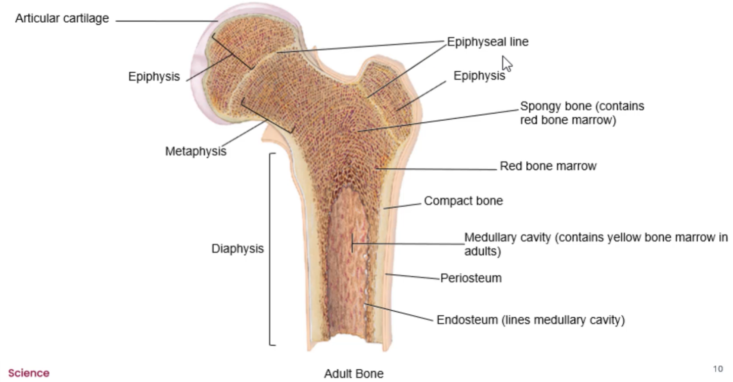
epiphyseal line
what epiphyseal plate calcifies into when full height is reached

osteogenesis/ossification
process of forming bone tissue
bone formation happens in embryo at about _ weeks
8
mesenchyme cells
stem cells that form all CT
form osteochondral progenitor cells
how does bone develop
8 weeks: blood vessels invade into areas where bone will be formed → stimulates mesenchyme cells to becomes osteochondral progenitor cells
either intramembranous ossification or endochondral ossification
intramembranous ossification
bone formation in CT membranes
e.g. skull bones, mandible, part of clavicle
mesenchyme cells create membrane of CT that’s primarily made of collagen fibres
osteochondral progenitor cells → osteoblast cells
osteoblast cells ossify membrane
endochondral ossification
bone formation in cartilage
e.g. base of skull, part of clavicle and remainder of bones
mesenchyme cells → osteochondral progenitor cells
osteochondral progenitor cells → chondroblasts
chondroblasts form hyaline cartilage skeleton
blood vessels invade into area where hyaline cartilage is → enter into perichondrium
stimulates osteochondral progenitor cells to → osteoblasts
perichondrium → periosteum
osteoblasts invade into area cartilage is formed + calcify it into lamellar bone
both __________ ossification and endochondral ossification create ____ bone first, then it gets remodelled into either spongy bone or ______ bone
intramembranous, woven, compact
bone (unlike cartilage), cannot undergo _______ bone growth. why?
interstitial
because the bone matrix is solid → osteocyte cells can’t divide and push each other
2 ways that bone growth can occur
bone length
bone thickness
growth in bone length
bone forms on diaphyseal side
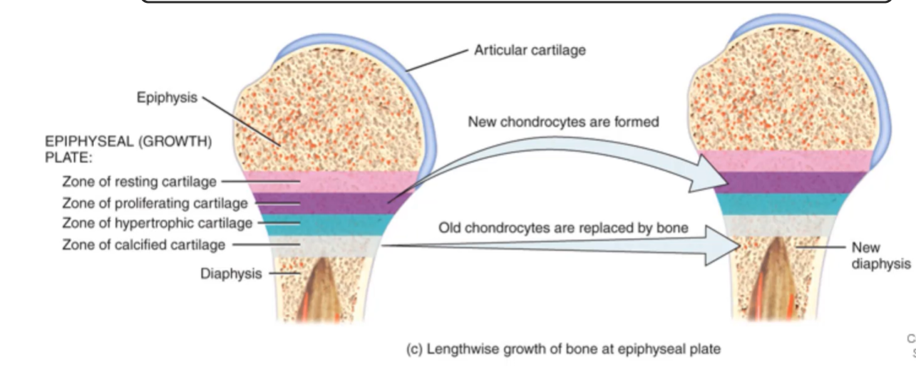
zone of resting cartilage
cartilage closest to the epiphyseal
has slowly dividing chondrocytes (via interstitial growth)
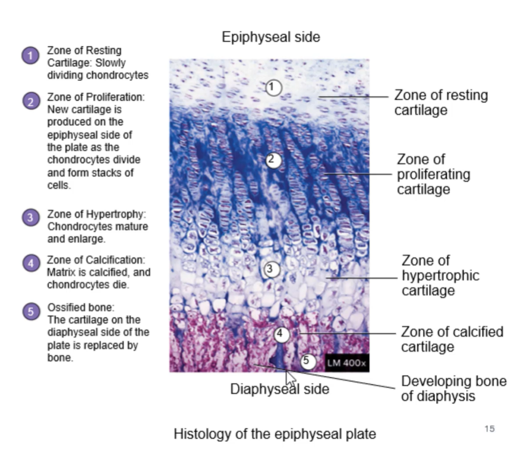
why is the zone of resting cartilage considered ‘resting?’
because it provides an anchor plate for the epiphysis so the epiphysis stays connected to the diaphysis

zone of proliferating cartilage
underneath the zone of resting cartilage
rapidly dividing chondrocytes (via interstitial growth)
chondrocytes divide into stacks that look like stacks of coins
newer cells end up closer to the zone of resting cartilage; older cells end up closer to the zone of hypertrophy

zone of hypertrophic cartilage
underneath the zone of proliferating cartilage
chondrocytes enlarging + maturing
secrete matrix vesicles that contain hydroxyapatite
begin to calcify matrix of the zone of calcified cartilage

zone of calcified cartilage
underneath the zone of hypertrophic cartilage
calcified cartilage layer
chondrocytes die off
blood vessels + osteoblasts invade into area from endosteum
osteoblasts develop bone on diaphyseal side

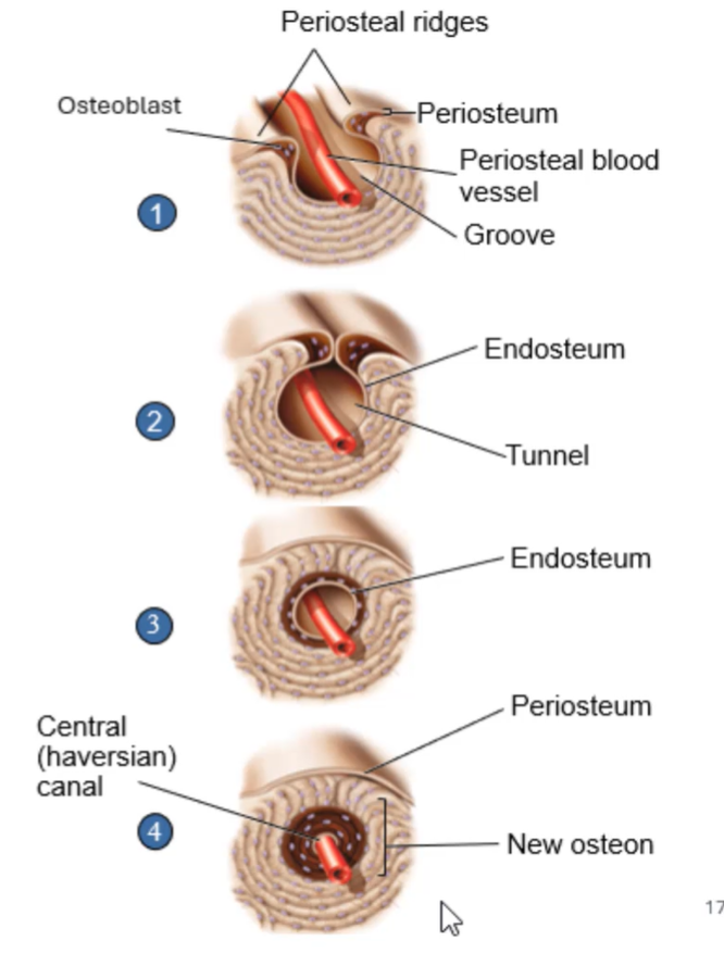
growth in bone thickness
periosteum surrounding outside of compact bone
osteoblast cells beneath the periosteum begin to lay down matrix/form bone (typically around periosteal blood vessels)
creates periosteal ridges around the blood vessel
ridges create a groove in which the blood vessel is sitting
ridges meet + form a tunnel
periosteum gets pinched off; layer on inside becomes endosteum
osteoblasts that are part of the endosteum fill in the concentric lamellae towards the blood vessel
osteoblasts form new layers of concentric lamellae (eventually creating a new osteon)
bone remodelling is a function of continuous ________ and _______ activity
osteoblast, osteoclast
bone remodelling
inside of bone being destroyed by osteoclast cells
outside of bone being formed by osteoblast cells
→ medullary cavity gets larger, thickness of bone stays relatively constant
in __ years, our entire skeleton will have been broken down + rebuilt by osteoblasts + osteoclasts
10
in what scenarios do we stimulate bone remodelling?
stress on bone
fracture
changes in minerals in body
exercise (picture refers to an athlete that plays tennis)
