11/6 Development 2: Fertilization, patterning, & morphogensis
 Call Kai
 Learn
 Practice Test
 Spaced Repetition
 Match
 Flashcards
 Knowt Play
1/21

There's no tags or description
Looks like no tags are added yet.
22 Terms
cell fate determination
most of the time one directional, but can be reversible
can be induced to de-differentiate from determined to totipotent/pluripotents state

adult cell to iPS cell
cell fate determination is reversible
start: fully differentiated adult cell (ex. fibroblast — connective tissue)
4 regulatory genes were identified by researchers (Oct3/4, Sox 2, Klf4, c-Myc)
introduce genes via plasmid/vector
dedifferentiation - new genes overtake current programming
result: iPS cell, back into pluripotent state
fertilization
sperm and egg come together; sperm has genetic material from dad/egg has from mom

stages of fertilization
sperm penetration of follicle cell layer & zona pellucida
membrane fusion
egg activation — activates signal transduction pathway
what do membrane fusion & egg activation stimulate
PLCzeta signaling, Ca2+ wave
PLCzeta
cleaves PIP2 to make DAG & IP3 (released (released from plasm membrane)

fertilization pathway
Trigger: A sperm fuses with the egg's plasma membrane, activating the egg.
Enzyme Activation: This fusion activates the enzyme Phospholipase C zeta (PLCzeta).
Cleavage: PLCzeta acts like molecular scissors, cutting a membrane lipid called PIP2 (phosphatidylinositol 4,5-bisphosphate).
Products: PIP2 is split into two molecules:
DAG (diacylglycerol), which remains stuck in the plasma membrane.
IP3 (inositol trisphosphate), which is released into the cytoplasm to act as a second messenger.
Calcium Release: IP3 travels to the Endoplasmic Reticulum (ER) and binds to IP3 receptors. This acts like a key opening a gate, causing the ER to dump a massive wave of stored Calcium (Ca2+) ions into the cytoplasm.
Cellular Response: The spike in Calcium triggers cortical granules (tiny vesicles inside the egg) to fuse with the membrane and release their contents outside. This alters the egg's outer layer, creating a "slow block to polyspermy" that physically prevents any additional sperm from entering.

Taste Transduction Pathway
Trigger: A tastant molecule (sweet, bitter, or umami) binds to a G-protein-coupled receptor (GPCR) on the outside of a taste bud cell.
Enzyme Activation: The receptor activates a G-protein, which then activates Phospholipase C (specifically the PLCβ2 version).
The Shared Middle Steps: Exactly like fertilization, PLC cleaves PIP2 into DAG and IP3. IP3 then binds to the ER, triggering a release of Calcium (Ca2+).
Cellular Response (Where they diverge): Instead of blocking sperm, the Calcium spike in the taste cell opens a specific ion channel (TrpM5) that lets Sodium (Na+) rush into the cell. This changes the cell's electrical charge (membrane potential) and causes the cell to release neurotransmitters (ATP via the Panx1 channel) to send a "taste" signal to your brain.
Why use amphibians?
The eggs develop outside the body, so it is easier to see the development
Morphogenesis
generation of ordered form & structure
Body begins to develop recognizable structure
Includes pattern formation (how does body pattern happen during body development)
Gastrulation
cell and tissue movements during which blastula folds inward, generating 3 germ layers (ectoderm, endoderm, mesoderm)
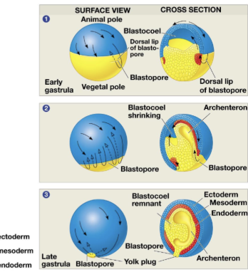
Layers within the embryo
Endoderm
Ectoderm –
Mesoderm
In the early embryo
Pattern formation, Positional information leads to changes in signaling & gene activity

Pattern formation
use of positional information for body pattern
We are 3D, can be on xyz axis
Front (ventral) & back (dorsal) & (anterior) & (posterior)
what approaches have been the primary importance in determining the regions of the embryo that act in pattern formation?
Embryology
the Spemann-Mangold experiment
1924
What part of the embryo plays an important role in that the dorsal side should develop into the dorsal side
What if you took dorsal tissue from an embryo and transplanted it into ventral → it would develop into a tadpole, but it has 2 dorsal sides & 1 ventral side
Indicates that there is something produced in dorsal tissue that instructs cells to take the fate of the dorsal structures
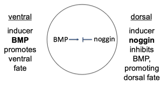
the dorsal inducer noggin antagonizes the ventral inducer bone morphogenetic protein (BMP)
Richard Harland – Noggin
Noggin – inhibitor of bone morphogenetic protein (BMP)
By inhibiting BMP, cells in the dorsal region can develop into dorsal structures
What type of signaling scheme? Paracrine
which approaches have been of primary importance in identifying the genes & corresponding proteins that function in pattern formation?
Genetics in model organisms
Note: just sequencing the genome will tell you all genes, but won’t tell you which genes are important
Drosophila melanogaster
model organism for studying A/P axis
Short lifespan
Develops quickly
Lots of progeny
Induce mutations → genetic cross (fly x fy) → egg → embryo → adult – look at altered body patterns
Dropsophilia homeotic mutants
class of mutations that lead to the appearance of normal body parts in the wrong locations
Ex. mutant w/ 4 wings, mutant w/ legs as antennae
Are in homeobox (Hox) gene clusters
Hox genes
control anterior/posterior pattern in animals, including in humans
genes/proteins conserved in animals
Contain homeodomain that binds to DNA
Act as transcription factors
Differences in Hox gene sequence, position, or regulation result in striking differences in animal form
Mutations in HOXA1
disrupt human brain development
Rare: but some people have a disorder in the HOXA1 gene
Why is it rare? Messing with these genes/expressions, won;’t be able to develop
4 clusters – 1 could sub for the other, maybe there is some redundancy