pnb block 4
 Call Kai
 Learn
 Practice Test
 Spaced Repetition
 Match
 Flashcards
 Knowt Play
1/114

There's no tags or description
Looks like no tags are added yet.
115 Terms
first-order neuron
cell body location: dorsal root ganglion (body) or sensory cranial nerve ganglion
function: carries information from receptor into the CNS
second-order neurons
location: spinal cord or brainstem
function: receives the first neuron’s signal and relays it, often crossing the midline.
third-order neurons
location: usually thalamus
function: relays information to the cerebral cortex
tracts
begin and end at the same place
the precise path may have slight anatomical variation because fetal environment is unique
no apparent advantage of one tract over another, perhaps a more direct path may be faster
all tracts develop in utero
clinically, the path taken is critical

dorsal column-medial lemniscus (DCML)
pathway that carries: fine touch, vibration, conscious proprioception, information important for two-point discrimination
step-by-step:
a sensory receptor detects a stimulus and first order neuron has its cell body in the dorsal root ganglion
its axon enters the spinal cord and ascends ipsilaterally
travels through dorsal columns to medulla, where it synapses in either the gracile nucleus or cuneate nucleus.
the second-order neuron crosses the midline in the medulla, where it ascends as the medial lemniscus.
it synapses with the third order neuron in the thalamus.
the third-order neuron projects to the primary somatosensory cortex.
fasciculus gracilis
carries information from lower body, especially legs and lower trunk. position in dorsal columns: medial.
fasciculus cuneatus
carries information from upper body, especially arms and upper trunk. position in dorsal columns: lateral.
spinothalamic pathway
important for pain, temperature, some crude touch and related sensations.
step-by-step pathway:
a nociceptor or thermoreceptor detects a stimulus
the first-order sensory neuron has its cell body in the dorsal root ganglion, where its axon enters the spinal cord.
it synapses with a second-order neuron in the dorsal horn, which crosses to the opposite side WITHIN the spinal cord.
it ascends through the contralateral anterolateral region.
it reaches the thalamus, where it synapses with a third-order neuron, which then projects to the somatosensory cortex.
olivary tracts
aid in muscle control of balance
tectal tracts
aid in head movement in response to cutaneous stimulation
reticular tract
conscious response to cutaneous stimulation, pain.
somatosensory cortex
site of the synapse of the 3rd neuron.
topographically mapped: spatial discrimination, sensation is physically mapped. subdivided into different stimulus modalities.
provided feedback from skin, muscles, tendons, and joints.
volume of cortex proportional to sensory input, and more stimulus collected sent to a larger area of cortex, proportional.
representation is drawn as homunculus

homunculus
a distorted representation of the human body across the primary somatosensory cortex. you will notice that the hands, lips, and face are disproportionately large compared with the trunk and legs because the amount of cortical representation devoted to a body region is related to its sensory input and processing requirements, rather than its physical size.

cortical plasticity
the brain’s ability to reorganize its sensory maps in response to changes in sensory input. this explains phantom limb pain, as the brain’s representation of the limb does not necessarily disappear immediately after amputation. changes in cortical organization, ongoing neural activity, and altered sensory processing can contribute to phantom sensations and pain.
haptic feedback
information provided through touch or physical sensation. examples:
raised bumps at the top of stairs
textured sidewalk edges
vibrations from iPhone
textures that communicate information without requiring vision.
nociceptors
free nerve endings detect cell damage
cell damage causes release of intracellular chemicals that act as a stimulus.
experienced pain depends on type of damage and type of nociceptor
fast, localized, and sharp pain - myelinated Adelta fibers
Full, slow, ache - unmyelinated C fibers
both impossible to ignore.
nociceptors are most abundant in skin, and may occur in many internal tissues. however, the brain tissue itself does not contain pain receptors in the same way that skin does. the brain, during headaches, dilates the cerebral and dural blood vessels, as well as inflames mediators.
CGRP
a migraine trigger. can promote vasodilation, participate in inflammatory signaling, and contribute to transmission of nociceptive information from intracranial structures.
acute pain
rapid onset, often related to an identifiable injury or stimulus; may resolve after the stimulus is removed
example: touching a hot surface
chronic
persistent or recurrent pain that continues beyond the expected period of healing
example: persistent neuropathic or musculoskeletal pain
visceral pain
originates from internal organs. generally, have less precise sensory localization than the skin, so it is harder to localize.
example: abdominal pain
referred pain
occurs when pain is perceived at a location different from the actual source of the problem. happens through a mechanism called convergence, which is when sensory fibers from different body regions can synapse on the same neurons in the spinal cord or brainstem.
examples:
myocardial infarction (heart attack): pain may be felt in the neck, shoulders, back, or arms rather than only in the chest
ice-cream headache: cold stimulation in the throat or palate can be associated with pain perceived in the head.
special senses
anatomically complex and process large amounts of information. requires substantial neural resources and considerable cortical processing.
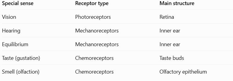
vision
one of the most complex sensory systems in the human body. involves: the eyeball, eyelids and brows, lacrimal glands and ducts, cornea, lens, retina, optic nerve, muscles that move the eyes, and extensive processing in the brain. some facts to know:
only 1/6th of eye is exposed
image formed on retina is inverted
the retina contains a large number of photoreceptors
the fovea has the highest density of cones
the optic disc contains no rods or cones
the cornea is avascular (the only tissue without blood vessels in body)
fibrous tunic
structures: schlera and cornea
functions: protection, shape, and light entry

vascular tunic
structure: choroid, ciliary body, iris
function: blood supply, lens adjustment, pupil control
nervous tunic
structures: retina
function: detects light and initiates visual signaling.
sclera
part of the fibrous tunic.
sclera is the white, tough connective tissue covering that helps protect the eye and maintain its shape.
cornea
part of the fibrous tunic. the cornea is the transparent front surface of the eye. it provides a major portion of the eye’s refractive power by bending incoming light. the cornea is avascular, and fed by tears. it is transparent due to keratan sulfate proteoglycans.
choroid
part of the vascular tunic. the choroid is part of the vascular layer that supplies nutrients to the retina and absorbs stray light.
ciliary body
part of the vascular tunic. the ciliary body contains the ciliary muscle, which changes the shape of the lens during accommodation
iris
part of the vascular tunic. the iris is the colored part of the eyes, and it controls pupil diameter.
retina
part of the nervous tunic. the retina contains photoreceptors and other neurons that convert light into electrical signals and begin visual processing.
pupil
the opening in the center of the iris. it is not a structure made of black tissue, but appears black because light entering the eye is largely absorbed rather than reflected back out. the iris changes pupil size: can dilate or constrict, which controls how much light enters the eye.
lens
biconvex, meaning it bulges outward on both sides. function is to refract light so that incoming rays are focused onto the retina.
what lens do to light:
a convex lens converges light rays
the light rays are brough toward a focal point
the eye must adjust its lens to focus light from different distances onto the retina.

accomodation
the process of hcanging lens shape to focus on objects at different distances. the ciliary muscle controls the tension on the suspensory ligaments (zonular fibers), which attach to the lens.

when looking at a nearby object…
the ciliary muscle contracts
tension on the zonular fibers decreases
the lens rebounds into a more rounded shape
the lens becomes more convex
its refractive power increases
light from the nearby object can be focused on the retina

when looking at a distant object…
the ciliary muscle relaxes
zonular fiber tension increases
the lens becomes flatter
its refractive power decreases
light from distant objects can be focused on the retina.

aqueous humor
fluid in the anterior portion of the eyes, in front of the lens.
vitreous humor
gel-like substance filling the large posterior cavity behind the lens.
schlemm’s canal
Aqueous humor is produced by the ciliary processes. It flows through the posterior and anterior chambers, then drains through the trabecular meshwork into Schlemm's canal.
This drainage helps regulate intraocular pressure

cataracts
clouding of the lens. when the lens becomes cloudy: light is scattered, images become blurry, glare can increase, color perception may become less vivid, and vision can become impaired.
differences between glaucoma and cataracts

fovea
region of the highest cone density; responsible for the sharpest central vision

peripheral retina
contains many rods, which are especially useful in low-light conditions

optic disc
where a ganglion cell axons leave the eye to form the optic nerve; contains no rods or cones.
rods
a photoreceptor in the retina. excels in low-light vision, as rods are extremely sensitive to light. they allow you to see in dim environments. Multiple rods can synapse onto shared downstream neurons. This means that even if each rod responds to a very small amount of light, their combined input can help activate the next neuron.
This increases sensitivity.
Trade-off: When multiple rods converge onto shared neurons, the brain has less information about exactly which rod was activated. Spatial acuity decreases.
cones
bright-light vision, best function in brighter conditions. they provide high visual acuity, color discrimination, and detailed central vision. In the fovea, cone pathways have very little convergence, allowing the brain to preserve detailed spatial information.
Trade-off: Less convergence improves acuity but means that cones are less sensitive to very dim light.
rods and cones tradeoff

scotopic vision
lighting conditions: dark
main photoreceptors: rods
visual experience: grayscale
photopic vision
lighting conditions: bright light
main photoreceptors: cones
visual experience: color vision
mesopic vision
lighting conditions: intermediate lighting
main photoreceptors: both rods and cones contribute
visual experience: mixed characteristics.
phototransduction
unlike many sensory receptors that depolarize when stimulated, rods and cones actually hyperpolarize in response to light. in darkness:
cGMP levels remain relatively high.
cGMP-gated (CNG) channels remain open.
Sodium and calcium enter the photoreceptor.
The photoreceptor remains relatively depolarized.
It continuously releases glutamate.
This ongoing release of glutamate helps inhibit certain bipolar cells.
phototransduction sequence
A photon is absorbed by retinal.
11-cis-retinal changes into all-trans-retinal.
The activated opsin changes conformation.
Opsin activates a G protein called transducin.
Transducin activates phosphodiesterase (PDE).
PDE breaks down cGMP.
Intracellular cGMP concentration decreases.
cGMP-gated sodium/calcium channels close.
Positive ion entry decreases.
The photoreceptor hyperpolarizes.
Glutamate release decreases.
how does information travel from the retina to the brain
rods/cones —> bipolar cells —> ganglion cells —> optic nerve. more specifically:
What happens in darkness?
photoreceptors continuously release glutamate in darkness.
This glutamate inhibits certain bipolar cells, depending on the bipolar cell's receptor type.
What happens in light?
Photoreceptors hyperpolarize.
Glutamate release decreases.
The inhibition of certain bipolar cells is relieved.
Bipolar cell activity changes.
Bipolar cells influence ganglion cells.
Ganglion cells generate action potentials.
Ganglion cell axons form the optic nerve.
horizontal cells
modify communication between photoreceptors and bipolar cells. contribute to lateral interactions that help the retina detect contrast and edges. important because the visual system needs to distinguish changes in brightness across neighboring areas, not merely detect the total amount of light.
amacrine cells
modify communication between bipolar cells and ganglion cells. help regulate visual information, including temporal changes and movement-related processing.
cell types of vision

binocular vision
the brain processes two images together to create depth perception
stereopsis
Each eye views an object from a slightly different angle.
The two retinas receive slightly different images.
The visual cortex compares the images.
The brain uses the differences to estimate depth.
helps you estimate how far away an object is, how far you need to reach, and where an object is located in 3D space.
myopia - nearsightedness
occurs when light from distant objects focuses in front of the retina.a too-long eyeball may be the cause.
as a result of this disease, nearby object appear relatively clear, but distant objects appear blurry. correction involves a concave lens
hyperopia - farsightedness
.occurs when light would focus behind the retina. a too-short eyeball may the cause. as a result, nearby objects may be difficult to focus on. correction involves a convex lens
presbyopia
Presbyopia is the age-related loss of accommodation.
As the lens becomes less flexible, it cannot round up as effectively when focusing on nearby objects.
This makes reading and other close-up activities increasingly difficult.
It is different from myopia and hyperopia because it primarily involves reduced ability to change lens shape.
astigmatism
Astigmatism occurs when the cornea or lens has an irregular curvature.
Instead of focusing light evenly onto one point, the optical system produces uneven focusing.
This can cause:
Blurry vision.
Distorted images.
Difficulty seeing fine details at different distances.
emphasis on malformed corneal shape
age-related macular degeneration (AMD)
The macula is the central region of the retina responsible for detailed central vision.
AMD affects this region and can cause progressive loss of central vision.
The lecture distinguishes two forms:
Dry AMD
Associated with thinning and degeneration of retinal tissues.
Generally progresses more gradually.
Wet AMD
Associated with abnormal growth of blood vessels beneath or within the retina.
These vessels can leak fluid or blood and damage retinal structures.
tunnel vision
The lecture describes tunnel vision as potentially resulting from a tumor pressing on the optic nerve.
The general idea is that damage or compression affecting visual pathways can cause loss of part of the visual field.
However, tunnel vision has multiple possible causes, and optic nerve compression is only one possibility.
sound
produced by air or water molecules. vibrations travel as pressure waves that can be detected by the ear. frequency and amplitude are two major properties that determine how we perceive sound.
frequency
physical measurement: Hertz (Hz) // we perceive pitch (high vs. low)
the number of wave cycles per second. A high frequency sounds high-pitched; a low frequency sounds low-pitched.
amplitude
physical measurement: size/intensity of pressure wave. we perceive loudness.
related to the strength of the pressure wave. greater amplitude is generally perceived as louder.
decibels
measure sound level on a logarithmic scale.
Outer ear
contains: pinna, auditory canal
function: collects and directs sound
middle ear
contains: tympanic membrane, maellus, incus, stapes, Estuachian tube
function: collects and directs sound
inner ear
contains: cochlea, vestibule, semicircular canals, hair cells
function: hearing and balance
pinna
the visible external part of the ear, it funnels sound waves into the auditory canal
auditory canal (auditory meatus)
directs sound toward the tympanic membrane.
tympanic membrane (eardrum)
vibrates when sound waves reach it
ossicles
three small bones in the middle ear
malleus (hammer): attached to the tympanic membrane
incus (anvil): receives vibrations from malleus
stapes (stirrup): transfers vibrations to the oval window.
sequence: tympanic membrane —> malleus —> incus —> stapes —> oval window
eustachian tube
connects the middle ear to the throat region and helps equalize air pressure on both sides of the tympanic membrane. this is why your ears may pop when you change altitude.
inner ear
The inner ear contains structures involved in both hearing and balance:
Cochlea: Hearing.
Vestibule: Detects head position and linear acceleration.
Semicircular canals: Detect rotational head movements.
The cochlea contains sensory hair cells that convert mechanical movement into electrical signals.
inner ear nested systems
The inner ear has two nested systems:
Bony labyrinth: Cavities carved into the temporal bone. (LOW sodium, HIGH potassium)
Membranous labyrinth: Membranous structures located inside the bony labyrinth. (HIGH sodium, LOW potassium)
The membranous labyrinth contains endolymph, a fluid with an unusual ionic composition.
The bony labyrinth surrounds the membranous labyrinth and contains perilymph in the spaces around it.
anatomy of cochlea
a spiral-shaped structure that contains three fluid-filled chambers
scala vestibuli: begins at the oval window. contains perilymph.
scala media: cochlear duct; contains endolymph
scala tympani: leads toward the round window. contains perilymph.
vestibular membrane
Separates the scala vestibuli from the scala media.
basilar membrane
Separates the scala media from the scala tympani and supports the organ of Corti, which contains the auditory hair cells.
oval window
receives vibrations from the stapes
round window
allows pressure changes in the cochlear fluid to be released. we need the round window because the cochlea is fluid-filled. since fluid is difficult to compress, movement at the oval window needs a flexible region where pressure can be displaced. the round window provides that flexibility.
how sound becomes an electrical signal.
sound waves enter the auditory canal and the tympanic membrane vibrates.
the malleus, incus, and stapes transmit the vibration.
the stapes pushes against the oval window.
movement of the oval window generates pressure waves in the cochlear fluid, which causes the basilar membrane to vibrate.
movement bends the stereocilia on the hair cells.
bending opens mechanically gated cation channels.
K+ enters the hair cell from the potassium rich endolymph.
the hair cell depolarizes and releases NT onto cochlear neurons
cochlear neurons generate APs that travel through cochlear nerve.
stereocilia
tiny projections in hair cells. arranged in bundles and connected by tip links
tip links
arranges the stereocilia in bundles.
tympanic reflec
two muscles contract: tensor tympani and stapedius. their contraction stiffens the middle-ear structures and reduces transmission of vibrations to the oval window.
this helps reduce the amount of sound energy reaching the inner ear.
inner vs. outer hair cells
Inner hair cells are the main sensory receptors. They communicate with cochlear neurons, whose axons form the cochlear nerve.
Outer hair cells change their length in response to electrical stimulation. This movement helps amplify and sharpen vibrations of the basilar membrane.
This is called the cochlear amplifier.
A useful way to remember the distinction:
Inner hair cells tell the brain what sound is present.
Outer hair cells help make the cochlea more sensitive and selective.

cochlear frequencies
The basilar membrane varies in physical properties along its length:
Base of cochlea: Narrower and stiffer; responds best to high-frequency sounds.
Apex of cochlea: Wider and more flexible; responds best to low-frequency sounds.
This arrangement is called tonotopic organization.
auditory pathway
cochlear nerve —> dorsal and ventral cochlear nuclei —> superior olive —> inferior colliculi —> medial geniculate body —> auditory cortex

how does the brain locate sounds?
interaural intensity differences (ILDs): A sound coming from one side of your head is generally louder in the ear closest to the source.
The lateral superior olive (LSO) compares sound intensity between the ears.
The lecture emphasizes that this mechanism is particularly useful for frequencies above approximately 3,000 Hz.
interaural timing differences (ITDs): A sound reaches the closer ear slightly earlier than the farther ear.
The medial superior olive (MSO) compares the timing of signals arriving from the two ears.
The lecture emphasizes that this is particularly useful for frequencies below approximately 3,000 Hz.
ostosclerosis
abnormal bone growth/remodeling restricts movement of middle-ear bones, especially the stapes
meniere’s disease
inner ear disorder associated with hearing loss, dizziness, and balance symptoms
tinnitus
perception of buzzing, ringing, hissing without an external sound source
ototoxicity
certain medications damage inner-ear structures, including hair cells.
vestibular system
detects information about
Head position.
Gravity.
Linear acceleration.
Rotational movement.
Orientation in three-dimensional space.
It helps you maintain balance and stabilize your gaze while moving.
The vestibular system uses hair cells similar to those in the cochlea, but the structures and mechanics differ.
static labyrinth
structures: urtricle and saccule
function: head position and linear accleration