Anatomy Exam #2
 Call Kai
 Learn
 Practice Test
 Spaced Repetition
 Match
 Flashcards
 Knowt Play
1/61

There's no tags or description
Looks like no tags are added yet.
62 Terms
Internal intercostals
Lie deep to the external intercostal muscles
function: work to retract the rib cage

Innermost Intercostals
The deepest of the intercostal muscles
Run parallel to the internal intercostals
function: assist in retraction of the rib cage

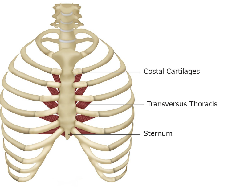
Transversus Thoraci
Exists on the inside surface of the rib cage
function: retracts the rib cage and resists elevation of the rib cage
Subcostals
Deep to the internal intercostals on the inferior interior surface of the posterior thorax
The length and number of subcostals vary from person to person
function: retract the rib cage

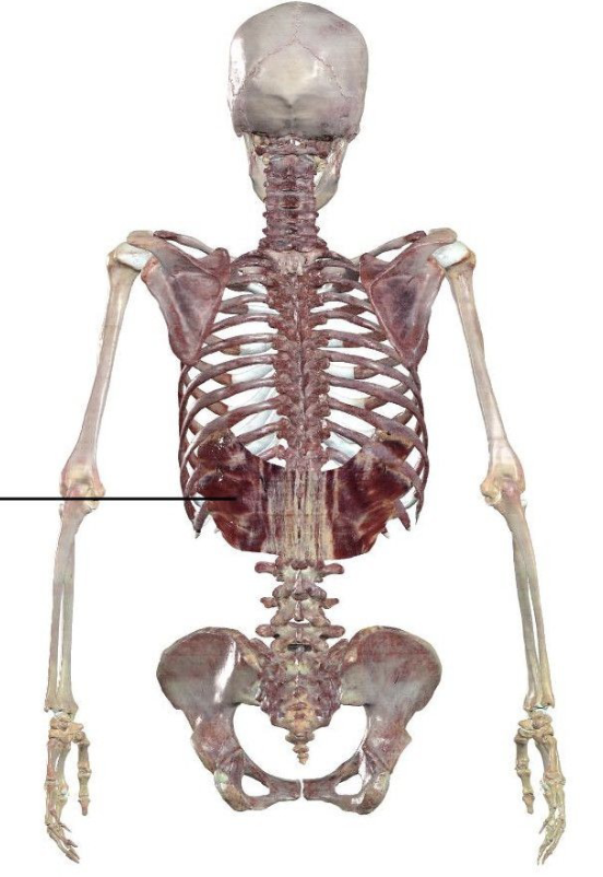
Serratus Posterior Inferior
Quadrilateral shaped muscle of the lower back
Has extensions that give the muscle its serrated appearance
Primary function: Stabilizes the trunk
Secondary function: assists in retracting the rib cage
Points of attachment for abdominal muscles
Abdominal aponeurosis: large flat tendinous structure
Forms the anterior wall of the abdomen
Deep to it are the abdominal muscles
Very few skeletal points of attachment for muscles in the abdomen
Instead, connect via tendinous aponeuroses:
o Linea alba
o Linea semilunaris
o Abdominal aponeurosis
o Lumbodorsal fascia
Transversus Abdominis
Deepest of the abdominal muscles
The fibers course horizontally, as is implied by the word transversus
Function: Compresses the abdomen, which stretches the diaphragm, reducing vertical thoracic volume


Internal Oblique Abdominis
Located between the external oblique abdominis and transverse abdominis
Smaller and thinner than external oblique abdominis
Pulls in the opposite direction to the external oblique abdominis
Function: Bilateral: Pulls the lower ribs down and compresses the abdomen
Unilateral: Assists in rotation and bending of the trunk towards the side of contraction.
External Oblique Abdominis
Most superficial and largest abdominal muscles
Function:
Bilateral contraction pulls the thorax down and compresses the abdomen
Unilateral contraction facilitates contralateral rotation of the trunk and ipsilateral bending of
the trunk

Rectus Abdominis
Paired flat muscle across the anterior of the abdomen
Divided into its paired right and left divisions at midline by the linea alba and laterally by other tendinous connective tissue
Functions:
To bend the torso anteriorly
To pull the thorax towards the pelvis as in the action performed during sit-ups
To compress the abdomen

Quadratus Lumborum
Deep posterior muscle of the abdomen
Roughly quadrilateral in shape
Function: Bilateral contraction pulls inferiorly on the last rib, retracting the rib cage

Vegetative Respiration
occurs automatically; Neural signals originate from the brainstem
Respiratory center receives information from the body and brain about oxygen and carbon
dioxide levels
This information is used to manage the rhythm, rate, and depth of respiration
Ensures homeostasis (balance) of oxygen and carbon dioxide in the body
If not enough oxygen during quiet respiration
Respiratory center signals to increase the speed and volume of respiration to take
in larger amounts of air faster and acquire greater levels of oxygen (more muscles - hyperpnea)
Speech Breathing
Forced respiration pattern that is used to volitionally commandeer the respiratory system
to produce speech sounds
Adduction of the vocal folds during expiration to generate phonation
Varying constriction of the vocal tract for articulation of phonemes
During speech breathing
10% inspiration/ 90% expiration
The inspiratory phase for speech breathing
shorter duration and greater inspiratory volumes
Inspiratory Checking
– Controlled, slowed rebound of thorax
■ During quiet respiration the thorax rebounds quickly
and unopposed by muscle activation
– Allows airstream to be controlled for speaking
■ Slows the airstream & maintains pressure beneath
vocal folds
■ Controlled contraction of expiratory muscles to
slowly push air out
Thorax expands and retracts in three dimensions
Anterior-posterior,Vertical,Transverse
Active force of muscular contraction
mostly involved in inspiration (diaphragm & accessory)
Passive forces that contribute
mostly to expiration
Passive Forces – Gravity (Standing)
Standing: gravity pulls base of the lungs and abdominal viscera inferiorly
– Increases the ease of inferior expansion of the thorax by the diaphragm, assists
inspiration
– Works against superior retraction of the lungs and diaphragm, against expiration
– For active respiration, works against elevation of the thorax for inspiration but facilitates
inferior retraction of the thorax during expiration
Passive Forces – Gravity (Supine)
Supine: gravity pulls down on ribcage and flattens abdominal viscera
– Pushes viscera towards the head against the base of the diaphragm
■ Makes expansion of the thorax for inspiration more difficult
■ Facilitates retraction of the thorax for expiration
Tissue Recoil/Elasticity
Degree to which the fibers of the body are able to passively retract to their original position
– Tissue elasticity is associated with the retraction of the thorax
Atmospheric pressure
Air pressure on the surface of the earth; consistent during respiration
Intraoral pressure
Air pressure within the oral cavity
Subglottal pressure
Air pressure below the glottis between vocal folds; responsible for phonation
Alveolar pressure
Air pressure within the alveoli of the lungs
Intrapleural pressure
Air pressure within pleural linkage
Brownian Motion
Within a closed container, molecules exhibit:
– Random, high speed, and constant motion
– Constant collisions with each other and walls
of the container
More frequent collisions = greater air pressure
Less frequent collisions = lower air pressure
Boyle’s Law
Relationship between volume and air pressure within a closed container
Container volume decrease = air pressure increase
Container volume increase = air pressure decrease
Respiratory Cycle
Thorax expands → lungs expand (increases lung volume) → pressure differential → air rushes in → gas exchange in alveoli → thorax shrinks → lungs shrink (reduce lung volume) → pressure differential → air rushes out of lungs → cycle repeats
Tidal Volume (TV)
the volume of air that is inspired and expired during a cycle of quiet respiration
Inspiratory Reserve Volume (IRV)
The maximal amount of air that can be inspired beyond a tidal inspiration
Expiratory Reserve Volume (ERV)
The maximal amount of air that can be expired beyond a tidal expiration
Residual Volume (RV)
Amount of air remaining in the lungs after maximal exhalation
• Cannot be used to generate breath to speak on
Total lung capacity (TLC)
TLC= TV+ERV+IRV+RV
Functional residual capacity (FRC)
FRV= RV + ERV
Inspiratory Capacity (IC)
IC= TV+ IRV
Vital Capacity (VC)
VC= IRV+TV+ERV
Developmental differences
Children and infants have faster rates of quiet respiration
– Smaller lungs = faster air exchange to
maintain appropriate oxygenation
■ TV and rate of respiration during quiet respiration changes as the body grows and ages
lungs fully mature
20-25 (declines after do not impair functional respiration in healthy adults)
Four primary changes with age
• Lung elasticity decreases (less dilated = less airflow; airways may collapse)
• Thorax stiffens (increases energy needed)
• Muscles weaken (increases energy needed)
• Gas exchange becomes less efficient (reduced SA for exchange, capillaries weaken)
Changes in Lung Volumes & Capacity During Aging
Expiratory reserve volume (ERV) declines
Residual volume (RV) increases
Vital Capacity (VC) progressively declines after early 20s
Total lung capacity (TLC) declines slightly with normal age but does not impair respiration
Respiration is usually measured along three parameters:
Rate
Cycles of respiration (inhale + exhale) per minute; breaths per minute; BPM
Pressure
Volume
Manometer
Measures expiratory pressure
● Simplest form: U-tube manometer
● Expiration pushes water upward
● Harder expiration = more water displaced
● Measured in cm H2O
● ~5 cmH2O needed to drive vocal folds for phonation
Wet Spirometer
Measures most lung volumes and vital capacity
• Person expires through the tube, air moves into the first container displaces water, causing it to rise
• Amount of displacement is measured to generate lung volumes and capacities
• Must calculate residual volume and total lung volume
Measures tidal volume, inspiratory reserve volume, and expiratory reserve volume
ALSO HANDHELD AND INCENTIVE
Pulse Oximeter
● O2 sat = oxygen-saturated Hgb ÷ total Hgb
● Normal: 95–100% (can drop to 80%)
● Too low = hypoxia
● can occur w/ normal HR/RR
● causes reversible cognitive/motor slowing
● Pulse oximeter uses infrared and red light
● Measure differences in oxygenated vs deoxygenated blood
● SLP relevance: monitor during feeding/swallowing (e.g., NICU, COPD therapy)
Laryngeal function
Protect the airway (e.g., during swallowing)
– Adduct (close) the vocal folds
– Cough/throat clearing mechanism
• Breathing
– Abduct (open) the vocal folds so air can move in and out
Secondary biological function
• Phonation
– Build up air pressure below the closed vocal folds so they can vibrate
– Convert air from the lungs into acoustic energy (sound)
False vocal folds (ventricular folds/vestibular folds)
not responsible for sound production
True vocal folds
sound production; close off glottis to protect airway
Sex differences
male larynx grows larger (about 23 mm vs. 18 mm)
Branches of CN X (vagus) supply the larynx:
Superior laryngeal nerve (SLN)
– Internal branch: sensation above the vocal folds
– External branch: motor to the cricothyroid muscle
Recurrent laryngeal nerve (RLN)
– Motor to all other intrinsic laryngeal muscles
– Sensation below the vocal folds
The left RLN loops under the aortic arch, so its path is longer

unpaired cartilages
epiglottis, cricoid, thyroid
paired
arytenoids, corniculates, cuneiform
cricoid cartilage
Hyaline cartilage; the only complete ring of cartilage in the airway
Located inferior to the thyroid cartilage and articulates with inferior horns
Superior to the trachea (about C6); attaches to first tracheal cartilage
Attachment site for multiple muscles, ligaments, and cartilages
Thyroid cartilage
Largest cartilage in the larynx
• At front of larynx, superior to the thyroid gland
– Shaped like a shield; its two laminae protect the front and sides of the larynx
• Superior thyroid notch: superior to the laryngeal prominence
• Cricothyroid joint: the inferior horns of the thyroid articulate with the sides of the cricoid cartilage
Epiglottis
Elastic cartilage - resembles a large spoon or leaf
Attached to the hyoid bone
The sides of the epiglottis are connected to the arytenoid cartilages via the aryepiglottic folds
The stem (bottom) of the epiglottis is attached to the anterior thyroid cartilage
valleculae
small pockets between base of tongue and epiglottis; space that SLPs observe during modified barium swallow studies
Pyriform sinuses
pockets on each side, between the aryepiglottic folds and the thyroid cartilage
– Together, the valleculae and pyriform sinuses are the pharyngeal recesses
Sensory nerves of the epiglottis
– CN IX (glossopharyngeal) → upper epiglottis → sensory side of the gag reflex
– CN X (internal branch of the superior laryngeal nerve) → lower epiglottis → sensory
side of the cough reflex
Arytenoids
Triangular (hyaline)
Located at the posterior-superior border of the cricoid cartilage
● Muscular process: projects laterally; attachment site for muscles that open
and close the vocal folds
● Vocal process: projects anteriorly; attachment site for the vocal ligament (the
vocal fold)
● Attached to the vocal folds and allow for movement of the folds
● Adjust tension/relaxation and position of the vocal folds
Corniculate Cartilages
Horn-shaped elastic cartilages
● Derived from the Latin root “cornu” which means “horn-like
projection”
● Located at the tip of each arytenoid cartilage
● Posterior to the aryepiglottic folds
● Provides structural support to the aryepiglottic folds