Protozoan of the Intestinal Tract
 Call Kai
 Learn
Learn Practice Test
 Spaced Repetition
 Match
Match Flashcards
 Knowt Play
1/37

There's no tags or description
Looks like no tags are added yet.
Name | Mastery | Learn | Test | Matching | Spaced | Call with Kai | Chat |
|---|
No analytics yet
Send a link to your students to track their progress
38 Terms
Epidemiology of Entamoeba histolytica
protozoan: Amoeba
Worldwide distribution with highest incidence in the tropical and subtropical regions (freshwater contaminated with human feces)
Endemic in Central/South America, Africa and Asia
10% of population carries the protozoan
~50 million with 100000 deaths/annually
Prevalence in the US is 1 to 2% of the population
Human and animal reservoir (dogs, cats, cattle and pigs)
Risk factors for Entamoeba histolytica
sexually active homosexually men
Immigrants
Tourists
Institutionalized persons
HIV positive individuals
Life Cycle of E. histolytica
infection occurs through ingestion of cysts (infective stage) in fecally contaminated food, water or hands (exposure to infectious cysts/trophozoites in fecal matter during sexual contact may also occur)
Cyst excyst in the small intestine and trophozoites are released which migrate to the large intestines
Trophozoites may remain in the intestines or invade the intestinal mucosa/blood vessels causing extraintestinal disease
Trophozoites multiply and produce cysts and both are passed in the stool (diagnostic stage)
Cysts survive in the external environment and are infectious while the trophozoites degrade (if ingested do not survive the gastric environment)

Clinical Syndrome of Entamoeba histolytica
mostly asymptomatic
About 10% become symptomatic = amebiasis
Mild symptoms: Loose stools, stomach pain, and stomach cramping
Severe symptoms: Amebic dysentery (stomach pain, bloody stools, and fever)
Rarely, E. histolytica invades the liver and forms an abscess; or spreads to other parts of the body, such as the lungs or brain
Diagnosis of Entamoeba histolytica
microscopy:
E. histolytica trophozoites and cysts in stools using trichrome stain
Trophozoites in tissue
Immunodiagnosis
Antigen detection
Antibody detection
Molecular analysis
treatment and prevention of Entamoeba histolytoca
metronidazole for the invasive trophozoites along with a lumenal amoebicide for those still in the intestine (paramomycin is the widely most used)
Prevention by eradicating fecal contamination of food and water through improved sanitation, hygiene, and water treatment
Amebic cysts are not killed by soap or low concentrations of chlorine or iodine; therefore, boil water and vegetables should be washed with a detergent soap and soaked in acetic acid or vinegar for 10-15 minutes before consumption
avoid sexual practices that involve fecal-oral contact
Epidemiology of Giardia lamblia
protozoan: Flagellate
Worldwide distribution; most common intestinal protozoan in the US (highest incidence rates in Northern states)
Developed countries: 2% adult and 6% children
Developing countries: 33%
Has been found in as many as 80% of raw water supplies from lakes, streams, and ponds and in as many as 15% of filtered water samples
animal reservoir
Risk factors for Giardia lambila
travel to countries where giardiasis is common
people in childcare settings
those who are in close contact with someone who has the disease
people who swallow contaminated drinking water
people who have contact with animals who have the disease
men who have sex with men
people with B cell immunodeficiency
Life Cycle of Giardia
infection occurs through the ingestion of cysts (infective stage) in fecally contaminated food, water or hands
cysts excyst in the small intestine and trophozoites are released which migrate to the large intestines
trophozoites multiply and produce cysts and both are passed in the stool (diagnostic stage)
cysts survive in the external environment and are infectious while the trophozoites degrade (if ingested do not survive the gastric environment)

Clinical Syndrome of Giardia lambia
infectious dose - ingestion of one or more cysts may cause disease
symptoms begin 1 to 3 weeks after becoming infected
50% of infected individuals can be asymptomatic carriers
mild diarrhea to severe malabsorption
profuse watery diarrhea and foul-smelling flatulence
greasy stool that can float
normally illness lasts 2 to 6 weeks, but there are cases of chronic infection lasting months to years
recurrent abdominal pain
Diagnosis of Giardia lambia
microscopic examination of stool specimens for cysts and trophozoites using:
trichrome stain or immunoflorescent stain
immunodiagnosis
molecular analysis
Giardia species may occur in “showers” with many organisms present in the stool on a given day and few or none detected the next day; test at least 3 stools separated a day or 2 apart
Treatment and prevention of Giardia
metronidazole is the drug of choice (for disease patients as well as asymptomatic carriers) (cure rate of 85-90%)
fluids to prevent dehydration
education and good hand hygiene
avoid contaminated water/food (boil and properly functioning filtration systems)
avoid sexual practices that involve fecal-oral contact
Epidemiology of Cystoisopora (Isospora) belli
protozoan: coccidian (sporozoa)
worldwide distribution; common infections in tropical and subtropical areas
unknown incidence rate
outbreaks in daycare centers and mental institutions'
human reservoir
Life Cycle of C. belli
infection occurs through ingestion of sporocysts-containing oocyts (infective stage) in fecally contaminated food/water
sporocysts excyst in the small intestine and release their sporozoites which invade the epithelial cells
both asexual and sexual reproduction occurs with production of oocysts
oocysts are excreted in the stool (diagnostic stage)
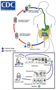
Clinical Syndrome of Cystoisopora belli
rare infections in immunocompetent individuals; more common in immunocompromised patients (AIDs patients)
asymptomatic carriers to mild or severe gastrointestinal disease (leading to dehydration) (rare tissue invasion possible)
Diagnosis of Cystoisospora belli
microscopic examination of concentrated stool sediment using modified acid-fast or safranin stain

Treatment and prevention of Cystisopora belli
trimethoprim-sulfmethoxazole
inhibits synthesis of dihydrofolic acid
use of trimethoprim-sulfamethoxazole for prophylaxis against Pneumonocystis in HIV-infected patients diminishes the likelihood of developing Cystoisoporiasis
education and sanitation
avoid sexual practices that involve fecal-oral contact
Epidemiology of Cryptosporidium sp.
protozoan: coccidian
worldwide distribution
presence in immunocompetent hosts: 1-3% in developed countries; 7-10% in developing countries (2.9 to 4.7 million cases in Africa/Asia within 202,000 deaths)
in the US, an estimated 748000 cases of cryptosporidiosis occur each year
human and animal reservoirs
Risk factors for Cryptosporidium sp
animal handlers
children in daycare centers (especially those under 2)
elderly patients
foreign travel
immunocompromised individuals
contact with positive patients
Life Cycle of Cryptosporidium
infection occurs through ingestion of sporocysts-containing oocysts (infective stage) in fecally contaminated food/water
sporocysts excyst in the small intestine and release their sporozoites which invade the epithelical cells
both asexual and sexual reproduction occurs with production of oocysts
oocysts are excreted in the stool (diagnostic stage)

Clinical Syndrome of Cryptosporidium sp
incubation period of 2-10 days
asymptomatic carriage to acute watery diarrhea for 1 to 2 weeks (healthy individuals); abdominal pain, nausea, mild fever
50 or more stools per day and tremendous fluid loss which can last for months to years (immunocompromised individuals); abdominal pain, nausea, mild fever
shedding of oocysts on average of 9 to 12 days post infection
extraintestinal infection (ex. gallbadder)
Diagnosis of Crytosporidium sp.
microscopic examination of stool specimens for oocysts using:
modified acid-fast/safranin stain or immunofluroscent stain
immunodiganosis
molecular anaylsis

Treatment and prevention of Crytosporidium sp.
nitazoxanide
fluid replacement
education and sanitation
treatment of contaminated water supplies
Epidemiology of Cyclospora cayetanensis
protozoan: coccidian
worldwide distribution
endemic in at least 27 countries with prevalence ranging from 3.3% in Mexico to 41.6% in Peru (average prevalence of 3.5%)
~16000 cases per year in the US
human reservoir
Life Cycle of Cyclospora
infection occurs through ingestion of sporocysts-containing oocysts (infective stage) in fecally contaminated food/water
sporocysts excyst in the small intestine and release their sporozoires which invade the epithelial cells
both asexual and sexual reproduction occurs with production of oocysts
unsporulated oocysts are excreted in the stool (diganostic stage)
oocysts sporulate in the environment and become infectious
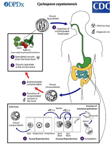
Clinical Syndrome of Cyclospora cayetansis
incubation period is 1-14 days
clinically indistinguishable from cryptosporidiosis and cystoisosporiasis
mild nausea and watery diarrhea that lasts 6-7 weeks (in immunocompromised the diarrhea may last for months)

Diagnosis of Cyclospora cayetanesis
microscopic examination of stool specimens for oocysts using:
modified acid-fast/safrainin stain
molecular analysis
Treatment and prevention of Cyclospora cayetanensis
trimethoprim-sulfamethoxalzole and fluid replenishment
improved personal hygiene and sanitation
care when visiting areas where the protozoan is endemic (generally tropical and sub-tropical regions such as Peru, Brazil, Haiti)
treatment of contaminated water supplies
Epidemiology of Dientamoeba fragilis
protozoan: flagellate (yet does not possess flagella)
worldwide distrubition
estimated prevalence in developed countries = 2-5%
higher rates (19-69%) in those living in crowded conditions
most common age at which infection has been reported in children is 5-10 years
as more common or more common than giardia
human reservoir
Life Cycle of D. fragilis
infection occurs through ingestion of trophozoite (infective stage) in fecally contaminated food/water (possible transmission via helminth eggs)
trophozoites multiply in the large intestine
trophozoites are shed in the stool (diagnostic stage)

Clinical Syndrome of Dientamoeba fragilis
most infections are asymptomatic disease (75-85% of infected individuals)
some patients develop abdominal discomfort, intermittent diarrhea, weight loss (duration 1 to 2 weeks)
Diagnosis of Dientamoeba fragilis
examination of stool specimens for trophozoites (cyst stage?)
Treatment and prevention of Dientamoeba fragilis
iodoquinoal
better sanitary conditions/reduce enterobius infections
Epidemiology of Blasocystis spp.
protozoan: stramenophiles-alveolates-rhizaria super group (ex. diatoms and water molds)
worldwide distribution
reservoirs: humans, farm animals, birds, rodents, amphibians, reptiles, fish, and cockroaches
very common!
Life Cycle of Blastocystis
infection occurs through ingestion of cyst (infective stage) in fecally contaminated food/water
replication occurs in the intestinal tract
cysts and vacualor forms are shed in the stool (diagnostic stage)

Clinical Syndrome of Blastocystis
debate on whether it causes gastrointestinal disease
water or loose stools, diarrhea, abdominal pain, anal itching, weight loss, and excessive gas
Diagnosis of Blastocystis spp.
examination of the cyst forms present in trichrome stained and unstained stool speciments
Treatment and prevention
metronidazole
education and sanitation