Anatomy Grand Slam
 Call Kai
 Learn
Learn Practice Test
 Spaced Repetition
 Match
Match Flashcards
 Knowt Play
1/384

There's no tags or description
Looks like no tags are added yet.
Name | Mastery | Learn | Test | Matching | Spaced | Call with Kai | Chat |
|---|
No analytics yet
Send a link to your students to track their progress
385 Terms
Olfactory Nerve
Cranial nerve 1
*purely sensory
*sense of smell

Optic Nerve
Cranial Nerve II
*purely sensory
*sense of sight

Oculomotor Nerve
Cranial Nerve III
*purely motor
*stimulates the ciliary smooth muscle - constrict pupil and bend lens (parasympathetic funcion
*moves eyeyball by controlling 4 eye skeletal muscles: medial rectus, inferior rectus, superior rectus, and inferior oblique
*attaches to the brainstem
*tested to check for brainstem activity (pupils fixed and dilated means that it is not functioning properly)

Trochlear Nerve
Cranial Nerve IV
*moves eyeball by controlling the superior oblique skeletal muscle

Trigeminal Nerve
*Cranial Nerve V
*responsible for sense of feeling on the face (three branches - forehead, upper jaw, and lower jaw)

Abducens Nerve
Cranial Nerve VI
*purely motor
*moves eyeball (abducts it) by controlling the lateral rectus eye muscle

Facial Nerve
Cranial nerve VII
*Sensory - sense of taste for the front of the tongue. *Somatic Movement of the facial expression skeletal muscles
*parasympathetic stimulation of salivary and lacrimal glands.

vestibulocochlear nerve
cranial nerve VIII
*purely sensory
*carries balance information and hearing to the temporal lobe for processing

glossopharyngeal nerve ("tongue and pharynx")
Cranial nerve IX.
*Sense of taste for the back of the tongue. *Somatic movement of the skeletal muscles for swallowing
*Parasympathetic stimulation of the parotid salivary glands.
*carries baroreptor information from the carotid sinuses

Vagus nerve ("wanderer")
Cranial nerve X.
*Sensory information from the internal organs -- particularly notable for connecting GI activity with the brainstem, and for carrying baroreceptor information from the heart to the brainstem.
*Parasympathetic motor for internal (visceral) organs - slows heart, dilates bronchi, stimulates GI organs, etc.

accessory nerve
Cranial Nerve XI
*purely somatic motor to move trapezius and sternocleidomastoid

hypoglossal ("under tongue")
Cranial Nerve XII
*purely somatic motor
*controls tongue skeletal muscles

vertebral body

spinous process

transverse processes

superior articular facets

lumbar inferior articular process
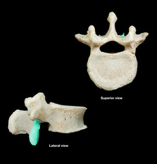
cervical articular facets

C1
(atlas) has no body and no spinous processes, holds up head. Articulates around the dens (C2)
C2
(axis) has a dens/odontoid process projecting superiorly from the body. allows head to rotate.
Thoracic
T1-12

Lumbar
L1-L5

sacrum
posterior wall of the pelvis, S1-S5 fused together
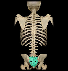
coccyx
tailbone, 3-4 fused sections

cervical vertebrae
C1-C7

Transverse foramina

vertebral canal

Intervertebral foramen
Hole between 2 vertebrae

intervertebral discs
separate and cushion the vertebrae
Trapezius
O: Occipital bone, ligamentum nuchae, spines of thoracic vertebrae
I: Spine and acromion process of scapula, part of clavicle
N: Accessory nerve (XI)
Fx: Fixes, elevates, rotates scapula; extends head

Latissimus Dorsi
O: Spines of T7-Sacrum, iliac crest, 2 ribs
I: Intertubercular sulcus of humerus
N: Thoracodorsal nerve
Fx: Extends, adducts, and medially rotates the arm

Levator Scapulae
O: Transverse processes of C1-C4
I: Medial border of scapula above the root of the spine
N: Dorsal Scapular nerve
Fx: Fixes and elevates the scapula; flexes the neck laterally
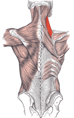
Rhomboid Minor
O: Spine of T1
I: Medial border of scapula
N: Dorsal Scapular nerve
Fx: Fix, retract, and rotate scapula

Rhomboid Major
O: Spines of T2-T5
I: Medial border of scapula
N: Dosal Scapular nerve
Fx: Fix, retract, and rotate scapula

Splenius Cervicis
O: Spines of T1-T6
I: Transverse processes of C1-C4
N: Middle and lower cervical nerves
Fx: Laterally flexes, rotates, and extends the head and neck

Splenius Capitis
O: Lower part of ligamentum nuchae
I: Lateral aspect of the mastoid process and lateral part of the superior nuchal line of the skull
N: Middle and lower cervical nerves
Fx: Laterally flexes, rotates, and extends the head and neck

Illiocostalis
O: Common origin
I: Lower 6 ribs, upper 6 ribs, transverse process of the lower cervical vertebrae
N: Spinal nerves
Fx: Extends and laterally flexes the spine
*Part of Erector Spinae

Longissimus
O: Common origin
I: Transverse processes of thoracic, lumbar, and cervical vertebrae to the mastoid process of the skull
N: Spinal nerves
Fx: Extend and laterally flex the spine; extent and rotates the head
*Part of Erector Spinae

Spinalis
O: Common origin
I: Spines of lumbar vertebrae to spines of upper thoracic vertebrae between T2 and L2 (occasionally up to cervical vertebrae)
N: Spinal nerves
Fx: Extends the vertebral column
*Part of Erector Spinae

Semispinalis
O: Transverse processes of C2-T12
I: Spines of superior vertebrae
N: Spinal nerves
Fx: Rotates and extends the vertebral column of the trunk

Semispinalis Capitis
O: Transverse processes of T1-T6
I: Medial part of the occipital bone between the superior and inferior nuchal lines
N: Spinal nerves
Fx: Rotates and extends the head and neck

Multifidus
O: Vertebral arches from C2-S4
I: Spines along the entire vertebral column from C2-S4
N: Spinal nerves
Fx: Laterally flexes, rotates, and extends the vertebral column

List the five types of vertebrae and indicate the number of vertebrae of each type
7 Cervical Vertebrae
12 Thoracic Vertebrae
5 Lumbar Vertebrae
5 Sacral Vertebrae
3-4 Coccyx Bones fused together
Specify the actions and innervation of the intrinsic back musculature
Intrinsic back muscles move the vertebral column and provide support. Innervated by the posterior rami of spinal nerves.
Explain the developmental basis of the medullary cone occurring at the L1 vertebral and its effect on the relationship between the spinal cord level of origin and location of vertebral canal exit for the lower lumbar and sacral spinal nerves
The vertebral column grows much faster than does the spinal cord. The result is a longer vertebrae than spinal cord. The conus medularis (end of spinal cord) is around L1-L2. The spinal nerves below will run down in the cauda equina and leave at their respective level.
Where does C1 articulate with the skull at?
Each lateral side articulates with the occipital condyle of the skull. Known as the 'atlanto-occipital' joint. This allows the head to nod up and down on the vertebral column.

What separates a cervicle vertebrae from other types?
Transverse foramen, which the vertebral artery and the vertebral plexus of veins will run through.

What separates a thoracic vertebrae from other types?
Demifacets and costal facets

What separates a lumbar vertebrae from other types?
Extra processes, mammillary body, large vertebral body

What is a zygapophyseal joint?
Joint formed by the inferior articulating process emanating from the level above a superior articulating process emanating from the level below.

Pars Interarticularis
The plate of bone that intervenes between the superior and inferior articular processes.

What are the extrinsic muscles of the back?
Trapezius, latissimus dorsi, Levator scapulae, Rhomboid muscle group.
What are the intrinsic muscles of the back?
Erector spinae muscle group: Iliocostalis, Longissimus, and Spinalis.
What are the names of the 3 transversospinalis muscles
Semispinalis Capitis, Splenius Capitis, and Splenius Cervicis. Splenius has a capitis and a cervicis component.

Pedicle

Lamina

Superior articular process

Inferior articular facet
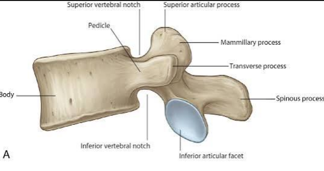
Uncinate process
Raised ring around cervical vertebral body

Costal demifacets

Mammillary process

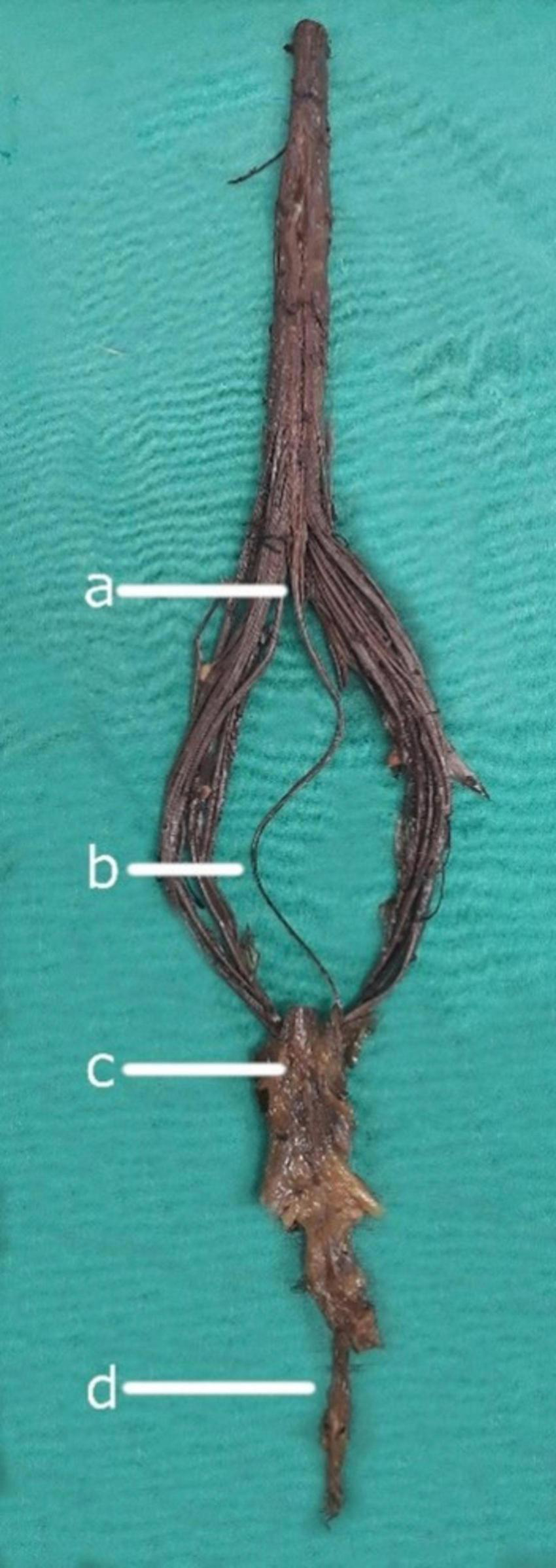
Denticulate ligaments

Filum terminale
C1 Dermatome
Top of skull

C2 Dermatome
Temple, forehead, occiput

C3 Dermatome
Neck, posterior cheek, temporal area, under mandible
Paresthesia: Cheek, side of neck

C4 Dermatome
Shoulder, clavicle area, upper scapular area
Paresthesia: Horizontal band along clavicle and upper scapula

C5 Dermatome
Lateral side of elbow
Deltoid, anterior whole arm to base of thumb

C6 Dermatome
Anterior arm, radial side of hand to thumb and index finger
Paresthesia: thumb and index finger

C7 Dermatome
medial arm and forearm to middle finger
Paresthesia: index and middle finger

C8 Dermatome
Ring and pinky, ulnar side of forearm, and medial arm
Paresthesia: Little finger, ulnar side of ring finger
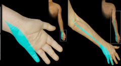
T1 Dermatome
Medial side of forearm/elbow
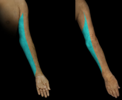
T2 Dermatome
Medial upper arm (pit), sub clavicle area

T4 Dermatome
Level of nipple line

T10 Dermatome
Level of belly button

L1 Dermatome
Upper groin and lower back
Inguinal ligament area
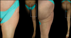
L2 Dermatome
Lower groin, anterior thigh and knees, upper buttocks
Paresthesia: anterior thigh

L3 Dermatome
Sacral area, knees, medial thigh from groin to knee
Above the knee

L4 Dermatome
Medial lower leg anterior and posterior
Check above inner ankle

L5 Dermatome
Anterior tibial region, toes
Shoelace area

S1 Dermatome
Lateral foot, plantar aspect of foot, gastroc/soleus area
Paresthesia: lateral foot and toes and plantar aspect of foot
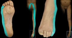
S2 Dermatome
Posterior thigh
Back of knee

S3 Dermatome
Buttock region

S4 Dermatome
Rectum/anal area, lower sacrum
Paresthesia: saddle area

C1, 2 Myotome
head flexion/extension

C3 Myotome
head Lateral Neck Flexion

C4 Myotome
Shoulder Elevation Depression

C3,4,5 Myotome
Keeps the diaphragm alive
C5 Myotome
shoulder abduction

C6 Myotome
elbow flexion, wrist extension
C7 Myotome
elbow extension, wrist flexion
C8 Myotome
Thumbs up and UD, finger/wrist flexion

T1 Myotome
abduction/adduction of fingers

L2 Myotome
Hip flexion
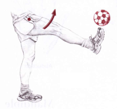
L3 Myotome
Hip abduction

L4 Myotome
knee extension

L5 Myotome
Dorsiflexion and great toe extension

S1 Myotome
Plantar flexion

S2 Myotome
Knee flexion

Sternum
