Female / Male Reproductive System
 Call Kai
 Learn
 Practice Test
 Spaced Repetition
 Match
 Flashcards
 Knowt Play
1/50

There's no tags or description
Looks like no tags are added yet.
51 Terms
tail of spence
aka axillary tail or axillary process
an extension of normal breast tissue that reaches from the upper outer quadrant of the breast into the armpit (axilla)
highest rate of breast cancer occurs here

female vs male breast
Gynecomastia is the term for excessive development of breast tissue in males. It's often caused by an imbalance of hormones, specifically an increase in estrogen relative to testosterone. This can occur naturally during puberty, infancy, or later in life, or it can be caused by certain medications, diseases, or other factors
Doesn’t typically increase the male’s risk for developing breast cancer
It depends on why it happened in the first place (like if they were taking steroids, then it is likely for them to develop breast cancer)
The glandular tissue is what produces the milk
Male breast tissue also has ducts but it doesn’t have the glands that actually produce the milk

breast anatomy and physiology
breast overlies pectoralis major
breast is a hormonally sensitive tissue
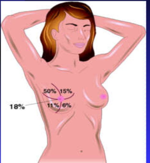
upper outer quadrant → site of more breast cancers
breast composed of glandular tissue, fibrous tissue including suspensory ligaments, adipose tissue
proportion varies with age, cycle, pregnancy, lactation and general nutrition
suspensory ligaments (Cooper’s ligaments) support breast tissue, contract in cancerous breast
produce pits or dimples in overlying skin
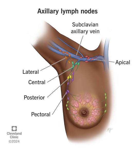
breast lymphatics
central
pectoral (anterior)
subscapular (posterior)
lateral
breast cancer risk factors
family history / genetics
menstrual history
pregnancy
age
gender
education and income
location
race
a good history is imperative
breast cancer risk is associated with surges of estrogen in the body
getting your period at a young age is a risk for breast cancer
also menopause later than 55
(becauase you are having more periods = more surges of estrogen
menarche
the first menstrual period in a female adolescent
usually occurs at age 12
the age in the US has decreased to about 8 yrs old
this is due to diet (hormones in the milk → the cows are pumped with steroids to produce more), same with chicken (but milk is consumed more than chicken in children)
menopause
the stage of a woman’s life when her menstrual periods stop permanently, and she can no longer get pregnant
not a disease or disorder — normal part of aging for women
the time leading up to this is called the menopausal transition or perimenopause
usually occurs between age 48 and 55
the absence of your menstrual cycle for 12 consecutive months
typically genetically based
amenorrhea
absence of period
could be caused by eating disorders, stress, polycystic ovarian syndrome, *pregnancy, sometimes breast feeding (but you can still ovulate)
polymenorrhea
many periods
average cycle 28-30 days
this would be getting your period in 21 days or less
also an increased risk of breast cancer because of the amount of periods that you have
dysmenorrhea
painful periods
heavy bleeding, nausea, headaches, etc.
painful or uncomfortable menstrual cramps. It is a common condition that affects many women, characterized by severe cramping in the lower abdomen, often accompanied by other symptoms such as nausea, vomiting, diarrhea, and fatigue.
pregnancy and breast cancer risk
decreases your risk because you aren’t getting your period for 9 months
a person who has had 1 pregnancy vs person who had 5 pregnancies
the person who had 1 pregnancy is more at risk (the person with 5 has had their period gone for longer time)
age and breast cancer risk
your risk goes up the older you are
over 60
women are more at risk because of their exposure to estrogen all their life
education / income and breast cancer risk
people with a higher income bracket are at higher risk (because people aren’t having children earlier → women are having less children because they are career focused)
but people with low income are more likely to die because of breast cancer (can’t afford screening or treatment)
urban areas are higher risk → pollution and other toxic environmental factors
race and breast cancer risk
white / caucasian have higher risk
African Americans have higher mortality rate due to more aggressive tumors
family history and breast cancer risk
first degree relatives (sister, mother, etc) who have had breast cancer increase your risk
genetics play a part in increased breast cancer
BRACA1 and BRACA2 are tumor suppressing genes
when they have a mutation, it could cause a SIGNIFICANT increase in breast cancer risk in females and ovarian cancer (increased breast cancer risk for male gender as well)
mutations are highest in the Ashkenazi Jewish population
what tissues have increased risk of breast cancer
upper outer is the most common
that’s why it is important to check the axilla region because that tissue goes up there
self breast exam
First starts in the shower
Lift up your left arm and exam left breast
Then do the same for the other arm
Mirror
Put your hands on your hips and look in mirror
Look for any dimpling (not stretch marks)
It’s like when you see a concavity (tumor pulling tissue in)
Are there any difference on your nipples
Lie down
Do the same thing that you did in the shower except you are lying down
Females should do a self exam 5 to 7 days after your period starts if you are female
Male gender → just choose a day to exam every month

clinical breast exam
same as self breast exam except someone does it for you
techniques of breast exam - inspection
pt sitting, disrobed to the waist, arms at sides
inspect breasts, note appearance of skin; color, erythema from infection or inflammatory cancer
the skin may have ridges or appear pitted like the skin of an orange (called peau d’orange)
if you touched it, it would be stiff and bumpy
size and symmetry of breasts
contour
drainage from the nipple would be like clear or milky
inspect nipples; note size and shape, direction in which they point, rashes or ulceration, discharge
check for dimpling or retraction
techniques of breast exam - palpation
ask pt to lie down
bring pts arm overhead of the breast being examined, use pads of first 3 fingers, compress tissue gently
use a pattern: vertical strip pattern
note: consistency of tissues, tenderness, nodules
do not forget the tail of Spence and axillary region
palpate each nipple
compress the areola with your index finger, watch for discharge
inspect nipple and areola for nodules, swelling, ulceration
palpate the areola for nodules
breast exam - if you find a nodule
location
look at breast like a clock for location and how far from the nipple it is
approximate size
shape
does it feel round, does it feel like a pea, is it disk shaped
consistency
is it soft or hard (soft is usually a cyst and goes away)
movable
movable is good but if it is fixed it might be cancer
nipple changes
lymphadenopathy
are the lymph nodes that are associated with the breast enlarged
breast cancer screening
encourage self breast exam (SBE) monthly, 5-7 days after onset of menses
they should continue after menopause
age 20-39 clinical breast exam (CBE) every three years
40 and older should get a clinical breast exam yearly along with mammography
for women at increased risk of cancer, mammography should be initiated at 30 years of age
after 70 the benefits of mammography is less well defined
male breast: SBE monthly, CBE every 1-3 years
breast cancer awareness
cumulative lifetime risk factor for developing breast cancer is
1 in 7
choose to be proactive
choose to live
external female genitalia (perineum)
labia majora
labia minora
vestibule
introitus → the external opening to the vagina, leading into the vaginal canal.
perineum → the sensitive region of soft tissue and skin located between the vaginal opening and the anus
urethral meatus
skene’s glands → two small glands located on either side of the urethral opening in female
They secrete a mucus-rich fluid that helps moisten the urethral and vaginal opening.
bartholin’s glands → are two small, pea-sized glands located on each side of the vaginal opening (near the 4 o'clock and 8 o'clock positions)
They secrete a small amount of clear, mucoid alkaline fluid that helps lubricate the vaginal opening, especially during sexual arousal

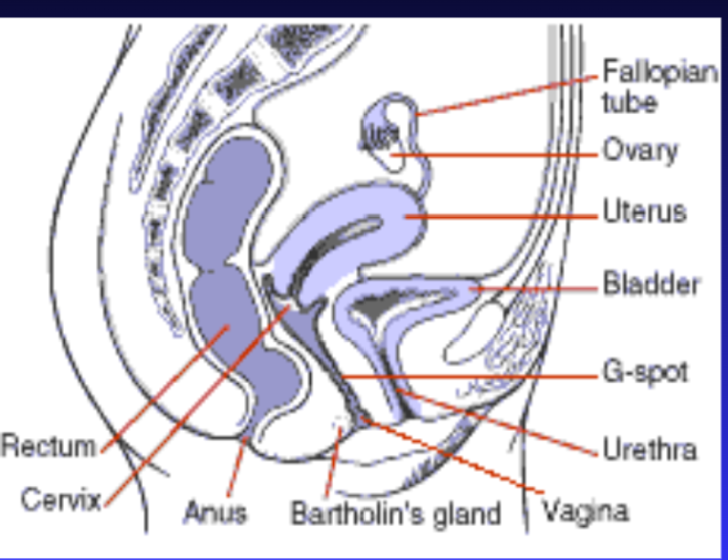
internal female genitalia
vagina
uterus
cervix
external Os → the external opening of the cervix that projects into the upper end of the vagina
fallopian tubes
ovaries
adnexa = fallopian tubes, ovaries and supporting structures
interview: review of systems (female)
menstrual history
sexual history
medical / surgical history
medication
any complaints
last CBE, SBE compliance, mammography, Papanicolaou exam (pap smear), DRE (digital rectal exam)
pap smear / pap test
a screening procedure used to detect abnormal cells in the cervix, the lower part of the uterus that opens into the vagina
the test helps identify cellular changes that could develop into cervical cancer if left untreated
during a pap smear
a speculum is inserted into the vagina to visualize the cervix
a small brush or spatula is used to collect cells from the cervix
the cells are sent to a laboratory for exam under a microscope
pap smears can detect
precancerous cervical cell changes
cervical cancer
some infections or other cervical abnormalities
pap testing is often performed during a routine gynecologic exam and may be combined with human papillomavirus (HPV) testing, since certain types of HPV are the primary cause of most cervical cancers
mammographies begin when you are 40
female external genitalia examination
remember safety → 2 professionals in room during exam with patient
rose of the RN → assist practitioner during gynecological exam, support patient
empty bladder
position and drape appropriately
inspect external genitalia; separate labia majora and inspect
labia minora
clitoris
urethral meatus
introitus
note: inflammation, ulceration, discharge, swelling, nodules, palpate any lesions
clear or milky white is normal or discharge
if it’s like cottage cheese consistency, then it’s an infection (like vaginal thrush or vulvovaginal candidiasis or a vaginal yeast infection)
around rectal area, look for hemorrhoids
an engorged vein
female internal genitalia examination
locate the cervix
assess support of the vaginal wall
cystocele
uterine prolapse
rectocele
insert speculum
inspect cervix and Os
pap smear
cystocele
when the wall between the bladder and the vagina weakens.
this can cause the bladder to drop or sag into the vagina.
it may be caused by things that increase pressure on the pelvic muscles.
treatment depends on the severity
rectocele
type of prolapse where the supportive wall of tissue between a woman’s rectum and vaginal wall weakens
without the support of these pelvic floor muscles and ligaments, the front wall of the rectum sags and bulges into the vagina
uterine prolapse
occurs when the muscles and tissue in your pelvis weaken
the weakness lets the uterus drop down into your vagina
sometimes, it comes out through your vaginal opening
nearly half of all women ages 50 and 79 have this condition
leukorrhea
refers to a whitish or yellowish vaginal discharge
common and typically normal physiological phenomenon that occurs in women of reproductive age
yeast discharge
white curd-like discharge from the vagina
refers to the thick, white, and often chunky discharge associated with a vaginal yeast infection (candidiasis)
it’s often described as having a cottage cheese-like consistency and usually lack a strong odor
bimanual exam
a clinical procedure where a healthcare provider uses two hands—one hand with lubricated fingers inserted inside the vagina and the other hand pressing down on the lower abdomen—to feel and evaluate the internal female reproductive organs
palpate the cervix
palpate the uterus
palpate each ovary
rectovaginal exam
fingers go back in, and press on abdomen to bring uterus down to feel if they feel anything abnormal
a physical examination where a healthcare provider inserts one lubricated, gloved finger into the vagina and another finger of the same hand into the rectum to assess the pelvic organs and the tissue between them
also press on the sides
digital rectal exam (DRE): female
recommended starting at 50 years of age or based on individual risk factors to evaluate for rectal masses, and other anorectal abnormalities
check for rectal sphincter tone
Guaiac stool for occult blood
check posterior portion of uterus
check for hemorrhoids
check for STD’s → herpes, venereal warts

DRE female process
Take a gloved hand (you should double glove)
Sphincter tone → put fingers in and you should feel rectal tone
When you feel the anterior portion of their rectum, you can also feel the posterior portion of the uterus
You can also check for occult blood → blood that is hidden from the naked eye
When you put your fingers in someone’s rectum, you’re going to have stool on your fingers, then you have a test that you can do to check (it’s like a card that you wipe the stool on) → there’s a picture of it in later slides
Occult blood could be a sign for colon cancer
Hemorrhoids could be internal or external
If there are hemorrhoids at the entrance to the rectum, ask the doctor if you should still do the exam (because if you accidentally poke one, then it can burst and it will be very bad)
hemorrhoid
swelling of the veins or blood vessels in and around your anus and lower rectum
this happens when there is extra pressure on these veins
either inside your anus (internal) or under the skin around your anus (external
about half of all people will have hemorrhoids by age 50
male genitalia
shaft of the penis
glans
prepuce/foreskin
urethra
urethral meatus
scrotum
testes
A circumcised penis is one in which the foreskin has been surgically removed, leaving the glans penis exposed.
An uncircumcised penis is a penis with the foreskin intact, covering all or part of the glans.

male genitalia assessment findings
Foreskin covers the glans when the penis is flaccid.
Foreskin should be retractable in most adolescents and adults, allowing visualization of the glans.
Observe for redness, swelling, lesions, discharge, or difficulty retracting the foreskin.
Left testicle should be lower
Left = Lower
This is because sperm are heat sensitive so if they were on top of each other, it would be producing heat
Testicular cancer is common in white males
15 to 40 years old
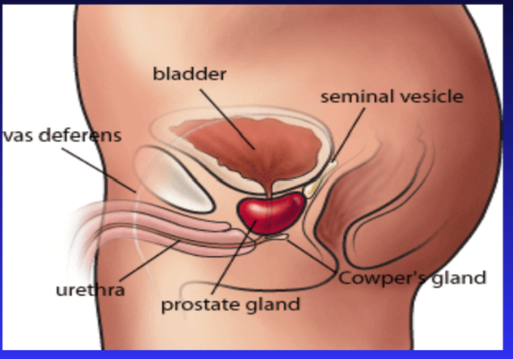
prostate
Prostate cancer risk increases after age 50yrs, most cases occur around 65 yrs of age
Around 50 men can have enlarged prostate gland
Benign prostatic hyperplasia (BPH) is a common condition in older men where the prostate gland enlarges, but it is not cancerous. This enlargement can cause various urinary symptoms as the prostate presses against the urethra and bladder.
Common Symptoms of an Enlarged Prostate
Frequent urination, especially at night (nocturia)
Urinary urgency (sudden, strong urge to urinate)
Difficulty starting urination (hesitancy)
Weak or slow urine stream
Intermittent urine stream (starts and stops)
Dribbling at the end of urination
Feeling that the bladder does not empty completely
Straining to urinate
An enlarged prostate may also be associated with:
Difficulty achieving or maintaining an erection
Reduced sexual satisfaction
Lower urinary tract symptoms that interfere with sexual activity
If you put your fingers on the anterior portion of their rectum, you would be able to palpate the prostate
hernias in males
the protrusion of an organ, tissue, or part of the intestine through a weak area or opening in the muscle or connective tissue that normally contains it
common signs and symps
visible or palpable bulge under the skin
bulge may become more noticeable when coughing, straining, lifting or standing
feeling of pressure, heaviness, or discomfort
pain or tenderness at the site of the hernia
symptoms may improve when lying down
common types
inguinal
femoral
umbilical
inguinal hernia
occurs in the groin
most common type
femoral hernia
occurs in the upper thigh near the groin
umbilical hernia
occurs near the navel (umbilicus)
interview: review of systems (male)
sexual history
medical/surgical history
medications
any complaints
last DRE, PSA blood test
PSA test measures the level of prostate-specific antigen in the blood. While there isn't a single universally accepted normal range, a PSA level of 0-4 ng/mL is often considered the general range for men, with levels above 4 ng/mL often prompting further investigation to rule out prostate cancer.
clinical testicular and breast exam
self testicular and breast exam compliance
exam of male genitalia
penis
inspection
foreskin, smegma
phimosis (condition in which tight foreskin in an uncircumcised male can't be pulled back over the head of the penis.)
paraphimosis (occurs when the foreskin is retracted behind the corona (or crown) of the penis and cannot be returned to the unretracted position. this can cause entrapment of the penis, impairing the drainage of blood, and may be serious)
glans
hypospadias (congenital condition in which the urethral opening (meatus) is located on the underside of the penis rather than at the tip of the glans)
scrotum / testicles
inspection and palpation
testicular cancer
indications: swollen, painful, hydrocele
transillumination of the scrotum
hydrocele (fluid filled sac that develops around one or both testicles, causing swelling in the scrotum. it’s common in newborns, often resolving on its own, but can also occur in older males due to injury or infection. while usually painless, large hydroceles can cause discomfort)
hernias: inguinal and femoral

cryptochidism
a condition in which one or both testicles fail to move from the abdomen, where they develop before birth, into the scrotum
may increase the risk for development of tentacular cancer
also called undescended testicles
prostate exam
role of nurse: assist practitioner, support patient
patient should stand and bend over or side lying
shape: heart with palpable central groove
size: 2.5 cm x 4 cm wide
consistency: elastic, rubbery
nodules if present could indicate cancer
tenderness: nontender to palpation
mobility: slightly mobile
should not protrude more than 1cm into the anterior wall of the rectum
during a DRE, the prostate can be felt through the anterior (front) wall of the rectum
digital rectal exam (male)
recommended starting at 50 years of age
check for rectal sphincter tone
guaiac stool for occult blood
check for prostate enlargement
check for hemorrhoids
check for STD’s → herpes, venereal warts
Recommended in males beginning around age 50 or based on individual risk factors to evaluate for rectal masses, prostate enlargement and other anorectal abnormalities.