PARASITES
 Call Kai
 Learn
 Practice Test
 Spaced Repetition
 Match
 Flashcards
 Knowt Play
1/19

There's no tags or description
Looks like no tags are added yet.
20 Terms
parasite (geo, exposure, diagnostic?, morphology)
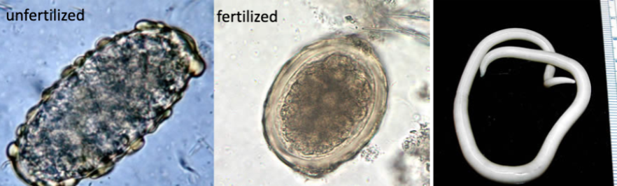
ascaris lumbricoides — ROUNDWORM
GEO: global, burden highest in tropical/subtropical regions & rural SE USA
most common helminthic infection
EXP: ingestion of fertilized ova
Diagnostic & Infectious
Ova:
Fertilized
45 - 60 um, cleavage of internal yolk
yellow/brown (bile)
oval/spherical
Thick hyaline shell
Unfertilized
80-90 um x 40 width
Difficult to recognize in stool – vegetable cell
Adult Worm:
15 - 35 cm
Male worms smaller, curved tail
smooth without striations

parasite (geo, exposure, diagnostic?, morphology)
Clonorchis sinensis, Chinese Liver Fluke OVA
•Trematode – Liver Fluke
•Geographic Distribution:
§Endemic to East Asia and Eastern Russia
•Exposure:
§Ingestion of freshwater fish
§Intermediate Host: Freshwater fish
•Diagnostic: Eggs in stool or duodenal aspirate
•Morphology:
§Size: 30 um x 15 um
§Convex operculum at shoulders of egg
§Knob/hook protrusion at abopercular end
§Indistinguishable from Heterophyes heterophyes and Metagonimus yokogawai ova

parasite (geo, exposure, diagnostic?, morphology)
Diphyllobrium latum (giant fish tapeworm) ova
cestode
GEO: freshwater in northern hemisphere & SA
Exposure: consuming undercooked fish
Diagnostic ova
OVA:
55-75 um x 40-55 um
Smooth shell, non-shoulder operculum (only operculate cestode)
Lack of shoulder differentiates paragonimus westermanii
Knob-like thickening at abopercular end
WORM:
Single proglottids rare in stool, distinct to organism
Diagnosis based on proglottid & ova morphology
Segments wider than longer
Uterus located in center of gravid proglottid
Contains coiled uterus in form of compact

parasite (geo, exposure, diagnostic?, morphology)
Enterobius vermicularis (pinworm) - roundworm nematode
Pinworm
worldwide
consuming ova & eggs can be aerosolized
infectious & diagnostic
Ova: collect early morning – tape prep
30 um x 50 um
Thin, smooth, transparent shell
Oval & asymmetrical with flattened side
Contains developed larvae
Infectious & diagnostic (eggs can be aerosolized)
fecal-oral
Adult Worm:
Female – 13 mm x 0.5 mm
Male – 2 - 5 mm
Thin flexible outer covering
Fin-like expansion at anterior end
Long pointed tail

parasite (geo, exposure, diagnostic?, morphology)
hymenolepis nana (dwarf tapeworm)
Cestode - tapeworm
GEO: worldwide, most common cestode infection
EXP: fecal-oral, only cestode with NO immediate host
Diagnostic
Ova: Eggs immediately infectious when passed in fecal material
30 - 50 um, oval
Thin outer shell with hexacanth (6 hooklets) & pair of polar filaments
Adult Worm:
15-44 mm in length, may be confused with pinworm
Associated with mucous threads, not common in stool
Small scolex with protruding rostellum with 20-30 hooklets

parasite (geo, exposure, diagnostic?, morphology)
hymenolepis diminute (rat tapeworm)
Cestode - tapeworm
GEO: worldwide, zoonotic
EXP: zoonotic
Diagnostic
Ova: not infectious person to person
70 - 80 um, round to oval
Striated outer membrane with space between 2 membranes is smooth
Oncosphere – hexacanth (6 hooklets)
Adult:
20 - 60 cm length, short & wide proglottids with 3 oval testes

parasite (geo, exposure, diagnostic?, morphology)
Ancylostoma duodenale & necator americanus (hookworms)
Ancylostoma duodenale (Old World) & necator americanus (hookworms) (New World)
Eggs are indistinguishable
Differentiate by mouth:
Ancylostoma = 2 pairs cutting teeth
Necator = cutting plate
Distinguished from strongyloides by pointed tip of tail
Cause anemia
Larvae passed in stool & penetrate skin, carried through bloodstream into lungs & swallowed
Adults attach to intestine causing blood loss & IDA
In stool
Ova:
2500-5000 eggs/day
60 um x 40 um
Oval, thin-shelled, smooth
Non-pigmented & transparent
Adult Worm:
Up to 1.5cm length
Upper intestine attached to mucosa by biting “hooks”
Differentiate by mouth:
Ancylostoma = 2 pairs cutting teeth
Necator = cutting plate

parasite (geo, exposure, diagnostic?, morphology)
Morphology of Taenia saginata (Beef Tapeworm) & Taenia solium (Pork Tapeworm) ova
GEO: endemic to SA, Africa, China
EXP: undercooked beef (saginata)/pork (solium)
Consumption of T. solium ova can cause cystericosis
Cannot differentiate ova
Ova:
30 - 35 um
Radial striated edge
Internal oncosphere contains 6 refractile hooks
Lab contaminant
Adult Worm: scolex & proglottid needed for ID
Gravid proglottids longer than wide
T. saginata:
4-10m
1000-2000 proglottids
12-40 lateral branches
Unarmed scolex
T. solium:
2-4m
800-1000 proglottids
7-13 lateral branches
Armed scolex

parasite (geo, exposure, diagnostic?, morphology)
Trichuris Trichuria, Whipworm – Adult Worm
§Size:
•Males: 30-45 mm x 1 mm, coiled posterior
•Females: 35-50 mm x 1 mm, straight posterior
§Long whip-like anterior
§Reside in large intestine, cecum, & appendix

parasite (geo, exposure, diagnostic?, morphology)
Trichuris Trichuria
Ova:
50-55 um x 20-25 um
Barrel - shaped, thick shell, polar plugs at each end
Unembryonated when passed in stool
Adult Worm:
Males = 30-45 with coiled posterior
Females = 35-50 mm with straight posterior
Whip-like anterior
In large intestine, ceum, appendix

parasite (geo, exposure, diagnostic?, morphology)
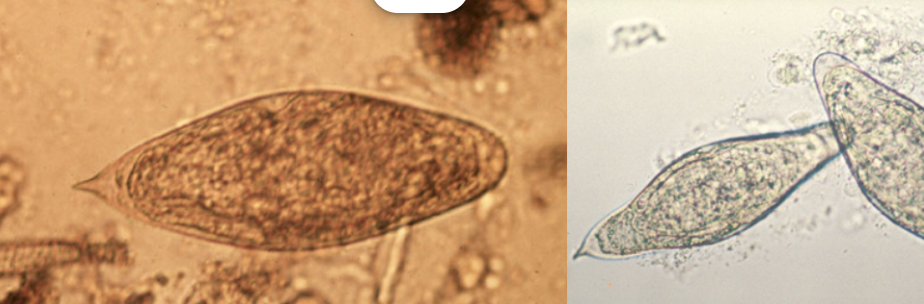
Schistosoma mansoni (Blood Fluke)
Fluke, trematode
GEO: sub-saharan Africa & some SA countries
EXP: laravel penetration through skin & freshwater
Diagnostic
Ova:
Large, 114-180 um x 45-70 um
Prominent lateral spine near posterior
Anterior is tapered & slightly curved
excrete d in stool
Contain mature miracidium

parasite (geo, exposure, diagnostic?, morphology)
Schistosoma japonicum (Human Blood Fluke)
Tematode – fluke
GEO: china, philippines, sulawesi
EXP: snail intermediate host, larvael penetration
Diagnostic
Ova:
Large & rounded
70-100 um x 55-64 um
Spine is smaller & less conspicuous than other species
Shed in stool

parasite (geo, exposure, diagnostic?, morphology)
Schistosoma japonicum, Blood Fluke – Male
§Size:
•Males: 1.2 cm x 0.5 mm
§Spines on oral & ventral suckers
§Flatworm with hooks

parasite (geo, exposure, diagnostic?, morphology)
Schistosoma japonicum, Blood Fluke – Female
Size:
•Females: 2 cm x 0.4 mm
§Spines on oral & ventral suckers
§Female integument is rigid with fewer spines than in oral/ventral sucker
§Flatworm with hooks

parasite (geo, exposure, diagnostic?, morphology)
Schistosoma japonicum, Blood Fluke – Male & Female in Copulum
Size:
•Males: 1.2 cm x 0.5 mm
•Females: 2 cm x 0.4 mm
§Spines on oral & ventral suckers
§Female integument is rigid with fewer spines than in oral/ventral sucker
§Flatworm with hooks

parasite (geo, exposure, diagnostic?, morphology)
Schistosoma haematobium (Urinary Blood Fluke)
GEO: Africa & ME
EXP: larvael penetration through skin
Diagnostic
Ova:
Large, 110-170 um x 40-70 um
Terminal spine
Eggs with mature miracidium
Shed in URINE

parasite (geo, exposure, diagnostic?, morphology)
Trichinella spiralis, Pork Worm – Encysted Larvae
•Geographic Distribution: Worldwide
•Exposure:
§Undercooked meat
•Infectious & Diagnostic
•Morphology:
§Size: 152 um x 117.5 um
§Coiled larvae (C shaped)

parasite (geo, exposure, diagnostic?, morphology)
Trichinella spiralis, Pork Worm – Adult Worm
•Geographic Distribution: Worldwide
•Exposure:
§Undercooked meat
§Neither? Infectious or Diagnostic
§Morphology:
§Size:
•Males: 1.2 mm - 1.6 mm length
•Females: 2.2 mm – 3.5 mm length
§Small, slender
§Tapers at anterior
§Collagenous cuticle with ridges & creases

parasite (geo, exposure, diagnostic?, morphology)
vegetable spiral fiber
parasite (geo, exposure, diagnostic?, morphology)
Schistosoma intercalatum
\Restricted to east-central Africa
Eggs similar to s. Haematobium in shape with terminal spine
Longer, 140-240 um
Equatorial bulge & shed in stool