Lesson 10 - Anatomy
 Call Kai
 Learn
 Practice Test
 Spaced Repetition
 Match
 Flashcards
 Knowt Play
1/121

There's no tags or description
Looks like no tags are added yet.
122 Terms
Spinal cord extends from hindbrain and terminates as ____
Conus medullaris at L1-2
From L1-2, spinal cord extends through S as the ___
Cauda equina
The spinal cord is covered by all 3 meninges down ____
S2
Only the pia mater extends to the posterior coccyx as the ____
Filum terminale
The spinal cord is secured to the vertebrae by the _____ of the pia mater.
denticulate ligaments

Conus medullaris
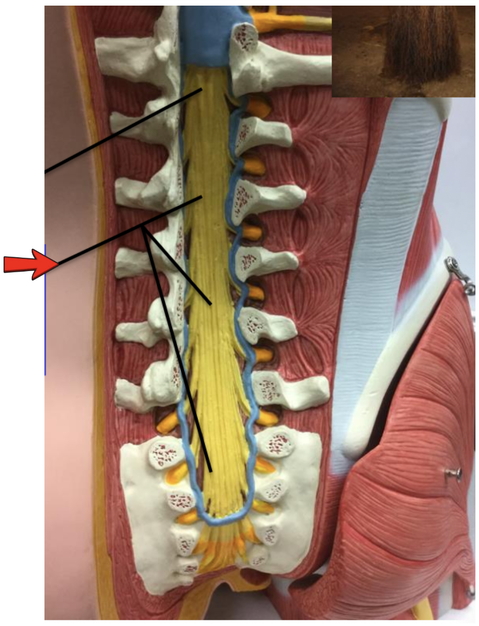
Cauda equina

Cauda equina
Gray matter:
location of neuronal cell bodies
Dorsal (posterior) horns:
Interneurons and sensory fibers from dorsal root ganglion (ie senses)
Ventral (anterior) horns:
Motor neurons of somatic nervous system that project fibers through the ventral root (ie movement)
Lateral horns:
Motor neurons of autonomic nervous system (sympathetic) which leaves cord through ventral root
Gray commissure:
Connects the left and right horns; surrounds the central canal

Epidural space (contains fat)

Subdural space

Subarachnoid space (contains CSF)

Spinal nerve
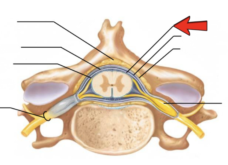
Pia mater

Arachnoid mater

Dura mater

Dorsal root ganglion

Gray commissure

Dorsal horn

Ventral horn
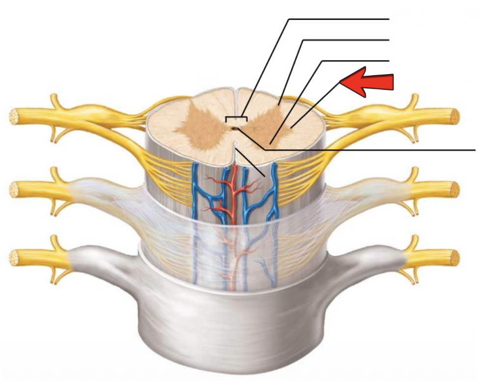
Lateral horn

Central canal

Gray matter
White matter:
myelinated nerve fibers, bisected by fissures (anterior median fissure and posterior median sulcus)
Posterior funiculus:
Carries information on joint/muscle position (proprioception) and fine touch
Lateral funiculus:
carries information on propriopception, pain,
temperature, pressure, coarse touch; also used in relaying motor
information to control skeletal muscles, posture, visceral muscle
functions, muscle tone, and balance
Anterior funiculus:
carries information on pain, temp, pressure,
coarse touch; also used in relaying motor information to control
skeletal muscles, posture, visceral muscle functions, muscle tone,
and balance
How to identify dorsal vs ventral:
Dorsal median sulcus is shallower than
the ventral median fissure (deeper than
the sulcus)
Dorsal root attached to the dorsal root
ganglion and the dorsal spinal cord
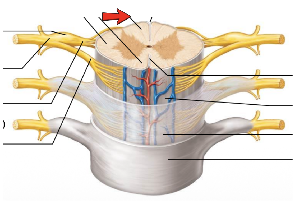
Dorsal funiculus

Ventral funiculus

Lateral funiculus
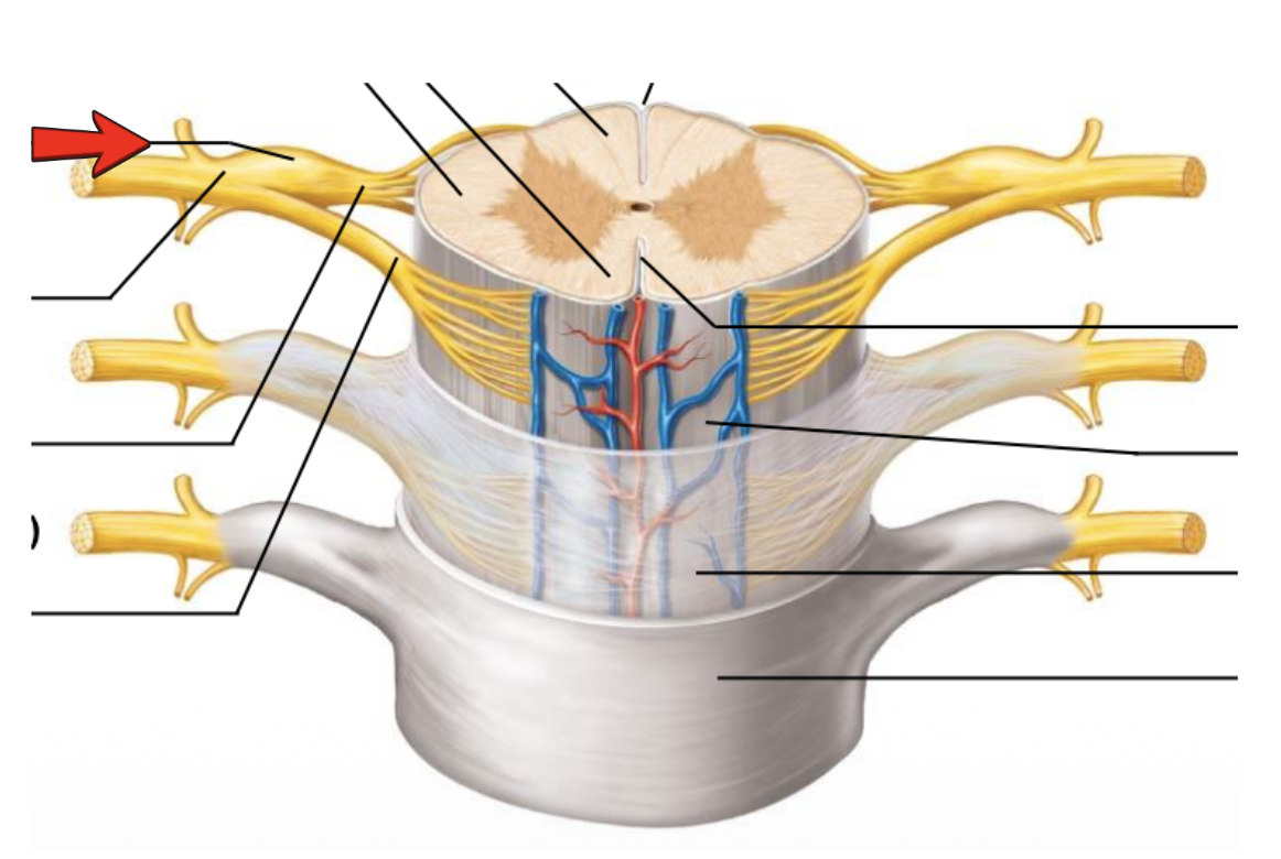
Dorsal root ganglion

Spinal nerve

Dorsal root (fans out into dorsal rootlets)
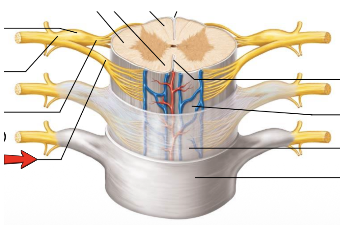
Ventral root (derived from several ventral rootlets)
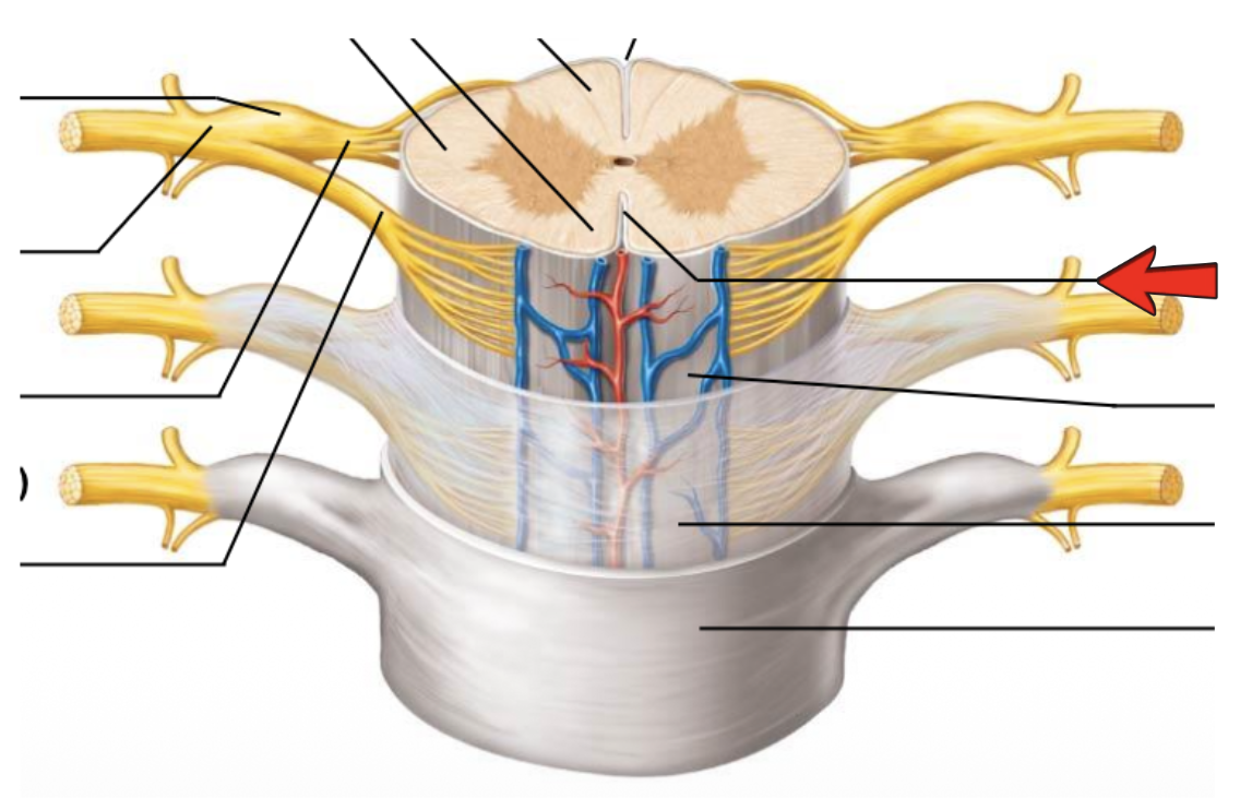
Vetral median fissure

Pia mater

Arachnoid mater

Spinal dura mater
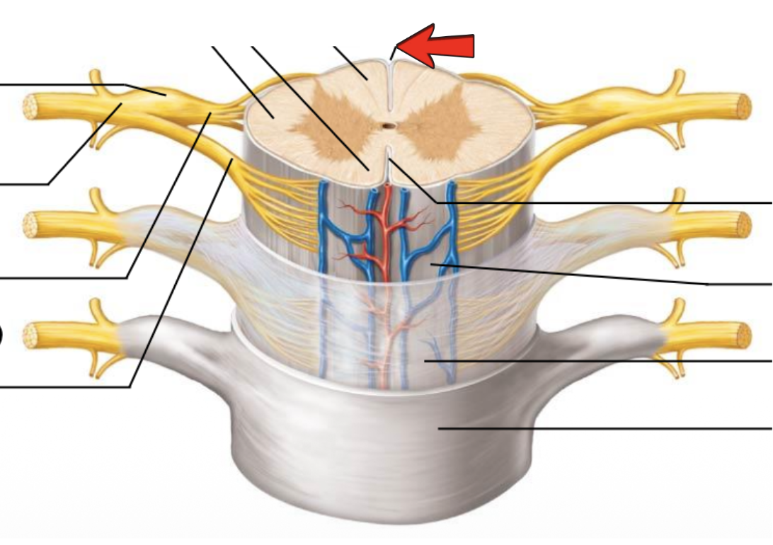
Dorsal median sulcus

White columns
How many pairs of spinal cords:
31 that leave through intervertebral foramina after C2
4 major nerve plexuses:
Cervical, brachial, lumbar, sacral

Cervical plexus C1-C5
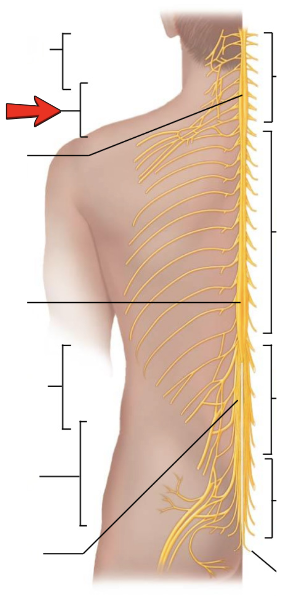
Brachial plexus C5-T1

Cervical enlargement
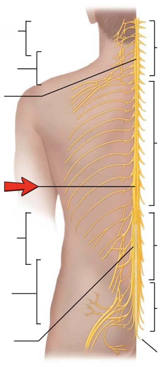
Lumbar enlargement

Lumbar plexus L1-L4

Sacral plexus L4-S4
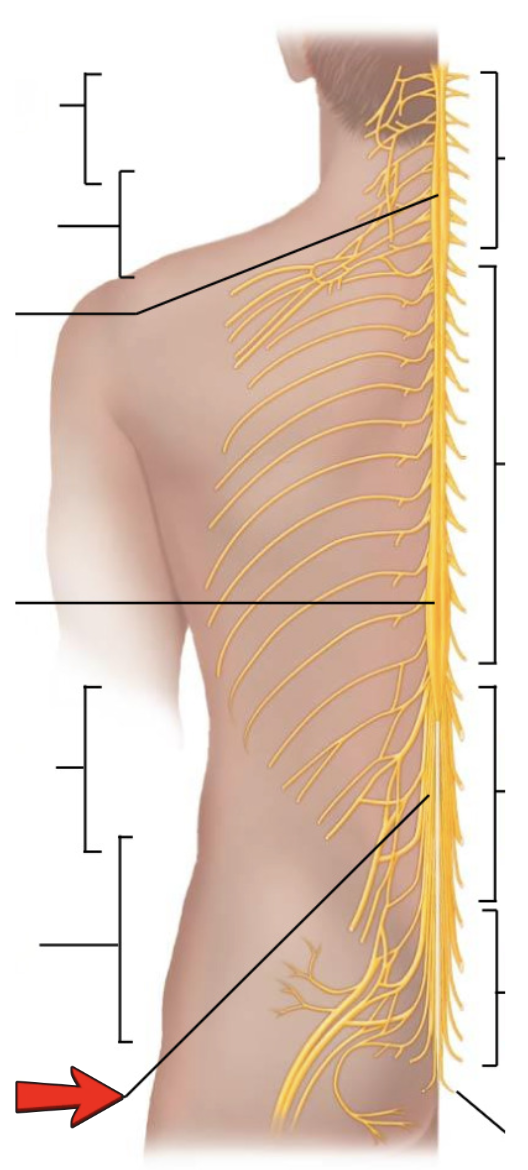
Cauda equina

Cervical nerves C1-C8
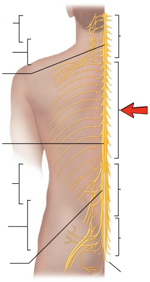
Thoracic nerves T1-T12

Lumbar nerves L1-L5
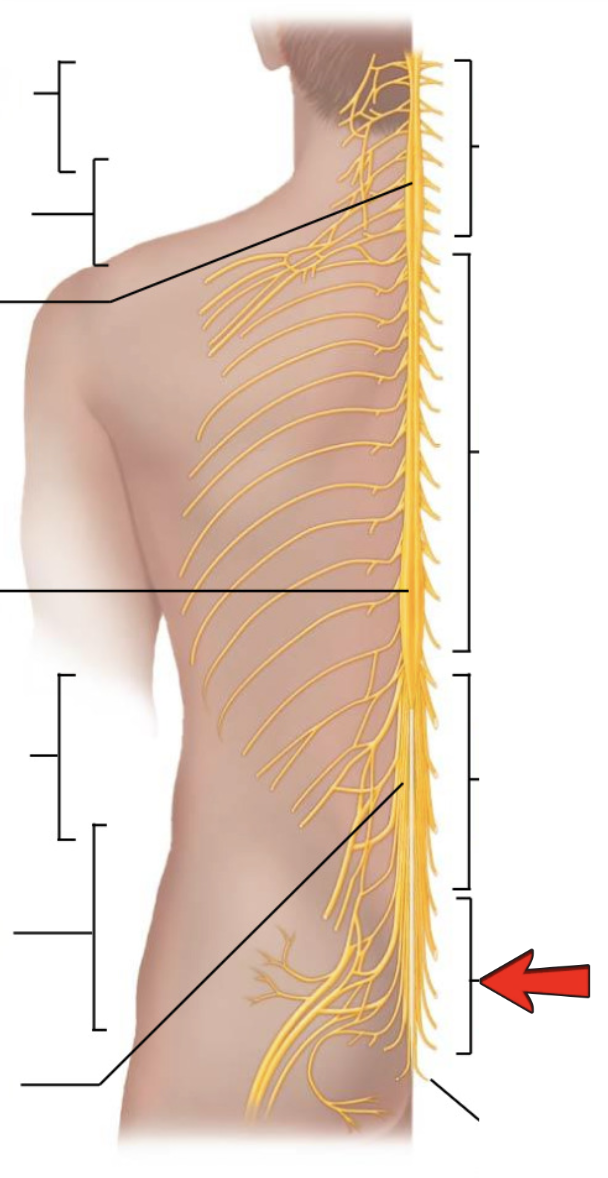
Sacral nerves S1-S5
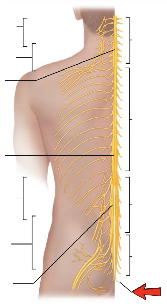
Coccygeal nerve Co1

Dorsal ramus

Ventral ramus

Spinal nerve

Dorsal root

Ventral root
[Cervical Plexus] Formed from:
Ventral rami of C1-5
[Cervical Plexus] Serves:
Neck and shoulder
[Cervical Plexus] Major motor branch:
Phrenic nerve (C3-4 and some of C5)
[Cervical Plexus] Innervates:
Diaphragm (C3-5 keeps you alive)
[Brachial plexus] Formed from:
Ventral rami of C5-8 and T1
[Brachial plexus] Serves:
Shoulder, arms, and hand
[Brachial plexus] Arranged into:
trunks, divisions, and cords and finally into major peripheral nerves
[Brachial plexus] Axillary Nerve:
(C5-6): serves shoulder muscle/skin shoulder area
[Brachial plexus] Radial Nerve:
(C5-8, T1): serves extensor muscles of arm, forearm, &
hand (on posterolateral surface of arm/forearm) thumb side
[Brachial plexus] Median Nerve:
(C8, T1; C5-7): serves flexor muscles of forearm, hand
(some), & skin on lateral surface of palm in the middle
[Brachial plexus] Musculocutaneous Nerve:
(C5-7): serves flexor muscles for forearm
flexion & skin of lateral surface of forearm in the
elbow/axillary area
[Brachial plexus] Ulnar Nerve:
(C8, T1): serves flexor carpi ulnaris, flexor digitorum,
other hand muscles (not served by median nerve) & skin of
medial third of anterior/posterior hand (at the “pinkie” side)
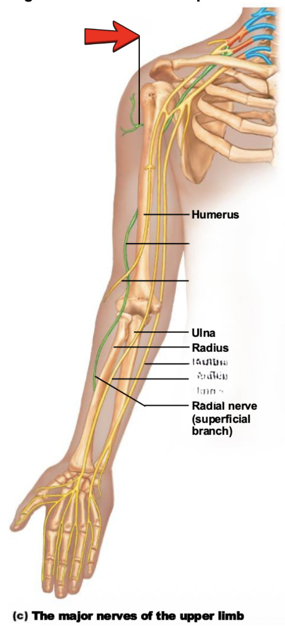
Axillary nerve

Radial nerve

Musculocutaneous nerve

Ulnar nerve

Median nerve

Ulnar nerve

Median nerve

Musculocutaneous nerve

Ulnar nerve

Median nerve

Radial nerve

Axillary nerve

Musculocutaneous nerve
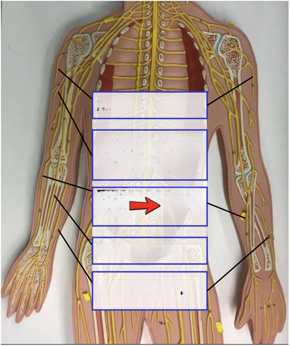
Radial nerve
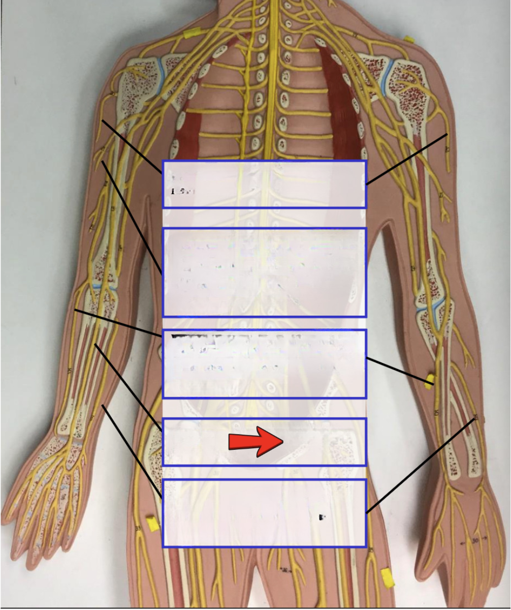
Median nerve

Ulnar nerve
Lumbar plexus from:
Ventral rami of L1-4 (sometimes T12)
Lumbar plexus serves:
Lower abdominopelvic region and anterior thigh
[Lumbar plexus] Femoral nerve:
(innervates anterior thigh), branches into the saphenous nerve
Sacral plexus from:
Ventral rami of L4-S4
Sacral plexus serves:
Buttock, posterior surface of thigh, leg, and foot
Sacral plexus major nerve:
Sciatic nerve (largest in body) which divides into common fibular and tibial nerve
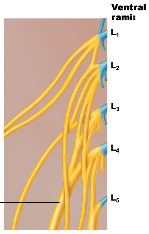
Femoral nerve

Femoral nerve