inflammatory and structural heart disorders
 Call Kai
 Learn
 Practice Test
 Spaced Repetition
 Match
 Flashcards
 Knowt Play
1/61

There's no tags or description
Looks like no tags are added yet.
62 Terms
cardiac muscle layers
Heart consists of 3 layers
•Epicardium
•Outermost layer of the heart
•Myocardium
•Middle layer
•Actual contracting muscle of the heart
•Endocardium
•Inner most layer
•Lines the inner chambers and heart valves

infective endocarditis (IE)
•Disease of the endocardium and the heart valves
•IE is associated with a poor prognosis and a decreased life expectancy
Main Causes include:
•IV drug use
•History of IE
•Having a prosthetic heart valve
•Healthcare-associated infection from an intravascular device (central line)
•Dialysis
Oral cavity risk increases if dental procedure in previous 3-6 months
Infections (cutaneous, genitourinary, gastrointestinal, and systemic)
IE patho
•Blood flow allows organisms to contact and infect previously damaged heart valves or other endothelial surfaces
•Mitral & aortic valves commonly affected
•Staphylococcus aureus most common bacteria
•Bacteria creates biofilms; protect against antibiotics
Typically develops in 3 stages:
◦Bacteremia
◦Adhesion
◦Vegetations
◦Primary lesions of IE
◦Often causes embolization
◦Left-sided move to organs and limbs
◦Right-sided move to lungs (PE)

IE clinical manifestations
Nonspecific, involve multiple organ systems
◦Fever, chills, weakness, malaise, fatigue, anorexia, clubbing
Heart failure is common
◦Most patients have a new or worsening systolic murmur in most patients

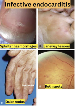
IE Vascular manifestations from microembolizations:
◦Splinter hemorrhages in nail beds
◦Petechiae
◦Osler’s nodes on fingertips or toes
◦Janeway lesions on fingertips, palms, soles of feet, and toes
◦Roth’s spots (retinal lesions)
IE diagnostics
Health history
- Recent surgeries
- Dental, urologic, gynecologic procedures (childbirth)
- IV drug use
-Heart disease
-Infection
Laboratory tests
-Blood cultures x3 in 60 minutes
-CBC with differential
-Erythrocyte Sedimentation Rate (ESR)
-C-reactive protein (CRP)
Echocardiogram
-Vegetation
IE Management Prophylactic Antibiotic Use
•Certain congenital heart diseases
•Heart transplant recipients who develop heart valve disease
•History of infective endocarditis
•Prosthetic heart valve or prosthetic material used to repair heart valve
•Dental procedures
•Oropharyngeal surgery
•Skin and/or musculoskeletal surgery
IE treatment
Long term IV antibiotics → picc line
Several weeks
Repeat blood cultures
Every 24-48 hours
Follow-up echocardiogram & inflammatory markers
Obtained once treatment complete
Complete at 1, 3, 6, and 12 months
Valve replacement
→ Completed in up to 60% of cases
Supportive care
Antipyretics
Fluids
Rest
IE pt teaching
Stress need to avoid people with infections
Avoidance of stress and fatigue
Plan rest periods
Good oral hygiene
Schedule regular dental visits
Prophylactic antibiotics
Rehabilitation for IV drug abuse
pericarditis overview
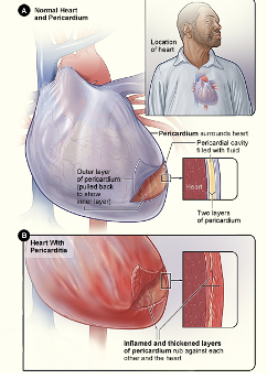
Pericardial sac encases and protects the heart from trauma and infection
◦5-20mL of fluid lubricates and cushions the heart
Has two layers:
•Parietal pericardium
◦Tough, fibrous outer membrane
•Visceral pericardium
◦Thin, inner layer that closely adheres to the heart

pericarditis patho
Inflammation of the pericardial sac
Leads to pericardial leak (effusion)
Most common causes are idiopathic or from a viral illness
pericarditis clinical manifestations
•Chest pain
•Progressive, severe, sharp
•May radiate
•Referred pain to the trapezius muscle
•Pain is worse with deep inspiration and when lying flat
•Pain is relieved when sitting up and leaning forward
•**Pericardial friction rub
◦Scratching, grating, high-pitched sound
◦Heard loudest at lower left sternal border with patient leaning forward
◦Have patient hold their breath for a few seconds
•Fever, chills, malaise, fatigue
complications of pericardial effusions
•Build-up of fluid in the pericardium
•Can occur rapidly or slowly
•Large effusions compress nearby structures
•Symptoms:
•Cough, dyspnea, tachypnea, hiccups (phrenic nerve), hoarseness
•Heart sounds are muffled and distant; bp remains constant
complications of cardiac tamponade
•Pericardial cavity fills with fluid and compresses the heart leading to decreased cardiac output
•Symptoms:
•Chest pain, JVD, Paradoxical pulse, Tachycardia, muffled heart sounds, hypotension, tachypnea
Medical Emergency!!
◦Pericardiocentesis
◦Pericardial window
◦Temporary pericardial drain

pericardial window
•May be required for recurrent tamponade or effusions
•Part (pericardial window) or all pericardiectomy) of the pericardium may be removed
•Allows for adequate ventricular filling and contraction

pericarditis diagnostic
•EKG
•May show ST elevation and/or PR segment depression
•Physical assessment
•Note friction rub, pulsus paradoxus
•Laboratory
•Elevated C-reactive protein
•Elevated sedimentation rate
•Elevated WBC count
•Pericardial biopsy
•Determine cause
•Echocardiogram
•Identify pericardial effusion or cardiac tamponade
•Other imaging
•Echocardiogram
•CT scan
•MRI
pericarditis treatment
Identify and treat underlying problem and symptoms
Bed rest
HOB at 45 degrees
Place overbed table for support
Pericardiocentesis
Pericardial window
Drug therapy
NSAIDs: control pain and inflammation
Corticosteroids: in select patients
Antibiotics: bacterial pericarditis
Aspirin: STEMI
Colchicine: anti-inflammatory
pericarditis nursing management
Keep patient on bedrest with HOB elevated 45 degrees
Administration of anti-inflammatory agents
Monitor for signs of cardiac tamponade and prepare for possible pericardiocentesis
myocarditis
•Inflammation of the myocardium
•Most often caused by a viral illness
•Causative agent invades the myocytes and causes cellular damage and necrosis
•Autoimmune response generated
•Further destruction of cardiac myocytes occurs
myocarditis clinical manifestations
•Symptoms vary from minor to severe
•Early symptoms:
•Fever, fatigue, malaise, myalgias, pharyngitis, dyspnea, lymphadenopathy, and nausea/vomiting
•Cardiac symptoms:
•Pleuritic chest pain, friction rub, signs of heart failure
•Appear 7 to 10 days after viral infection
myocarditis diagnostics
CBC
•Mild to moderate leukocytosis
•Inflammatory markers
•Increased ESR and CRP
Cardiac enzymes
•Elevated troponins
Endomyocardial biopsy
•Provides histologic confirmation of myocarditis
•Obtain within 6 weeks of illness
12 lead EKG
•ST segment changes
myocarditis management
Drug therapy
ACE Inhibitors
Beta blockers
Diuretics
Digoxin
Anticoagulants
Myocarditis with an autoimmune cause: given immunosuppressants and antivirals
Supportive measures
Oxygen therapy
Bed rest
High-fowler’s position
Restrictive activity
⭐️Goal is to decrease workload of the heart
rheumatic fever
•Rheumatic fever (RF) is an acute inflammatory disease that can involve all layers of the heart
•Occurs as a complication 2-3 weeks after an URI with group A streptococcal pharyngitis
•Strep throat

rheumatic heart disease overview
•Rheumatic heart disease is chronic scarring and deformity of the heart valves resulting from RF
•Results from an abnormal immune response to streptococcus bacteria
•Mitral and aortic valves most affected
•Valvular swelling and erosion of the leaflets
◦Vegetation forms on leaflets leading to calcification and impaired mobility (stenosis)
◦Valves unable to close completely causing regurgitation
rheumatic heart disease clinical manifestations
•Carditis (inflammation of the heart)
•New murmur or change in current murmur
•Cardiomegaly
•Signs of HF
•Friction rub, chest pain, muffled heart sounds
•Monoarthritis or Polyarthritis
•Joint pain, swelling, heat, redness, limited ROM of large joints
•Knees, ankles, elbows, and wrists
•CNS manifestations
•Uncontrolled movement of face and limbs
rheumatic heart disease diagnostics
Evidence of existing streptococcal infection
EKG change
◦Prolonged PR interval
◦Delayed AV conduction
Chest x-ray
◦Enlarged heart
Echocardiogram
◦Evaluate valve involvement
rheumatic heart disease treatment
Penicillin is antibiotic of choice
Cephalexin, Clindamycin, or Azithromycin effective is an alternative for PCN allergy
Teach patient to complete full 10-day course of antibiotic
Manage fever with hydration and antipyretics
Plenty of rest
Prophylactic antibiotics are necessary lifelong to prevent infective endocarditis
rheumatic heart disease management
•Bed rest or limited activity
•Drug therapy
•Antibiotics
•NSAIDs
•Corticosteroids
Prevention is KEY!!
Treat patients with antibiotics for:
◦Moderate-to-high fever
◦Abrupt onset of sore throat
◦Reddened throat with exudate
◦Enlarged and tender lymph nodes
stenosis vs regurgitation
Stenosis: Constriction or narrowing of valve leaflets opening
Regurgitation: Incomplete closure of the valve causing backward flow of blood

Mitral Valve Stenosis
•Most common cause is RHD
•Scarring of valve leaflets and chordae tendineae from RHD
•“Fish mouth” shape
•Mitral valve narrows preventing normal blood flow from the left atrium to the left ventricle
•Left atrium dilates, pulmonary artery pressure increases, right ventricle hypertrophies

Mitral stenosis clinical manifestations
Dyspnea on exertion
Loud S1
Low-pitched, diastolic murmur
Symptoms of right-sided heart failure as condition progresses
Hoarseness (atrial enlargement)
Hemoptysis (pulmonary hypertension)
Atrial fibrillation
Fatigue
Palpitations
mitral regurgitation
•Change in structure of mitral valve causes inability to fully close during systole
•Blood backflows into from the left ventricle into the left atrium
•Increase in blood volume and pressure causes the left atrium and left ventricle to dilate and enlarge

mitral regurgitation clinical manifestations
Acute onset:
Thready peripheral pulses
Cool, clammy extremities
New systolic murmur
Pulmonary edema
Cardiogenic shock may occur
Chronic:
May be asymptomatic for years
Palpitations, weakness, fatigue, dyspnea
May progress to right and left-sided heart failure symptoms
mitral prolapse
Abnormality of the mitral valve leaflets and surrounding structures
Mitral valve prolapses back into the left atrium during systole
Hereditary link
◦Marfan syndrome or other hereditary conditions that affect collagen structure

mitral prolapse clinical manifestations
Most patients are asymptomatic for their entire lives
About 10% of population become symptomatic
Regurgitation murmur that is louder during systole
◦Other manifestation:
◦Atypical chest pain
◦Palpitations
◦Exercise intolerance
◦Dizziness
◦Syncope
mitral prolapse pt teaching
Take cardiac medications as prescribed
Adopt healthy eating habits
Avoid caffeine & other stimulants
Begin or maintain adequate exercise program
Contact HCP or EMS for worsening symptoms
aortic stenosis
•Most common cardiac valve dysfunction
•Most common valvular disorder of the aging population
•Disease of “wear & tear”
•Aortic valve orifice narrows and obstructs left ventricular outflow during systole
•Ventricular hypertrophy occurs

aortic stenosis clinical manifestations
May be asymptomatic
Angina
Syncope
Dyspnea on exertion
Symptoms of heart failure
Systolic murmur
aortic regurgitation
•Aortic valve leaflets do not close properly during diastole
•Blood flow from the aorta backflows into the left ventricle during diastole
•The left ventricle dilates to accommodate the greater blood volume causing the LV to hypertrophy
◦Pulmonary hypertension and RV heart failure occur

aortic regurgitation clinical manifestations
Asymptomatic for years
Acute onset:
Severe dyspnea
Chest pain
Hypotension
Cardiogenic shock
Chronic onset:
◦Exertional dyspnea, orthopnea, and paroxysmal nocturnal dyspnea
◦Palpitations when laying on left side
◦Nocturnal angina with diaphoresis
aortic regurgitation diagnostic
History and Physical
Echocardiography (Echo)
Transesophageal echocardiography (TEE) → pictured
◦See next slide
Exercise tolerance testing (ETT)
Chest x-ray
Cardiac catheterization
EKG

aortic regurgitation management
Prophylactic antibiotics
Rheumatic fever and infective endocarditis
Sodium restriction
Drug therapy to treat or control HF
Vasodilators, positive inotropes, diuretics, beta blockers
Anticoagulation therapy
Anti-dysrhythmics
Percutaneous transluminal balloon valvuloplasty
Percutaneous valve replacement
percutaneous transluminal balloon valvuloplasty
◦Invasive nonsurgical procedure for mitral/aortic stenosis
◦Guidewire is inserted through femoral vein or artery
◦Balloon is inserted into affected valve and inflated to enlarge orifices and leaflet mobility
◦Immediate symptom relief usually occurs
◦Not a permanent fix for stenosis

valve repair
Preferred over replacement when possible
Often used for mitral or tricuspid valve disease
•Valvuloplasty
•Repair of the valve by suturing the torn leaflets or surrounding structure
•Annuloplasty
•Reconstruction of the annulus with or without the aid of prosthetic rings

valve replacement
Mechanical valves
Made from artificial material
More durable; last longer
Increased risk for thromboembolism
Long-term anticoagulant therapy necessary
Biologic valves
Made from bovine, porcine, or human heart tissue
Less durable; early calcification
Must be replaced every 7-10 years
Long-term anticoagulants not needed
transcatgeter aortic valve replacement (TAVR)
Option for patients with severe, symptomatic aortic stenosis
Transfemoral approach
◦Small incision is made in the groin to allow for bilateral transfemoral access
Bedrest for 6 hours
Dual therapy of aspirin and Plavix for at least 6 months
◦Aspirin daily for life
valve repair/ replace pt teaching
May require home health for medication management, wound care
Prophylactic antibiotics
◦Antibiotics are required before any dental procedures
◦Dental procedures should wait at least 6 months after surgery due to risk of endocarditis
Periods of rest
Understand medications
High risk for infective endocarditis
Notify all providers of heart valve history
Prosthetic heart valve recipients need lifelong anticoagulants
◦Educate about bleeding tendencies
◦Dietary considerations for Warfarin (Coumadin) therapy
Medical alert bracelet
Carry manufacturer’s valve information card
Avoid MRI for prosthetic valves
Report changes in cardiovascular status to provider
Cardiomyopathy
A group of diseases that directly affect myocardial structure and function
Classified as primary or secondary
Primary: idiopathic conditions involving only the heart muscle
Secondary: another disease process causes myocardial disease
Three major types of cardiomyopathy
Dilated cardiomyopathy
Hypertrophic cardiomyopathy
Restrictive cardiomyopathy
Dilated Cardiomyopathy
Most common type of cardiomyopathy
Diffuse inflammation and rapid degeneration of heart fibers
◦Results in ventricular dilation, impaired systolic function, atrial enlargement, and blood stasis in the left ventricle
◦Ventricular dilation causes cardiomegaly
◦HEART MUSCLE TOO THIN

Dilated Cardiomyopathy manifestations
May develop acutely after an infection or slowly over time
Decreased exercise capacity
Fatigue
Dyspnea at rest
Paroxysmal nocturnal dyspnea
Orthopnea
Disease progression:
Dry cough
Palpitations
Abdominal bloating
Nausea/vomiting/anorexia
Symptoms of RV/LV heart failure
Poor Prognosis
Dilated Cardiomyopathy diagnostic
Patient history
Echocardiogram
Chest x-ray
EKG
Increased BNP
Heart catheterization w/Endomyocardial biopsy
Dilated Cardiomyopathy treatment
Focus on controlling HF be enhancing heart contractility and decreasing preload and afterload
Treatment guidelines are based on the specific stage of disease progression
Nitrates & loop diuretics→ Decrease preload
Beta blockers and Aldosterone Antagonist
ACE inhibitors→ Decrease afterload
Digoxin (presence of atrial fibrillation
Anticoagulants
Decrease risk of systemic embolization
Cardiac Resynchronization therapy
Automatic Implantable Cardioverter Defibrillators (AICD)
Ventricular Assist Device
Cardiac transplant
Hospice
Hypertrophic Cardiomyopathy
•Genetic disorder that causes asymmetric left ventricular hypertrophy without ventricular dilation
•More common in males
•Most common cause of sudden cardiac death among healthy young people
•1st degree relatives of patients with condition should be screened
◦HEART MUSCLE TOO THICK

Hypertrophic Cardiomyopathy patho
4 main characteristics
Massive ventricular hypertrophy
Rapid, forceful contraction of the left ventricle
Impaired relaxation (diastole)
Obstruction to aortic outflow (not present in all patients)
Ventricular hypertrophy results in poor filling of stiff ventricle
Decreased cardiac output
Hypertrophic Cardiomyopathy clinical manifestations
May be asymptomatic
Many are asymptomatic until late adolescence or early adulthood
Atypical chest pain not relieved by nitrates, shortness of breath with exertion, syncope, dizziness, and palpitations
Many athletes that suffer sudden cardiac death may have had hypertrophic cardiomyopathy
Death is usually the first sign
Hypertrophic Cardiomyopathy diagnostics
Echocardiogram- main diagnostic tool
Physical exam may be unremarkable
EKG
Cardiac catheterization
Nuclear stress test
Hypertrophic Cardiomyopathy treatment
⭐️Goal is to Improve ventricular filling by reducing ventricular contractility & relieving LV outflow obstruction
B-Blockers
Calcium channel blockers
Amiodarone
Digitalis contraindicated unless used to treat A-fib
Implantable cardioverter-defibrillator→ Those at risk of SCD
AV pacing
Ventriculomyotomy→Surgically remove piece of thickened septal muscle along with removing some of the ventricular muscle→ Allows for better ventricular wall movement→ Improve cardiac output
Restrictive Cardiomyopathy
•Rarest of the cardiomyopathies
•Stiff ventricles restrict diastolic filling and stretch
•This inability to relax and fill with blood results in a “back up” of blood into the atria, lungs and body
•Symptoms are similar to right/left-sided heart failure
•Can be a primary or secondary disease
•Prognosis is poor

Restrictive Cardiomyopathy clinical manifestations
Fatigue
Exercise intolerance
Dyspnea on exertion
May develop signs of both left-sided & right-sided heart failure
Other symptoms:
◦Angina
◦Syncope
◦Orthopnea
◦Palpations
Restrictive Cardiomyopathy diagnostics
CXR: Normal or cardiomegaly. Pleural effusions & pulmonary congestion in more severe disease
ECG: Heart block, A-fib, SVT
ECHO: Thicken ventricular wall, small ventricular cavities & dilated atria
biopsy, CT & nuclear imaging may be helpful
Restrictive Cardiomyopathy management
Interventions are aimed at improving diastolic filling and underlying disease process
Conventional therapy for HF
Treat arrhythmias
Heart transplant
cardiomyopathies pt teaching
Take all drugs as prescribed and regularly follow up with HCP
Follow a low-sodium diet (if prescribed) and read all product labels (food and over-the-counter drugs) for sodium content
Drink 6 to 8 glasses of water a day unless fluids are restricted
Achieve and maintain a reasonable weight and avoid large meals.
Avoid alcohol, caffeine, diet pills, and over-the-counter cold medicines that may contain stimulants.
At risk for infective endocarditis, required prophylactic antibiotics prior to invasive procedures
Balance activity and rest periods
Avoid heavy lifting or vigorous isometric exercises and check with HCP for exercise guidelines
Use stress management techniques: relaxation breathing, guided imagery
Report any signs of HF to HCP, including weight gain, edema, shortness of breath, and increased fatigue
Encourage caregivers to learn CPR because of the potential for cardiac arrest
Access emergency response system (ERS) according to the HCP’s instructions