619 9/28 GLA Cellular Injury, Adaptation, and Death
 Call Kai
 Learn
 Practice Test
 Spaced Repetition
 Match
 Flashcards
 Knowt Play
1/71

There's no tags or description
Looks like no tags are added yet.
72 Terms
3 Cellular Outcomes
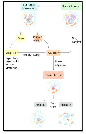
adaptation
reversible injury
irreversible injury
Homeostasis
normal healthy steady state maintained by the cell
Cellular Adaptations
allow for the survival and continued function of a cell in an altered steady state
stimuli may be either pathologic or physiologic
generally reversible if the stimulus is removed
can be one cell or all the cells in an organ
Cell Adaptations and Mechanism Table

3 Types of Cellular Adaptations
increased demand or stimulation
decreased nutrients and/or stimulation
chronic irritation
Increased Demand or Stimulation: Adaptive Mechanism
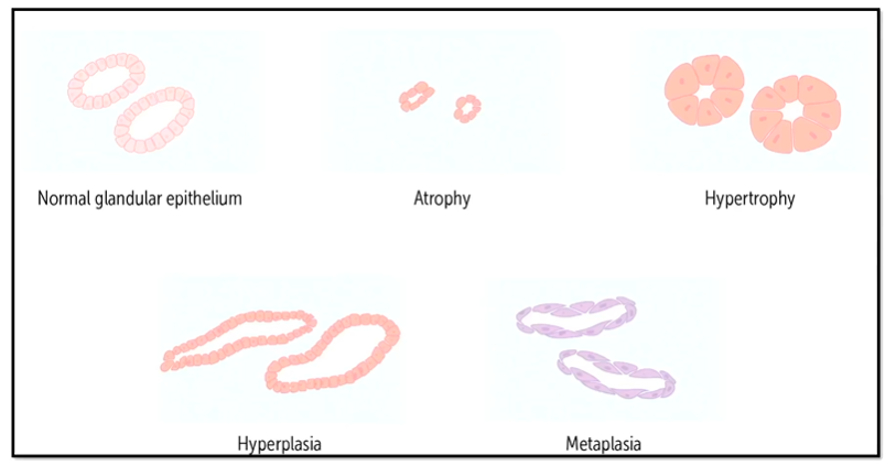
hyperplasia (increase in number of cells)
hypertrophy (increase in size of cells)
Hypertrophy
increase in size of cells due to increased demand, leads to an increase in size of affected organ
physiologic or pathologic
Mechanism of Hypertrophy
due to the synthesis/assembly of additional intracellular structural componenets
Where does hypertrophy occur?
typically in cells that are not capable of self-replication, such as skeletal and cardiac muscles
Physiologic Uterine Hypertrophy
normal growth of uterus during pregnancy, caused by hypertrophy of smooth muscle cells that make up the walls of the uterus

Pathologic Cardiac Hypertrophy
cariomyocytes respond to increased demand by undergoing hypertrophy, and hypertrophy makes it more susceptible to ischemia or heart failure
although function initially improves, over time this adaptation often fails leading to heart disease

Boxcar Nuclei
seen in hypertrophy myocardium, larger boxy shaped nuclei
Hyperplasia
increase in number of cells, can lead to increase in size of the affected organ
occurs in tissues composed of cells that have the capacity to divide
What drives hyperplasia?
increased stimulation by hormones or growth factors
Decreased Nutrients/Stimulation: Adaptive Mechanism
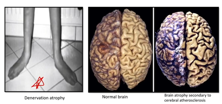
atrophy (decrease in size and number of cells)
Atrophy
decrease in size and number of cells that leads to a decrease in organ size due to a decreased demand
physiologic or pathologic
Physiologic Cause of Atrophy
less/decrease in hormonal stimulation
ex: post-menopausal breast atrphy and uterine atrophy following parturition
Pathologic Causes of Atrophy
decreased workload
loss of innervation
diminished blood supply
inadequate nutrition
Pathologic Atrophy
can also be unilateral renal atrophy, where one kidney shrinks because of reduced blood flow
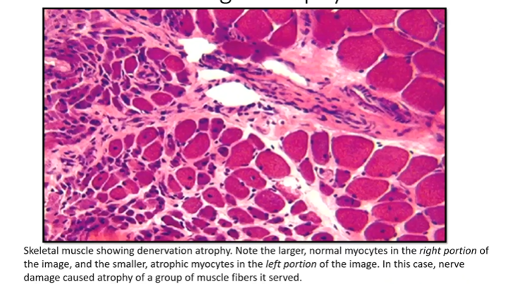
Histological Pathologic Atrophy
skeletal muscle showing denervation atriphy, where the cells on the right are normal and the cells on the left are atrophic myocytes
Chronic Irritation: Adaptive Mechanism
metaplasia (change in cell type)
Metaplasia
change in cell type, often in response to change in environment or stress
metaplasic cell type is better able to withstand the adverse environment
Adaptive Mechanisms Gland Example
Atherosclerosis
a buildup of lipid materal in the wall of our blood vessels
Physiologic Hyperplasia
occurs when functional capacity needs to be increased
also seen in some tissues in response to damage/deficiency
Physiologic Hyperplasia: Increase Functional Capactiy Example
proliferation of breast glandular epithelium during puberty or pregnancy
Physiologic Hyperplasia: Response to Damage/Deficiency Examples
compensatory hyperplasia during liver regeneration
bone marrow in response to deficiency of blood cells
Pathologic Hyperplasia
occurs when the actions of hormones or growth factors are excessive or innapropriate (begning prostatis hyperplasia, endometrial hyperplasia)
also seen in response to certain viral infections (skin warts seen in hpv infection)
Pathologic Hyperplasia Indications
may stabilize or regress if stimulus is eliminated
some of these proliferations are precursors to cancer
Pathologic Hyperplasia Due to HPV Infection
hpv causes skin cell proliferation, some areas of the skin become thicker because the keratinocytes undergo hyperplasia

Metaplasia Mechanism
occurs through reprogramming of stem cells to induce replacement of the native cell type with another that is better suited to handle the new stress
Columnar to Squamous Metaplasia
commonly occurs in respiratory tract in response to chronic irritation (ex- smoking)
respiratory cells are usually cilliated pseudocolumnar, but become squamous cells that are more hardy (but we have a loss of normal structure)

Price of Columnar to Squamous Metaplasia
predisposes to infection because mucous secretion and ciliary action lost
cancers can arise from metaplastic epithelium
Squamous to Comulnar Metaplasia
reflux of gastric acid causes squamous cells to become columnar epithelium that are similar to digestive cells with goblet cells (secrete mucus) so they can resist acids
also a precursor to cancer

Cell Changes Overall Diagram
Causes of Cell Injury
oxygen deprivation, genetic derangements, infectious agents, immunologic reactions, nutritional imbalances, chemical agents and drugs
Progression of Cell Injury and Death

2 Types of Cell Death
necrosis
apoptosis
Reversible Cell Injury Process
cell injury common occurs due to ischemia (inadequate blood flow) which causes hypoxia and local reduction in oxygen
without oxygen, mitochondria are unable to create atp by oxidative phosphorylation
insufficent atp production leads to an intracellular ion imbalance
as ions accumulate in the cell, water preferentially diffuses in from the extracellular space, leading to cellular swelling, the first manifestation of cellular injury
cellular swelling can be difficult to discern via light microscopy and often is more noticeable when looking at the entire organ
What is the first manifestation of cellular injury?
cellular swelling
Reversible Injury - Ultrastructural Changes
generalized swelling of the cell and organelles (ER and mitochondria)
plasma membrane alterations (blebbing and loss of microvilli)
Reversible Injury - Light Microsope Changes
cellular swelling caused by failure of energy-dependent ion pumps in plasma membrane, vacuoles appear in cytoplasm
increased eosinophilia (redness) due to decreased cytoplasmic RNA (increases with progression to necrosis
fatty change in tissues involved with fat metabolism
Hydropic Change/Vacuolar Degeneration
appearance of vacuoles in the cytoplasm of reversible cell injury, visible through light microscopic changes
Fatty Change
in the setting on hypoxia or toxin exposure, the cell cannot break down fat which accumulates within cells and is visible in reversible injury
droplets of fat are visible as clear round spaces
Ischemic Heart Disease
the cardiac cells need lots of oxygen from the coronary arteries
if the coronary artery is occluded, initially the cardiomyocytes are reversibly injured
but with time it will become more severe and irreversible, causing muscles cells to die resulting it myocardial infarction
this is why it is important to start treatment quickly, because restoration of blood flow during the window of time when cell injury is still reversible will prevent myocardial infarction
Necrosis
a pathologic process that is the end result of something that went wrong, cell falls apart
commonly associated with ischemia, exposure to toxins, various infections, and trauma
Apoptosis
may be physiologic or pathologic, may be a result of disease/injury or a normal process
leverages a proleaded cellular “death script” which is activated by certain signals
What should we associate membrane damage with?
necrosis
What should we associate protein misfolding and DNA damage with?
apoptosis
Principle Forms and Sites of Damage in Cell Injury

What do ROS trigger?
it can lead to necrosis or apoptosis, depending on what the pathologic effect is
3 Nuclear Changes in Irreversible Injury (Occur in Step-Wise Direction)
pykosis
karyorrhexis
karyolysis
Pyknosis
nuclear shrinkage and increased basophilia, used to diagnosie irreversible injury
Karyorrhexis
fragmentation of the nucleus, used to diagnosie irreversible injury
Karyolysis
fading of basophilia due to DNA degredation, necrosis only, used to diagnosie irreversible injury
4 Characteristics of Necrosis
denaturation of cellular proteins
leakage of cellular contents through damaged membranes
local inflammation
enzymatic digestion of the injured cell
4 Light Microscopic Changes of Necrosis
increased eosinophilia due to loss of cytoplasmic rna and accumulation of denatured proteins
cytoplasm becomes more vacuolated due to continued swelling
pyknosis, karyorrhexis, karyolysis
can have various morphologic patterns
Neutrophils
polylobic nuclei, attracted to necrosis so they will be seen in histological sections of it
6 Patterns of Tissue Necrosis
coagulative
liquefactive
gangrenous
caseous
fibrinoid
fat
Coagulative Necrosis
results in every organ (besides the brain) from blood loss
architecture of the tissue is preserved for a period of time, and the affected area may be pale with firm texture and wedge-shaped

Histology of Coagulative Necrosis
retention of eosinophilic, anucleate cells
cell architecture is preserved for a span of days, but is lost as inflammation increases

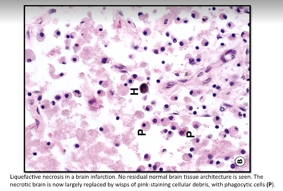
Liquifactive Necrosis
seen in infection (bacterial or fungal) and brain infarctions
enzymes digest dead cells and the tissue becomes liquified

Histology of Liquifactive Necrosis
cavity becomes filled with liquid cells, no structural integrity remains
Caseous Necrosis
cheese-like appearance, liquified tissue that has become thickened by an infectious agent (common with tuberculosis)
associated with granuloma formation

Caseating Granuloma
pink necrosis in the center of the granuloma, macrophages, giant cells, and small lymphocytes (mostly at periphery)

Why do we form granulosas?
when there is a bacteria we have a hard time getting rid of, we form circular structures around them to wall it off, making granulosas
Non-Caseating Granuloma
has giant cells, macrophages, and lymphocytes
NO necrosis in the middle

Fibrinoid Necrosis
seen in autoimmune reactions involving blood vessels (vasculitis), antigen-antibody complexes and fibrin deposit into blood vessel wall causing damage
appears in pink ring due to large amount of proteinaceous substances accumulated in blood vessel wall (arrow)

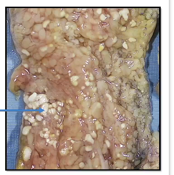
Fat Necrosis
focal areas of fat desctruction, most commonly associated with acute pancreatitis becasue lytic enzymes leak out and digest adjacent fat cells
grossy appears white and chalky
What gives fat necrosis its chalky white apperance?
fatty acids combining with calcium, aka fat saponification
Gangrenous Necrosis
ischemic tissue death with coagulative and liquefactive elements, usually applied to a limb that has lots its blood supply

Dry vs Wet Gangrene
dry: usually coagulative
wet: superimposed infection causes liquefactive necrosis