Neuro Seminar 1 - Brain & Multipolar Neuron
 Call Kai
 Learn
Learn Practice Test
 Spaced Repetition
 Match
Match Flashcards
 Knowt Play
1/35

There's no tags or description
Looks like no tags are added yet.
Name | Mastery | Learn | Test | Matching | Spaced | Call with Kai | Chat |
|---|
No analytics yet
Send a link to your students to track their progress
36 Terms
What is the cerebral cortex?
The cerebral cortex is the thin layer of the brain that covers the outer portion (1.5mm to 5mm) of the cerebrum.
What is the gyri and sulci of the brain?
Gyri = the raised ridges or bumps on the brain
Sulci = the shallow grooves or indentations on the brain
Can you name all 6 of the lobes of the cerebrum?
Frontal Lobe
Parietal Lobe
Temporal Lobe
Occipital Lobe
Insular Lobe
Limbic Lobe
What is the cerebrum?
The cerebrum is the largest part of the brain.
Where is the corpus callosum located and what does it do?
It is located in the middle of the brain.
It is a large bundle of more than 200 million myelinated nerve fibers and connects the two hemispheres permitting communication between the two sides.
What is the Basal Ganglia and where is it located?
It is located deep inside the cerebral hemispheres and upper brainstem.
A group of structures deep within the brain that help control voluntary movement, learning and emotions.
What is the Thalamus and where is it located?
It is located in the middle of the brain at the base of the cerebrum, below the cerebral cortex and above the midbrain.
Sorting sensory information before it connects to the cortex. It also relays motor information to the cortex and receives information from the cortex
Where is the Cerebellum located and what is it?
It is located at the back of the skull just below the cerebral hemispheres and behind the brainstem.
It also helps to conduct movement
Where is the brainstem located and what is it? What are the 3 parts of the brainstem?
It is located at the bottom of the brain and connects directly to the spinal cord and has 3 main parts:
Midbrain - The top section that controls eye movements, vision and hearing reflexes.
Pons - The middle section that coordinates facial movements, balance and sleep cycles.
Medulla Oblongata - The bottom section that connects directly to the spinal cord and control survival tasks.
What are the brain ventricles and where are they located? What are the 4 ventricles?
The ventricles consist of four cerebrospinal fluid filled cavities within the brain. The cerebrospinal fluid surrounds and cushions the brain.
Ventricles 1 and 2 projects the fluid towards the cerebrum
Ventricle 3 sits between the thalamus
Ventricle 4 lies between the brainstem and cerebellum
Can I name the 10 main parts of a multipolar neuron?
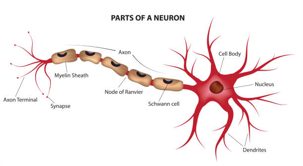
What is the Initial Segment?
Initial segment - a specialized region at the base of a neuron’s axon where electrical impulses, called action potentials, begin.
What is the Axon?
Axon - a long, slender projection of a nerve cell (neuron) that conducts electrical impulses away from the cell body to other neurons, muscles or glands.
What are the Dendrites?
Dendrites - the branched, tree-like extensions of a neuron that receive electrochemical signals from other nerve cells and carry them toward the cell body.
What is the Nucleus of a Multipolar Neuron?
Nucleus - a large, spherical and centrally located structure inside the cell body (soma) that contains the cell’s genetic material (chromosomes).
What is the Schwann Cell?
Schwann Cell - these wrap around the long axons of multipolar neurons in the peripheral nervous system to form an insulating myelin sheath
What is the Node of Ranvier?
Node of Ranvier - a small, regular gap in the myelin sheath of a myelinated axon where the nerve fiber is exposed to the extracellular space.
What is the Axon Terminal?
Axon Terminal - the small, bulb-like swelling at the very end of a nerve fiber (axon) that converts electrical signals into chemical messages to pass information to other cells.
What is the Myelin Sheath?
Myelin Sheath - a fatty, protective layer that wraps around the axons of nerve cells to speed up and insulate electrical signals.
What is the Synapse?
Synapse - a microscopic gap or junction where the axon terminal of one neuron communicates - usually chemically via neurotransmitters - with the dendrite or cell body of another target cell
What is the Cell Body?
Cell body - the central, bulbous part of the nerve cell that contains the nucleus and gives rise to a single axon and multiple branding dendrites
What is the Axon Hillcock?
Axon hillock - the specialized, cone shaped region of a neuron’s cell body (soma) that connects to the axon.
Can you name Phases 1 to 5 of an Action Potential?
Resting Potential
Threshold Phase
Depolarisation
Repolarisation
Hyper-polarisation

Where does action potential happen?
Action potential happens at the axon hillock (cone shaped region of a neuron’s cell body) and travels down the axon.
What happens at Phase 1 (Resting Potential) of action potential?
Concentration of sodium (Na+) is greater outside the cell, whilst the concentration of potassium ion (K+) is greater inside the cell.
It’s resting state, a neurons membrane potential, is around -70mv
The minus sign indicates the inside of the cell is negative relative to the outside of the cell.
What happens at Phase 2 (Threshold Phase) of action potential?
If a threshold of -55mv is reached, voltage gated sodium (Na+) ion channels will open.
It then allows Na+ ions to flow into the cell
What happens at Phase 3 (Depolarisation) of action potential?
As voltage-gated sodium (Na+) channels open Na+ flows rapidly into the cell, bringing their positive charge with them.
The voltage changes from -55mV to +30mV.
What happens at Phase 4 (Repolarisation) of action potential?
At 30 mV the voltage-gated Na+ channels are refractory/inactive. No more Na+ can enter the cell.
The K+ voltage gated channels open and K+ leaves the cell.
Slowing of Na+ inflow and acceleration of K+ outflow causes the membrane potential to be driven back towards its resting state.
What happens at Phase 5 (Hyper-Polarization) of action potential?
Voltage gated potassium gates are slow to close.
This can result in an overshoot of the normal resting membrane potential.
Diffusion and sodium potassium ATPases redistribute K= and Na+ ions until the membrane potential is back to its resting potential of -70mV again.
How does a cell reach threshold?
When incoming signals cause enough positive ions to enter the cell.
Graded potentials travel through the neurone until they reach the trigger zone at the axon hillock.
When multiple signals arrive at the trigger zone, they summate (add up). Spatial summation - multiple signals arrive simultaneously. Temporal summation - signals arrive at different times. These impulses are incremental and may be excitatory or inhibitory.
If the graded potential is large enough (reaches -55mv) an axon potential will propagate down the axon according to the ‘all or nothing’ principle.
What does mV stand for?
Millivolts
What is the Refractory Period - Absolute?
The time during which no new action potential can be triggered, no matter how strong the stimulus.
What is the Refractory Period - Relative?
The time after the absolute period when a new action potential can happen, but only if the stimulus is stronger than usual.
Explain what propagation down the axon means?
The sodium channels inside the axon segment have a refractory period, they cannot open again causing the action potential to only go in one direction towards the axon terminal.
Explain what saltatory conduction means?
The rapid way an electrical impulse or action potential “jumps” from one gap (node of ranvier) to the next along a myelinated axon (axon which is covered by myelin sheath).
Electrical activity and ion exchanges happen only at the exposed nodes. The electrical signal triggers the next node instantly, making the impulse appear to skip down the axon
What happens when the action potential reaches the presynaptic terminal inside the synapse?
Depolarization of the presynaptic terminal opens ion channels, allowing calcium (Ca2+) into cells.
Ca2+ triggers release of neurotransmitter from synaptic vesicles.
Neurotransmitter binds to receptor sites on postsynaptic membranes
Opening and closing of channels cause change in postsynaptic membrane potential.
Action Potential propagates through the next cell
Neurotransmitter is inactivated or transported back into presynaptic terminal.
