Effector mechanisms of humoral immunity
 Call Kai
 Learn
Learn Practice Test
 Spaced Repetition
 Match
Match Flashcards
 Knowt Play
1/16

There's no tags or description
Looks like no tags are added yet.
Name | Mastery | Learn | Test | Matching | Spaced | Call with Kai | Chat |
|---|
No analytics yet
Send a link to your students to track their progress
17 Terms
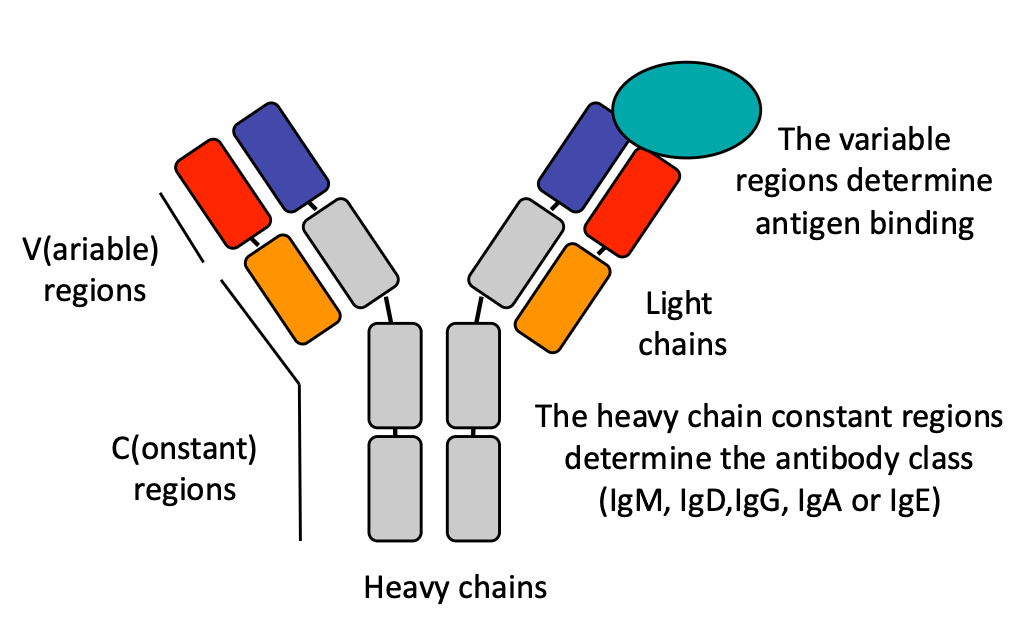
Describe the basic structure of an antibody molecule, including heavy chains, light chains, variable regions, and constant regions.
An antibody consists of:
Two heavy chains (shown in red) and two light chains (shown in blue)
Variable region - Located at the top of the antibody; this is the region that binds to the antigen. Both heavy and light chains have variable regions
Constant region - Located at the bottom; this region is longer in the heavy chain than in the light chain. The heavy chain constant region determines the antibody isotype (IgG, IgM, IgA, IgD, or IgE)
The variable region contains loop regions (CDRs - Complementarity Determining Regions) that directly contact the antigen. These loops are flexible and tolerate mutations well, unlike the beta sheet structures in other parts of the antibody
Where are different antibody types found in the body, and what are their primary functions in each location?
IgG in circulation: IgG is the most abundant antibody in the blood serum. It is excellent at all effector functions (neutralization, opsonization, ADCC, complement activation) and can cross the placenta to provide immunity to the fetus
IgA in mucosal surfaces: IgA is the most abundant antibody overall in the body, but most of it is NOT in circulation. Instead, IgA is found in massive quantities on mucosal surfaces (gut, lungs, mouth, genital tract) - anywhere in contact with the outside environment. In the gut alone, there are grams of IgA excreted daily in feces. IgA is present as a dimer (two molecules joined by a J chain) on mucosal surfaces, and it is the only antibody type that can be transported across mucosal barriers thanks to a special receptor
IgM in circulation: IgM is produced early in the immune response and is very good at complement activation. It circulates as a pentamer (five molecules joined by a J chain), which increases its avidity. IgM can also be found in milk
IgE in tissues: IgE is present in very low amounts in serum (3 × 10^-5) because it binds with extremely high affinity to mast cells and basophils in tissues. All IgE is essentially bound to these cells and never circulates freely. IgE is important for allergic responses and parasitic infections

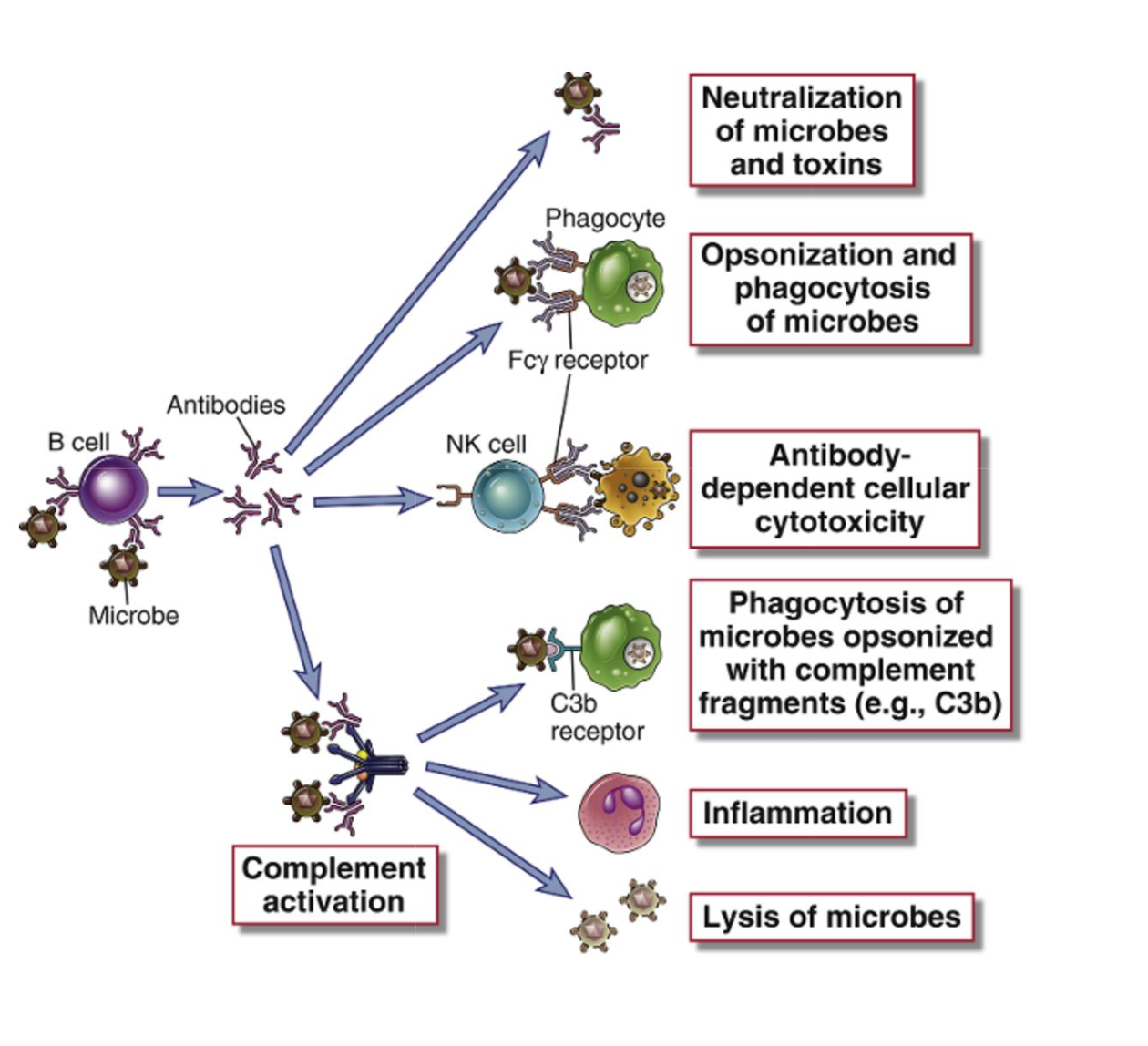
What are the four major effector functions of antibodies?
The four major effector functions are:
Neutralization - The antibody acts as a physical barrier, binding to pathogens or toxins and preventing them from binding to cell receptors
Opsonization - Antibodies bind to the surface of pathogens and flag them for phagocytosis by macrophages and other phagocytic cells'
ADCC (Antibody-Dependent Cellular Cytotoxicity) - Antibodies bind to infected cells or parasites and trigger NK cells or mast cells to release cytotoxic granules that kill the target
Complement Activation - Antibodies activate the complement cascade, leading to opsonization, inflammation, and formation of the membrane attack complex
How many IgG subtypes exist in humans, and which ones are most effective at effector functions?
There are four IgG subtypes: IgG1, IgG2, IgG3, and IgG4. These subtypes differ slightly in their constant regions, which affects their effector functions:
IgG1 and IgG3 - These are very good at effector functions. They are excellent at opsonization, ADCC, and complement activation because their constant regions are well-recognized by Fc receptors on macrophages, neutrophils, and NK cells
IgG2 and IgG4 - These are not very good at effector functions. They have lower affinity for Fc receptors and are therefore less efficient at activating phagocytes and complement
Explain the neutralization mechanism and describe why IgG is better at neutralization than IgM, despite IgM being a pentamer.
Neutralization mechanism: The antibody binds to a pathogen, toxin, or viral protein and acts as a physical barrier, preventing the pathogen from binding to its receptor on the cell. This is called steric hindrance. Importantly, neutralization does not require the Fc (constant) region - only the antigen-binding portion is needed.
Why IgG is better than IgM:
IgG - Comes from the germinal center after affinity maturation, so it has high affinity and binds very tightly to the pathogen. This strong binding makes it excellent at neutralization
IgM - Produced early in the immune response before affinity maturation, so individual IgM molecules have low affinity. However, IgM circulates as a pentamer (five antibodies joined together) by a J chain. This increases the overall binding strength through avidity - even if one IgM loses its grip on the pathogen, another one nearby can catch it. So while individual IgM has low affinity, the pentamer can still neutralize effectively through combined binding strength
It hinders Influenza virus and tetanus toxin

What is opsonization, and how do Fc receptors on macrophages recognize antibody-coated bacteria?
Opsonization definition: Opsonization is the process where antibodies bind to the surface of a pathogen (such as bacteria) and create a "flag" that marks the pathogen for destruction. This increases the efficiency of phagocytosis by phagocytic cells like macrophages.
How it works:
Antibodies (especially IgG1 and IgG3) bind to multiple sites on the bacterial surface'
Macrophages have Fc gamma receptors on their surface that recognize and bind to the constant region of these antibodies
Critical point: Macrophages do NOT recognize free antibodies floating in circulation. They only recognize antibodies that are bound to the pathogen, and they require multiple antibodies clustered together for efficient recognition
When multiple Fc receptors bind to multiple antibodies on the bacteria, this clusters the receptors together, activates phagocytic signals, and triggers the formation of a phagolysosome that internalizes and kills the microbe

Explain ADCC and describe the two different pathways by which it can occur.
ADCC definition: Antibody-dependent cellular cytotoxicity is a mechanism where antibodies bind to infected cells or parasites and trigger immune cells (NK cells or mast cells) to release cytotoxic granules containing perforin and other killing substances, resulting in death of the target cell.
Two ADCC pathways:
IgG-mediated ADCC (via NK cells): IgG1 and IgG3 antibodies bind to viral proteins on the surface of virus-infected cells. NK cells recognize these antibodies through their Fc gamma receptor III (FcγRIII). The NK cells then release cytotoxic granules that kill the infected cell. This is used for targets that are too large to be phagocytosed, such as virus-infected cells
IgE-mediated ADCC (via mast cells/eosinophils): IgE antibodies bind to the surface of large parasites (like worms) or infected cells. Mast cells or eosinophils recognize these IgE antibodies through their Fc epsilon receptors. The mast cells release granules containing cytotoxic substances that damage the parasite or infected cell. This pathway is particularly important for extracellular parasites that are too large to be phagocytosed

What are the three complement pathways, and which one is mediated by antibodies?
The complement system has three activation pathways:
Classical Pathway - This is the pathway mediated by antibodies. It was called "classical" because it was the first complement pathway discovered. IgG1, IgG3, and IgM antibodies activate this pathway when they bind to antigens on pathogens
Alternative Pathway - This is the evolutionarily more ancient pathway. It is conserved not only in humans but also in invertebrates and other animals that do not have antibodies. This pathway can be activated directly by bacterial surfaces without requiring antibodies
Lectin Pathway - This pathway is similar to the classical pathway in its steps, but the initial activation is not mediated by antibodies. Instead, it is activated by specific proteins (like mannose-binding lectin) that bind to bacterial sugars
Key point: All three pathways converge on the formation of C3 convertase, which is a critical amplification enzyme that cleaves more complement proteins and creates an exponential cascade of complement activation.

Describe how the alternative pathway is initiated and what happens to C3 protein during this process.
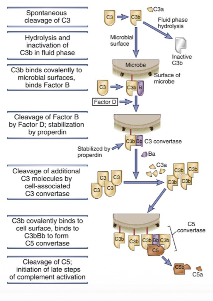
The alternative pathway is initiated by spontaneous and continuous cleavage of C3 protein in the plasma. C3 is present at high concentration and is constantly cleaved into C3b and C3a. C3b is the larger fragment that normally gets rapidly hydrolyzed and becomes inactive if it doesn't bind to anything. However, if C3b binds to a microbe surface, it forms a covalent bond and can recruit Factor B, starting the amplification cascade.
When C3b binds to a microbe surface, it recruits Factor B. Factor B is then cleaved by Factor D to form C3bBb, which is the C3 convertase of the alternative pathway. This C3 convertase is stabilized by properdin (Factor P), which is the only positive regulator of the complement system. On the body's own cells, Factor H prevents this process by degrading C3b.
The C3 convertase (C3bBb) can then cleave more C3 protein, and then they get cleaved to C3b and bind covalently to the cell surface and C3 convertase to form C5 convertase. Then C5 convertase cleaves C5 into C5a and C5b. Which them lead to three outcomes.
Describe how the classical pathway is activated and what antibodies can initiate it.
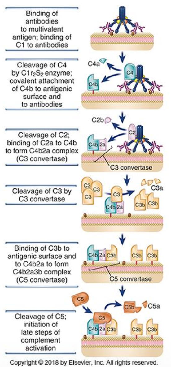
The classical pathway is initiated by antibodies bound to antigens. It can be activated by either one IgM molecule or two IgG molecules (specifically IgG1 or IgG3). When antibodies bind to antigen, they undergo conformational changes that expose the Fc portion, allowing the C1 complex to bind. IgM is particularly effective because it is a pentamer. The binding of C1 to antibodies triggers the cascade.
The C1 complex (made of C1q, C1r, and C1s) binds to antibodies on the microbe surface. C1r then cleaves C1s, which becomes active and cleaves C4 into C4a and C4b. C1s then cleaves C2 into C2a and C2b (note: this is the exception where "a" stays and "b" goes).
The resulting C4b2a complex is the C3 convertase of the classical pathway. This C3 convertase can then cleave C3 into C3a and C3b, and when C3b binds to it, it forms the C5 convertase (C4b2aC3b) and then cleave C5 and the initiation of late steps of complement activation which leads to three outcomes.
Describe the lectin pathway, including what initiates it and how it differs from the classical pathway.
The lectin pathway is initiated by plasma proteins such as mannose-binding lectin (MBL) and ficolins that bind to sugars present only on bacterial surfaces. When these proteins bind to bacterial sugars, they undergo conformational changes that expose proteases (similar to C1r and C1s). These proteases then cleave C4 and C2, forming the same C3 convertase (C4b2a) as the classical pathway. The key difference is the initiation mechanism—the lectin pathway is activated by sugar binding rather than by antibodies.
Once C5 convertase is formed (either C4b2aC3b in classical/lectin pathways or C3bBbC3b in alternative pathway), it cleaves C5 into C5a and C5b. C5b then recruits C6, C7, C8, and finally C9 in sequential order. C9 is the pore-forming protein that inserts into the bacterial membrane, creating holes. This sequential recruitment of proteins forms the membrane attack complex, which causes osmotic lysis of the bacteria by creating pores in the cell membrane.
MBL= Mannose binding lection - binds to mannose residues om polysaccharides
Ficolins= It is similar to MBL but binds to fibrinogen rich domains and bind to oligosaccharides containing acylated sugars

Describe the three main effector functions of the complement system.
The three effector functions are:
(1) Opsonization - C3b binds to bacteria and acts as a flag for macrophages that have complement receptors, marking the bacteria for phagocytosis.
(2) Inflammation - The "a" fragments (C3a, C4a, C5a) are released and act as pro-inflammatory molecules and chemotactic factors. They stimulate mast cell degranulation, increase vascular permeability, and recruit immune cells. C5a is particularly pro-inflammatory because it is downstream in the cascade.
(3) Membrane Attack Complex Formation (MAC) - Creates holes in the bacterial membrane leading to osmotic lysis. (C9 is the one who forms the hole in the pathogens membrane). (C5b binds to C6 and C7 and then tehy all together bind to the membrane and becomes a high affinity receptor for C8. Then the C5b-C8 complex binds to C9 and the C9 assembles at the site to form plasma membrane pores, allowing water and ions to flow freely.)
Describe how the complement system is regulated and how bacteria evade complement activation.
Factor H is present at high concentration and binds to C3b on the body's own cells, removing Bb to prevent complement deposition.
C1 inhibitor dissociates C1 to prevent excessive classical pathway activation.
C59 blocks C9 binding and prevents membrane attack complex formation on our own cells. Regulation is critical to prevent excessive complement activation that could cause autoimmune disease, to localize complement activation to the infection site, and to avoid low-level systemic activation.
Bacterial Evasion: Many bacteria produce proteins that mimic complement proteins to evade the system. Some bacteria block C1q assembly to prevent classical pathway activation. A famous example is Neisseria meningitidis, which produces a factor H binding protein that binds to factor H and coats the bacterial surface, preventing complement from working effectively. This evasion strategy was so successful that it led to vaccine development targeting the factor H binding protein directly, allowing the immune system to block this evasion mechanism and restore complement-mediated bacterial degradation.
Describe the consequences of complement deficiencies and what happens with under-reactive versus over-reactive responses.
Under-reactive Response: When complement doesn't work well and cannot be properly activated, patients experience recurrent and severe bacterial infections. This is particularly problematic because complement is one of the major defense mechanisms against extracellular bacteria. Without functional complement, the body cannot effectively opsonize, lyse, or clear these bacteria.
Over-reactive Response: When complement is always activated and cannot be properly regulated, patients develop severe autoimmune disease. The constant complement activation causes tissue damage and inflammation in the body's own cells.
General Principle: Complement deficiencies are generally quite severe, and the complement system requires very tight regulation by the body to maintain the balance between effective pathogen defense and protection of self-tissues.
Describe how IgA is transported across the gut epithelium and what role the secretory component plays.
IgA Production: IgA is produced in dimeric form by long-lived plasma cells located in the gut lamina propria. These plasma cells are continuously generated from germinal centers that are specific to gut bacteria, ensuring a constant supply of IgA tailored to the local microbiota.
The Gut Environment: The gut lumen contains many bacteria that must be kept out of the body. The epithelium forms a tight barrier with a mucus layer, but additional protection is needed—this is where IgA comes in.
Transport Mechanism: Dimeric IgA binds to the poly-IgA receptor (pIgR) on the basolateral side of epithelial cells. The receptor then undergoes receptor-mediated transcytosis, transporting the IgA across the epithelium from the basolateral side to the apical side (lumen).
Secretory Component: During transcytosis, a portion of the poly-IgA receptor remains attached to the IgA after transport. This attached portion is called the secretory component, and it wraps around the dimeric IgA, becoming an integral part of the secretory IgA complex.
Protection Function: The secretory component protects the IgA from proteolytic cleavage in the harsh lumen environment where many proteases are present and proteins get cleaved. This protection keeps the IgA stable and functional.
Final Defense: Once in the lumen, the secretory IgA can bind to microbes and toxins, preventing them from infecting epithelial cells. If a lesion occurs and a microbe-antibody complex enters the body, the bacteria are already coated with antibodies, facilitating their destruction by the immune system.

How is IgG is transported?
FcRn (Fc Receptor Neonatal): FcRn is located on the placenta and is responsible for transporting IgG across the placental barrier. Unlike other antibody transport mechanisms, FcRn operates bidirectionally, meaning it can transport IgG in both directions across the placenta.
Maternal IgG Transfer: IgG is transported from the mother to the fetus across the placenta via FcRn. As a result, babies are born with maternal IgG antibodies that provide passive immunity during the early months of life.
Comparison to Poly-IgA Receptor: This is different from the poly-IgA receptor in the gut, which is strictly unidirectional—it only transports IgA from the basolateral side to the apical side (into the lumen). FcRn's bidirectional transport capability makes it unique among antibody transport mechanisms.
Describe how IgE leads to mast cell activation and the immediate hypersensitivity reaction.
Upon first exposure to an allergen, a TH2 response is triggered that produces IL-4, which stimulates class switching to IgE. IgE is produced and released into circulation where it binds to mast cells through the high-affinity Fc epsilon receptor.
The affinity is so high that IgE remains bound to mast cells. Upon second exposure to the same allergen, the IgE on the mast cell surface immediately binds the antigen, triggering rapid degranulation. The granules release histamine and other vasoactive substances, TNF-alpha, prostaglandin, leukotriene and secretion of cytokines causing an immediate hypersensitivity reaction with vasoconstriction and inflammation.
