medical audiology midterm
 Call Kai
 Learn
 Practice Test
 Spaced Repetition
 Match
 Flashcards
 Knowt Play
1/113

Earn XP
Description and Tags
SLHS 570
114 Terms

what plane is this?
axial

what plane is this?
coronal
what plane is this?
saggital

CT or MRI?
CT

CT or MRI?
MRI (T-1 weighted)

CT or MRI?
MRI (T-2 weighted)

CT or MRI?
MRI (T-1 weighted with old lesion)

CT or MRI?
MRI (T-1 weighted with gadolinium and new lesions)

CT or MRI?
CT (axial view of a typical cochlea)
what does a CT (computed tomography) show best?
bone structures
what does an MRI (magnetic resonance imaging) show best?
soft tissues and nerves (brighter colors)
which type of MRI is the imaging study of choice for retrocochlear pathology (e.g. vestibular schwannoma)?
GAD-enhanced T1 MRI- contrast enhancement reliably detects even small, intracanalicular tumors that unenhanced MRI can miss. CT lacks the soft tissue/nerve resolution needed.
For a suspected vestibular schwannoma, which imaging sequence is most useful for detecting the abnormal tissue itself?
GAD-enhanced T1-weighted MRI
what are T2 MRI’s best for?
detecting fluid-filled structures and lesions. (SNHL, vertigo, secondary tinnitus)
what are unenhanced T1 MRI’s best for?
anatomy, fat, blood products, certain masses (lipoma, vascular trauma, high-protein fluid)
Which finding on the audiologic test battery is a ‘red flag’ that should prompt a referral for MRI to rule out retrocochlear pathology?
Asymmetric or sudden sensorineural hearing loss with unilateral tinnitus
A patient with a cochlear implant needs to undergo an MRI. What is the key device-safety consideration?
Many current CIs are MRI-conditional up to a specified field strength; some require magnet removal or a protective head wrap first
A patient is being worked up for suspected otosclerosis with possible stapes fixation, while another is being worked up for a suspected vestibular schwannoma. Which imaging modality is preferred for each?
CT (including HRCT) for the suspected otosclerosis; MRI for the suspected schwannoma
A patient is found to have bilateral vestibular schwannomas on GAD-enhanced T1 MRI. What syndrome does this confirm, and what does the patient’s device candidacy hinge on if hearing loss progresses?
Neurofibromatosis Type II (NF2); candidacy hinges on cochlear nerve preservation – CI if intact, ABI if sacrificed
A patient presents with a painful, swollen, fluctuant purple mass on the upper third of the auricle immediately after a wrestling match. What is the most important immediate management priority?
Prompt drainage (aspiration or incision) with a compression dressing
A newborn is found to have a preauricular pit. Based on the epidemiologic data presented in lecture, what is the most appropriate audiologic next step?
Consider a hearing screen, given the substantially higher prevalence of permanent hearing impairment in these infants
Which combination of features is most classic for Herpes Zoster Oticus (Ramsay Hunt syndrome) when facial paralysis is also present?
VZV reactivation in the geniculate ganglion, causing severe otalgia, vesicular rash, and CN VII (facial) paralysis
A newborn has small indentations anterior to the helix and superior to the tragus (preauricular pits), while a different newborn has small skin/cartilage nodules anterior to the ear along the line toward the mouth (preauricular tags). How do the syndromic associations of these two findings differ?
Pits may be the first sign of branchio-oto-renal (BOR) syndrome, the most common inherited syndrome causing hearing loss; tags may appear as part of the oculo-auriculo-vertebral spectrum
An older adult develops rapid growth of long, fine, lanugo-type hair around the ears, eyebrows, and forehead. Based on its associations, what should this finding prompt the clinician to consider?
Possible underlying malignancy (e.g., lung, colon, or uterine cancer), among other causes, warranting further medical evaluation
An older adult is noted to have an oblique crease in the earlobe during an otoscopic exam. What is the audiologic significance of this finding?
It may serve as a potential systemic health marker (studies link it to coronary artery disease) and should be documented, with referral if appropriate
A newborn with macroglossia, an umbilical hernia, and creases in the ear lobes is diagnosed with Beckwith-Wiedemann syndrome (BWS). What should the audiologist specifically watch for?
Craniofacial features that may affect ear canal or middle ear status, or complicate earmold fitting
A patient has chronic, untreated auricular hematomas that progressed to cauliflower ear. Beyond the cosmetic deformity, what additional complication is this patient at risk for?
Sudden or gradual sensorineural hearing loss, presumed related to vasculitis of the internal auditory artery or its branches, in up to 40% of patients
An elderly, fair-skinned patient with chronic sun exposure presents with a slow-growing, pearly nodule with rolled edges on the helix. Which type of pinna malignancy does this best describe, and how does it differ from the more aggressive type?
Basal cell carcinoma (BCC) presents as pearly, rolled-edge, slow-growing; squamous cell carcinoma (SCC), by contrast, presents as a scaly, crusted, ulcerative, more aggressive lesion
An older male patient is noted to have coarse hair growth on the tragus during an otoscopic exam. What is the audiology relevance of this common, benign finding, and how does it differ from the much rarer hair-growth condition linked to malignancy?
It may contribute to cerumen impaction and affect otoscopy, earmold impressions, or hearing aid fitting.
An infant has bilateral congenital aural atresia with normal cochleae on imaging. What is the most urgent audiologic priority?
Early amplification with a bone-conduction hearing aid to support speech and language development
A child has unilateral congenital aural atresia and normal hearing in the other ear. What is the most appropriate counseling for the family?
Speech and language usually develop normally; expect trouble with localization and hearing in noise, and protect the good ear
A child has conductive hearing loss and repeated cerumen impaction caused by a stenotic external auditory canal (diameter of 1 mm or less). What is the surgical treatment?
Meatoplasty to widen the ear canal
Where in the external auditory canal are the cerumen glands and hair follicles found?
The lateral one-third (cartilaginous portion)
Which patient is at the greatest risk for cerumen impaction?
An 85-year-old nursing home resident who wears hearing aids every day
A 45-year-old with no history of cold-water swimming has a single, unilateral bony mass near the canal opening, attached at the tympanosquamous suture. What is the most likely diagnosis?
Osteoma
A competitive swimmer has ear pain that gets worse when you press on the tragus. The ear canal is red and swollen, and the TM is intact. What is the most likely diagnosis and first-line treatment?
Acute otitis externa; topical antibiotic/steroid ear drops
A patient has had itchy ears for months with little or no pain. The canal skin is thickened, dry, and flaking. What is the most likely diagnosis and the goal of management?
Chronic otitis externa; restore healthy, cerumen-producing canal skin
A patient has a TM perforation from a past ear infection. Which statement about the expected hearing loss is most accurate?
The conductive loss grows with the size of the perforation and is largest at the low frequencies
A child with a history of repeated ear infections and PE tubes has a chalky white plaque on the TM, and the tympanogram is within normal limits. What is this finding, and how does it usually affect hearing?
Mature tympanosclerosis; seldom causes a measurable hearing loss when limited to the TM
A patient with confirmed stapedial otosclerosis asks about stapes surgery. Which finding is generally considered a contraindication to surgery?
The ear to be operated on is the patient’s only hearing ear
A child has a persistent, foul-smelling white mass visible medial to a retracted or perforated TM, along with progressive unilateral hearing loss. Why is this condition considered dangerous if left untreated, beyond the hearing loss itself?
Cholesteatoma can enlarge and erode surrounding bone, potentially spreading infection into the inner ear or intracranially
A 5-year-old has had fluid in the middle ear for over 3 months, with a retracted, opaque TM and decreased mobility on pneumatic otoscopy, but no fever, pain, or erythema. How should this be classified, and what type of hearing loss is expected?
Chronic otitis media with effusion (COME); conductive hearing loss
A child has had four episodes of acute otitis media in the past 6 months, and OME has persisted for more than 3 months. What is the most appropriate next step in management?
Surgical placement of pressure equalization (PE) tubes
A patient reports pulsatile tinnitus synchronized with their heartbeat, and otoscopy reveals a reddish-bluish mass behind an intact TM. What is the most likely diagnosis, and what should it be differentiated from?
Glomus tympanicum; differentiated from a hemotympanum
what are the red flags for an MRI referral to rule out retrocochlear pathology?
symmetric or sudden SNHL, unilateral tinnitus, abnormal interaural ABR latency, absent/asymmetric acoustic reflexes
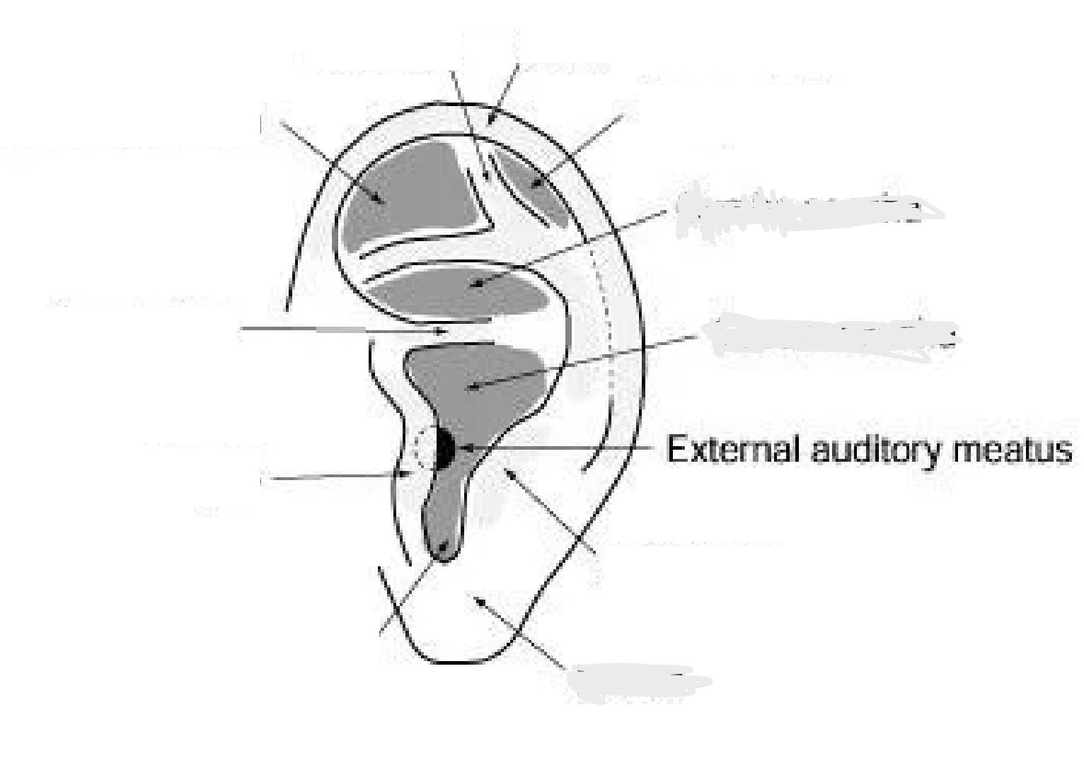
label the landmarks
clockwise from EAM: antitragus, lobule, intertragal notch, tragus, crus of helix, triangular fossa, helical crura, helix, scaphoid fossa, cymba concha, cavum concha
canal resonance of the EAC
Resonating tube
2.5cm x 0.7cm
Resonance frequency 2.6 kHz
contributes peak gain 10 dB @ 2.6 kHz
ideal for speech
self-cleaning EAC
epithelial migration
migration is centrifugal — skin grows outward from the eardrum toward the canal opening
keratinocytes (the skin’s outermost cell layer) are the migratory cells
exact mechanism driving the migration is still not well understood
sheds continuously as it migrates outward — about 0.07 mm/day (~2 mm/month) and this migration decreases frequency with age
cartilaginous EAC
lateral 1/3: Thick skin
Hair follicles
Elevates cerumen off skin
Cerumen glands
Cerumen
Hydrophobic – it can protect the ear from the negative effects of trapped water
Acidic – helps it fight bacteria
Lysozyme, i.e., mildly antibacterial, - it may decrease a risk for ear infections like otitis externa.
bony EAC
medial 2/3: Thin skin
Directly adherent to periosteum
Periosteum – a membrane that covers the outer surface of all bones, except at the joints of long bones. It consists of a dense layer of vascular connective tissue.
No sweat or cerumen-secreting glands
do pits/tags call for a hearing screeing?
yes, they indicate a higher risk for permanent hearing impairment
what outer ear disorders call for earmold caution?
hairy tragus and pinna, BWS, poor aplasia outcomes
do creased lobules warrant referral? why?
There is a correlation between a diagonal crease down the earlobe and coronary artery disease.
In children, consider BWS.
does hypertrichosis lanuginosa acquista warrant referral? why?
caused by some medications (ex. streptomycin, minoxidil), metabolic conditions (e.g. porphyria, pregnancy, hyperthyroidism, and malnutrition/anorexia). Uterine/lung/colon cancer.
do pits warrant referral? why?
Preauricular pits may be first indication of brachio-oto-renal (BOR) syndrome, the most common inherited syndrome causing hearing loss.
do tags warrant referral? why?
May appear as part of the oculo-auriculo-vertebral spectrum (a rare disorder involving malformations of the eyes, ears, and spine)
auricular hematoma
A collection of blood within the cartilage of the auricle following trauma. Typically result from direct blunt trauma to the auricle. Often observed in contact sports, such as boxing and wrestling. Painful, swollen, red or purple external ear. Fluctuant mass on pinna. Chronic/untreated: Scarring, thickening, or deformity of the cartilage (Cauliflower ear)

auricular hematoma management & treatment
Prompt drainage (aspiration or incision) + compression dressing to prevent reaccumulation. Antibiotics sometimes given to prevent infection. Surgery (otoplasty) for chronic deformity.
cauliflower ear
Repeated episodes of blunt trauma. Untreated hematoma. caused by fibrocartilage overgrowth that occurs when an auricular hematoma is not fully drained, recurs, or is left untreated. May cause EAC narrowing.
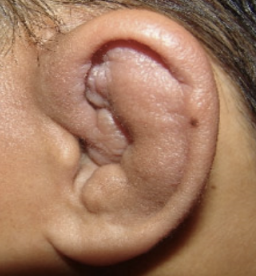
cauliflower ear assessment/diagnostic findings
Hearing loss will only occur if EAC is blocked.
Often conductive, secondary to edema of the EAC
Sudden or gradual SNHL presumably resulting from vasculitis of the internal auditory artery, or its branches, may occur in up to 40% of patients.
cauliflower ear management & treatment
Prevention is key → prompt drainage of hematomas + compression.
Established deformity: surgical reconstruction (otoplasty) if desired.
Cosmetic concern is the main issue; functionally stable if canal remains open.
carcinoma
Type of cancer that develops from epithelial cells (skin). It makes up the majority of the cases of malignancy of the breast, uterus, intestinal track, skin, and tongue.

carcinoma cause
Malignant. Chronic sun exposure (UV radiation), fair skin, old age, chronic inflammation, trauma
carcinoma signs & symptoms
Sore that never heal by itself, usually painful. 6% of all malignant lesions are found on or near the pinna.
basal & squamous cell
basal vs squamous cell carcinoma
Basal cell Carcinoma (BCC): pearly nodule, rolled edges, may ulcerate; slow growing
Squamous cell carcinoma (SCC): scaly, crusted, ulcerative lesion; more aggressive
herpes zoster oticus
Viral infection of the ear. Related to shingles and chicken pox. It manifests as sever otalgia (ear pain) and associated cutaneous (relating to skin) vesicular eruption, usually of the pinna in the regio of the concha bowl, pinna, along the skin of the external canal, and occasionally, on the tympanic membrane.
Ramsay Hunt Syndrome
Caused by the reactivation of pre-existing Varicella-zoster virus in the geniculate ganglion, a nerve cell bundle of the facial nerve. Incidence in males and females is equal. Incidence increases in patients older than 60. When associated with facial paralysis (CN7). Accounts for up to 12% of all facial paralysis (Robillard et al., 1986) and generally causes more severe symptoms and has worse prognosis than Bell’s Palsy.
Ramsay Hunt Syndrome signs & symptoms
Typically, inability to move facial muscles, pain in the ear, vertigo, tinnitus, taste loss on the front of the tongue (chorda tympani), dry eyes and mouth, and eruption of a rash.
RHS vs. HZO
They are both from shingles, or an activation of the varicella-zoster virus. RHS is when it’s associated with facial paralysis.
microtia
a congenital deformity affecting the pinna where the ear does not fully develop (small or missing) during the first trimester of pregnancy. usually small or peanut shaped. may occur in isolation or in association with other malformations, including facial clefts, cardiac defects, renal anomalies, etc.

microtia: type of hearing loss
conductive HL
microtia: reconstruction
nearly always associated with middle ear abnormality
microtia: unilateral vs bilateral management
In unilateral cases, the right side is affected more commonly than the left (approximately 5:3), and the reported incidence of bilateral microtia is approximately 10%.
anotia
pinna is absent at birth. may occur in isolation or in association with other malformations, including facial clefts, cardiac defects, renal anomalies, etc.

anotia: type of hearing loss
maximum conductive loss
anotia: reconstruction
nearly always associated with middle ear abnormality. atresia reconstruction process.
anotia: unilateral vs bilateral management
unilateral: surgery is delayed until 5-10yrs old. until then, hearing rehab isn’t required but evals should be consistent.
bilateral: surgery is delayed until 3-6yrs old. until then, hearing rehab is immediate and child should be fitted with non-surgical soft band hearing devices or BAHAs.
stenosis
occlusion of the EAC. EAC diameter of 1mm or less.
stenosis: type of hearing loss
impacted cerumen/buildup, naturally narrower passage. conductive hearing loss.
stenosis: reconstruction
surgical meatoplasty (to widen the EAC). must be older for procedure (4-6yrs).
atresia
the absence or closure of the EAC

atresia: type of hearing loss
conductive hearing loss.
after surgery:
- Conductive hearing loss – graft failure, middle ear adhesions, perfs etc
- Canal stenosis – 25% develop acquired stenosis
- Facial nerve injury – paresis in <1%
- Labyrinthine injury – may have high freq SNHL due to drilling etc. but rare
atresia: reconstruction
Microtia repaired first.
- Rib harvested and carved into a cartilaginous structure resembling a pinna.
- Fitted into a pocket under skin slowly. The skin is folded and formed around this scaffolding.
2nd surgery to create an ear canal
- Drill meatus
- Explore and repair middle ear structures
- Graft skin to form a TM
- Meatus is created to be 2 times larger than normal to allow for
swelling/stenosis
- skin graft
- Packed with ear wicks (small piece of expandable sponge that expands when it gets wet) that are hydrated with Cortisporin (a combination antibiotic medicine including neomycin)
Surgical results vary w/ experience of team and degree of aplasia
- Better than 30 dB HL thresholds – 50-75% with severe aplasias
- Better than 20 dB HL in 15 – 50%
- If you end up with a poor result, will be difficult to wear an earmold in this canal.
Alternative Rehabilitation strategy
- prosthetic ear w/ BC aid (BAHA)
atresia: unilateral vs bilateral management
Unilateral atresia: bilateral atresia -> 3:1
bilateral: urgent for speech and language development
unilateral: speech & lang will develop normally, expect difficulty with sound localization and emphasize preferential seating
cerumen impaction
Obstruction of the eardrum due to cerumen
cerumen impaction: removal methods
Removed via suction, wire loop, curette, or irrigation.
- Irrigation
Body temp
Aimed at the roof of the canal till the bolus extrudes.
cerumen impaction: when irrigation is contraindicated
TM perforation, ossicular disarticulation
ostemoa vs exostoses: causes
osteoma: cause unknown, occurs most frequently in middle age
exostoses: may result from chronic exposure of the EAC to cold water
ostemoa vs exostoses: appearance
osteoma: usually unilateral, solitary, and laterally placed. often found attached to the tympanosquamous or tympanomastoid suture
exostoses: bony growths in deep (medial) canal, often bilateral
ostemoa vs exostoses: management
osteoma: surgical if they block canal. skin graft is rarely necessary.
exostoses: surgical if they block canal. earplugs (in the water) to prevent reoccurrence
ostemoa vs exostoses: effect on earmolds
if they are removed, earmolds will look very different- in essence they complicate impressions. especially given the amount of occlusion, earmolds may not be possible.
AOM: causes
can result from bacterial or viral infection (usually follows a URI/may b associated with ET dysfunction/craniofacial abnormalities)
incidence decreases dramatically after age 7 for children with normal craniofacial structures
hereditary, daycare/large family, bottle feeding, smoking in the home, low socio-economic status
AOM: signs
rapid otaglia/irritability, hearing loss, fever, decreased appetite, otorrhea (if TM perforated), ear tugging is NOT reliable indicator
purulent effusion: thick and infected effusion
serous effusion: thin/water effusion (air bubbles), in addition to HL they experience aural fullness/pressure and clicking and popping when swallowing. retraction
mucoid effusion: thick/sticky. retraction
diagnosis criteria: moderate/severe bulging of the TM, new-onset otorrhea not due to otitis externa. mild bulging + otalgia <48 h or intense TM erythema, vessels in drum burst due to pressure/ TM perforation. diagnose with TYMP
AOM: management
observation-
most ME resolves spontaneously
watchful waiting with documented follow-up-
recommends against antibiotics. reevaluate every 3-6 months until effusion resolves, HL is found, or TM changes appear
surgery-
PE tubes
risk of anesthesia/surgery
autoinflation-
low cost/risk, requires a cooperative child
COME: causes
effusion persisting >3 months
infection of the ME space causes mucosal cells that line the ME to become secretory. acute inflammation resolves but changes in the cellular structure of ME and ET mucosa remain and effusion persists
COME: signs
accumulation of liquid in the middle ear cleft without other signs of inflammation or infection
COME: management
offer bilateral tubes for blateral OME > 3 months and documented hearing difficulty
can be offered before 3mnths with other sx (balance, school performance, ear discomfort)
at risk children- developmental, sensory, syndromic- a lower threshold applies
obtain hearing eval if persists/ before surgery
fungal otitis externa: causes
relatively rare, fungal infection of the EAC
high humidity (swelling and absorption of water into inflamed tissue), water exposure (removal of ear wax protection for bacteria), trauma, laceration, allergy, stress, excessive cleaning or scratching, hearing aids/headphones/ear plugs
fungal otitis externa: signs
erythematous (red)/ edematous (swollen)/ pruritus (itching)/ painful skin, clear otorrhea (discharge), otalgia (pain) at tragus manipulation
fungal otitis externa: management