4: Muscular & Musculoskeletal System (+ Imaging Modalities)

0.0(0)
Studied by 0 people
call kaiCall Kai
learnLearn
examPractice Test
spaced repetitionSpaced Repetition
heart puzzleMatch
flashcardsFlashcards
GameKnowt Play
Card Sorting

1/118

encourage image

There's no tags or description

Looks like no tags are added yet.

Last updated 3:58 AM on 10/7/26
Name
Mastery
Learn
Test
Matching
Spaced
Call with Kai
Chat

No analytics yet

Send a link to your students to track their progress

119 Terms

1
New cards

muscle tissue

able to contract + produce mechanical force

  • made of myocytes (muscle cells/fibers)

    • electrically excitable → conduct propagating electrical signals (action potentials)

    • creates tension force

  • 3 types:

    • skeletal

    • cardiac

    • smooth


<p>able to contract + produce mechanical force</p><ul><li><p>made of <strong>myocytes </strong>(muscle cells/fibers) </p><ul><li><p>electrically excitable → conduct propagating electrical signals (action potentials)</p></li><li><p>creates tension force</p></li></ul></li></ul><ul><li><p>3 types:</p><ul><li><p>skeletal</p></li><li><p>cardiac</p></li><li><p>smooth </p></li></ul></li></ul><p></p>
2
New cards

skeletal muscle

muscles moving skeleton

  • long, cylindrical cells arranged in parallel

  • multinucleate,

  • striated appearance

  • innervated by somatic motor neurons

  • uses ATP to generate tension force

  • has connective tissue packaging

    • provides blood to muscle fibers for O2 + nutrients


<p>muscles moving skeleton</p><ul><li><p><em>long, cylindrical cells </em>arranged in parallel</p></li><li><p><em>multi</em>nucleate,</p></li><li><p>striated appearance</p></li><li><p>innervated by <em>somatic </em>motor neurons</p></li><li><p>uses ATP to generate tension force</p></li><li><p>has connective tissue packaging</p><ul><li><p>provides blood to muscle fibers for O2 + nutrients </p></li></ul></li></ul><p></p>
3
New cards

cardiac muscle

muscles pumping blood through heart

  • short, branched fibers lined up end-to-end via intercalated discs (specialized cell junctions)

  • mononucleated

  • striated appearance

  • innervated by autonomic (visceral) motor neurons


<p>muscles pumping blood through heart</p><ul><li><p><em>short, branched fibers</em> lined up end-to-end via <em>intercalated discs </em>(specialized cell junctions)</p></li><li><p><em>mono</em>nucleated</p></li><li><p>striated appearance </p></li><li><p>innervated by <em>autonomic (visceral)</em> motor neurons </p></li></ul><p></p>
4
New cards

smooth muscle

muscles in blood vessels, digestive tract, etc.

  • spindle-shaped cells → thick in middle, tapered at ends

  • mononucleated

  • no striations

  • innervated by autonomic (visceral) motor neurons


<p>muscles in blood vessels, digestive tract, etc.</p><ul><li><p><em>spindle-shaped</em> cells → thick in middle, tapered at ends</p></li><li><p><em>mono</em>nucleated </p></li><li><p>no striations</p></li><li><p>innervated by <em>autonomic (visceral) </em>motor neurons</p></li></ul><p></p>
5
New cards

epimysium

dense irregular connective tissue

  • surrounds entire muscle organ

  • connects to tendon


<p>dense irregular connective tissue </p><ul><li><p>surrounds <em>entire muscle </em>organ</p></li><li><p>connects to tendon</p></li></ul><p></p>
6
New cards

perimysium

dense irregular connective tissue

  • surrounds fascicle (bundle of muscle fibers


<p>dense irregular connective tissue </p><ul><li><p>surrounds fascicle (bundle of muscle fibers </p></li></ul><p></p>
7
New cards

endomysium

loose/areolar connective tissue

  • surrounds each muscle fiber/cell


<p>loose/areolar connective tissue</p><ul><li><p>surrounds each muscle fiber/cell </p></li></ul><p></p>
8
New cards

sarcolemma

plasma membrane of myofiber/myocyte

<p>plasma membrane of myofiber/myocyte</p>
9
New cards

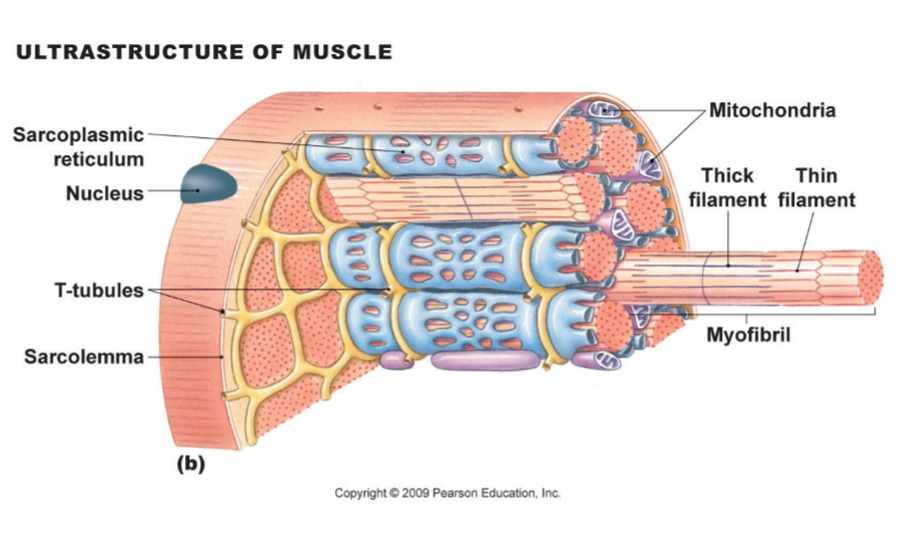
transverse tubules (T-tubules)

invaginations of sarcolemma that extend into interior of myocyte → creates tunnels filled w/ ECF

  • at right angle to cell surface

  • connects sarcolemma to myofibril

  • brings in sarcolemma AP into vicinity of sarcoplasmic reticulum → triggers Ca2+ release

<p>invaginations of sarcolemma that extend into interior of myocyte → creates tunnels filled w/ ECF</p><ul><li><p>at right angle to cell surface</p></li><li><p>connects sarcolemma to myofibril </p></li><li><p>brings in sarcolemma AP into vicinity of sarcoplasmic reticulum → triggers Ca<sup>2+</sup> release</p></li></ul><p></p>
10
New cards

sarcoplasm

cytoplasm of myofiber/myocyte

  • contains specialized cytoskeletal + membranous organelles

    • glycogen

      • chains of glucose → broken down to release glucose → used to make ATP

    • myoglobin

      • protein only in myocytes → binds O2 + releases O2 when needed by mitochondria

    • creatine phosphate

      • transfer ADP to reconstitute ATP when additional ATP needed for myocyte contraction

<p>cytoplasm of myofiber/myocyte</p><ul><li><p>contains specialized cytoskeletal + membranous organelles</p><ul><li><p>glycogen</p><ul><li><p>chains of glucose → broken down to release glucose → used to make ATP</p></li></ul></li><li><p>myoglobin</p><ul><li><p>protein <em>only in myocytes</em> → binds O2 + releases O2 when needed by mitochondria</p></li></ul></li><li><p>creatine phosphate </p><ul><li><p>transfer ADP to reconstitute ATP when additional ATP needed for myocyte contraction</p></li></ul></li></ul></li></ul><p></p>
11
New cards

myofibers (myocytes)

muscle fibers/cells

  • contains:

    • myofibrils

    • myosatellite cells

    • many superficial nuclei

    • T-tubules

    • sarcoplasmic reticulum

  • stretches from muscle origin to insertion → tendon to tendon

  • associated w/ support cells + myofibers

  • produced by fusion of many myoblasts


12
New cards

myofibrils

specialized organelle found within myofiber

  • contractile protein fibers arranged in parallel that extend along length of myocyte → allow muscle contraction

  • made of overlapping thin + thick filaments (actin + myosin)


<p>specialized organelle found within myofiber</p><ul><li><p>contractile protein fibers arranged in parallel that extend along length of myocyte → allow muscle contraction</p></li><li><p>made of overlapping thin + thick filaments (actin + myosin)</p></li></ul><p></p>
13
New cards

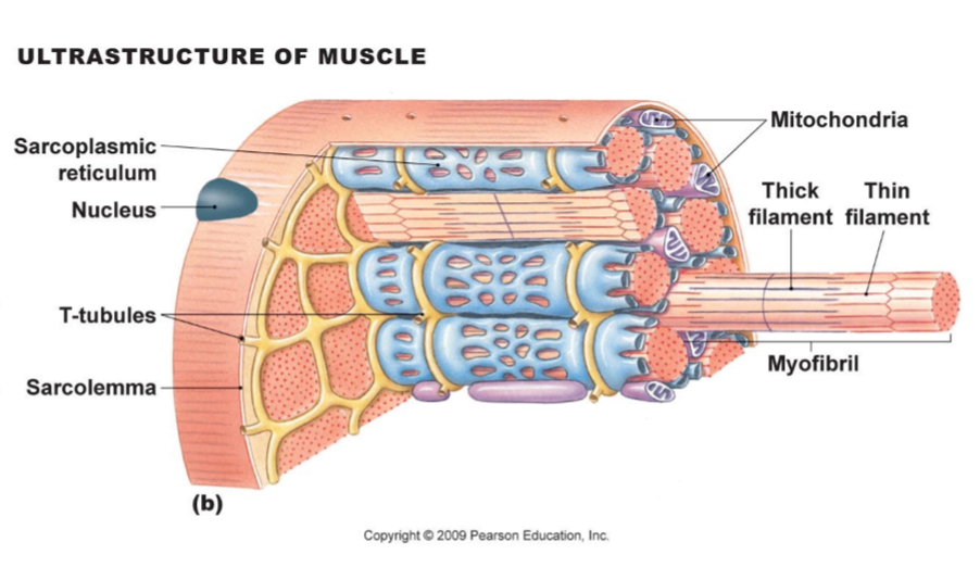
sarcoplasmic reticulum

endoplasmic reticulum of myofibers

  • specialized smooth ER

  • wraps around myofibril

  • stores + releases Ca2+

  • ends = terminal cisternae (closely associated w/ T-tubule)

  • 2 terminal cisternae + T-tubule = triad


<p>endoplasmic reticulum of myofibers</p><ul><li><p>specialized smooth ER </p></li><li><p>wraps around myofibril </p></li><li><p>stores + releases Ca<sup>2+</sup></p></li><li><p>ends = terminal cisternae (closely associated w/ T-tubule)</p></li><li><p>2 terminal cisternae + T-tubule = triad</p></li></ul><p></p>
14
New cards

triad

2 terminal cisternae + T-tubule

  • T-tubule (yellow internal structures)

    • tubes bringing in sarcolemma

  • cisternae (blue internal structures)

    • enlargement of sarcoplasmic reticulum


<p>2 terminal cisternae + T-tubule</p><ul><li><p>T-tubule (yellow internal structures)</p><ul><li><p>tubes bringing in sarcolemma</p></li></ul></li><li><p>cisternae (blue internal structures)</p><ul><li><p>enlargement of sarcoplasmic reticulum</p></li></ul></li></ul><p></p>
15
New cards

thick myofilament

composed of myosin protein

  • double thickness of thin filaments

  • myosin heads project to ends of myofilament

  • myosin tails attached at middle

  • contains titin → tethers filament to Z-line + allows muscle contraction/retraction


<p>composed of myosin protein </p><ul><li><p>double thickness of thin filaments </p></li><li><p>myosin heads project to ends of myofilament </p></li><li><p>myosin tails attached at middle </p></li><li><p>contains titin → tethers filament to Z-line + allows muscle contraction/retraction</p></li></ul><p></p>
16
New cards

myosin

has mobile head + neck that can bind actin

  • hinge → moves head

  • free head → binds to actin

    • head = ATPases

      • cleave ATP = ADP + Pi energy → used to form cross-bridge b/w myosin head + actin


<p>has mobile head + neck that can bind actin</p><ul><li><p>hinge → moves head</p></li><li><p>free head → binds to actin</p><ul><li><p>head = ATPases </p><ul><li><p>cleave ATP = ADP + P<sub>i</sub> energy → used to form cross-bridge b/w myosin head + actin</p></li></ul></li></ul></li></ul><p></p>
17
New cards

thin myofilaments

twisted strands of actin, troponin + tropomyosin

  • actin contains myosin-binding sites

    • at rest = blocked by tropomyosin

  • troponin holds tropomyosin in place → contains Ca2+ binding sites
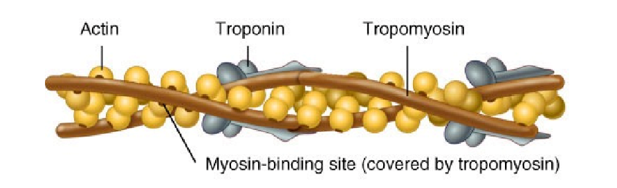

<p>twisted strands of actin, troponin + tropomyosin </p><ul><li><p>actin contains myosin-binding sites </p><ul><li><p>at rest = blocked by tropomyosin</p></li></ul></li><li><p>troponin holds tropomyosin in place → contains Ca<sup>2+</sup> binding sites </p></li></ul><p></p>
18
New cards

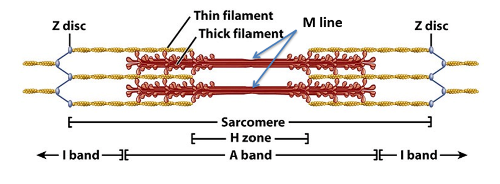
sarcomere

functional, repeating units of myofibrils

  • made of overlapping thick + thin myofilaments

  • line up end-to-end along length of myofibril

  • myofibrils within myocyte arranged so that they align w/ e/o = striated appearance in muscle!


<p>functional, repeating units of <em>myofibrils</em></p><ul><li><p>made of overlapping thick + thin myofilaments</p></li><li><p>line up end-to-end along length of myofibril</p></li><li><p>myofibrils within myocyte arranged so that they align w/ e/o = striated appearance in muscle!</p></li></ul><p></p>
19
New cards

H band

sarcomere region w/ only thick filaments

  • decreases in size when sarcomere contracts
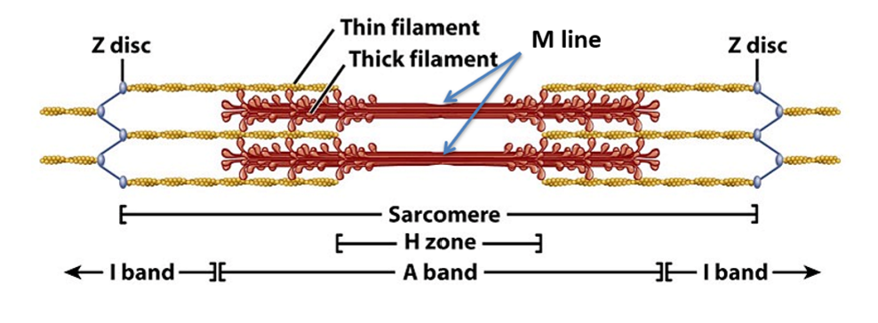

<p>sarcomere region w/ only thick filaments</p><ul><li><p>decreases in size when sarcomere contracts</p></li></ul><p></p>
20
New cards

A band

overlapping sarcomere region of thick + thin filaments

  • darker band

  • myosin w/ some overlapping actin


<p>overlapping sarcomere region of thick + thin filaments</p><ul><li><p><em>darker </em>band</p></li><li><p>myosin w/ some overlapping actin</p></li></ul><p></p>
21
New cards

I band

sarcomere region w/ only thin filament

  • lighter band

  • includes actin of next filament


<p>sarcomere region w/ only thin filament</p><ul><li><p><em>lighter </em>band</p></li><li><p>includes actin of next filament</p></li></ul><p></p>
22
New cards

M line

thick band of thick filaments in center of sarcomere

<p>thick band of thick filaments in center of sarcomere </p>
23
New cards

Z disc

end part of sarcomeres w/ proteins that anchor + hold actin-myofilaments in place

  • dark line in middle of I band

  • only actin


<p>end part of sarcomeres w/ proteins that anchor + hold actin-myofilaments in place</p><ul><li><p>dark line in middle of I band</p></li><li><p>only actin</p></li></ul><p></p>
24
New cards

tropomyosin-troponin complex

wraps around actin helix

  • when bound w/ Ca2+ reveals myosin-binding site on actin


25
New cards

sliding filament mechanism

  • myocyte contraction → myosin heads of thick myofilaments bind thin myofilament

  • myosin heads pull thin myofilaments toward M line (sarcomere center)

  • ⬆overlap = ⬇ sarcomere length (shortens)

    • all sarcomeres of all myofibrils shorten at same time = entire myocyte shortens
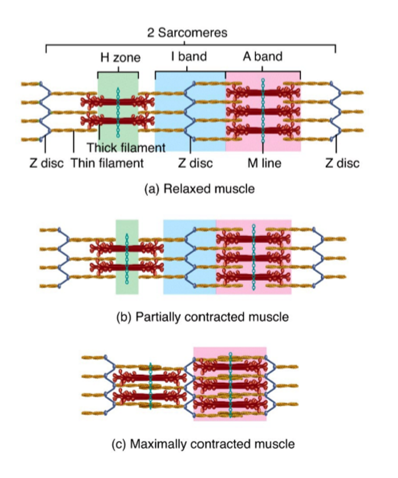

<ul><li><p>myocyte contraction → myosin heads of thick myofilaments bind thin myofilament </p></li><li><p>myosin heads pull thin myofilaments toward M line (sarcomere center)</p></li><li><p><span data-name="arrow_up" data-type="emoji">⬆</span>overlap = <span data-name="arrow_down" data-type="emoji">⬇</span> sarcomere length (shortens)</p><ul><li><p>all sarcomeres of all myofibrils shorten at same time = entire myocyte shortens</p></li></ul></li></ul><p></p>
26
New cards

muscle excitation

sarcolemma generates electrical signal that precedes contraction

  1. electrochemical signal from somatic motor neuron

    1. motor neurons release NTs into synaptic cleft

  2. electrical excitation of NMJ

    1. NTs bind to ligand-gated ion channels on sarcolemma

    2. Na+ flows into myofiber → charges move across sarcolemma = electrical signal

  3. propagation of electrical signal

    1. sarcolemma depolarization from Na+ influx opens nearby VGNCs

    2. VGNCs along sarcolemma open as more VGNCs open = AP!

  4. calcium release

    1. T-tubules bring sarcolemma AP to sarcoplasmic reticulum

    2. AP triggers release of Ca2+ from sarcoplasmic reticulum

  5. Ca2+ presence → binds to troponin → pushes tropomyosin aside = exposes myosin-binding sites on actin = muscle contraction


<p>sarcolemma generates electrical signal that precedes contraction </p><ol><li><p>electrochemical signal from somatic motor neuron</p><ol><li><p>motor neurons release NTs into synaptic cleft </p></li></ol></li><li><p>electrical excitation of NMJ </p><ol><li><p>NTs bind to ligand-gated ion channels on sarcolemma </p></li><li><p>Na+ flows into myofiber → charges move across sarcolemma = electrical signal</p></li></ol></li><li><p>propagation of electrical signal </p><ol><li><p>sarcolemma depolarization from Na+ influx opens nearby VGNCs</p></li><li><p>VGNCs along sarcolemma open as more VGNCs open = AP!</p></li></ol></li><li><p>calcium release</p><ol><li><p>T-tubules bring sarcolemma AP to sarcoplasmic reticulum </p></li><li><p>AP triggers release of Ca2+ from sarcoplasmic reticulum </p></li></ol></li><li><p>Ca2+ presence → binds to troponin → pushes tropomyosin aside = exposes myosin-binding sites on actin = muscle contraction</p></li></ol><p></p>
27
New cards

neuromuscular junction (NMJ)

point of connection b/w motor neuron + myofiber

  • made up of:

    • axon terminal (of motor neuron)

    • synaptic cleft (in b/w)

    • motor end plate (of myofiber)


28
New cards

motor end plate

specialized region (midpoint) of myofiber that forms NMJ

  • has slightly different proteins + integral membrane proteins

  • has extra folds to ⬆ SA for communication


29
New cards

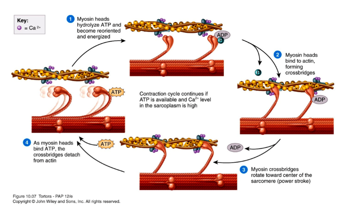
muscle contraction

myofibers + myofibrils becoming shorter = tension

  • begins w/ Ca2+ release after excitation

  • continues as long as Ca2+ remains elevated

  • myosin + actin move relative to e/o

  1. calcium release → active site exposed

  2. binding

  3. power stroke

  4. ADP release

  5. ATP binding

  6. ATP hydrolysis


<p>myofibers + myofibrils becoming shorter = tension </p><ul><li><p>begins w/ Ca<sup>2+</sup> release after excitation </p></li><li><p>continues as long as Ca<sup>2+</sup> remains elevated</p></li><li><p>myosin + actin move relative to e/o</p></li></ul><ol start="0"><li><p>calcium release → active site exposed</p></li><li><p>binding </p></li><li><p>power stroke</p></li><li><p>ADP release</p></li><li><p>ATP binding </p></li><li><p>ATP hydrolysis </p></li></ol><p></p>
30
New cards

contraction cycle step 0 (calcium release)

  1. Ca2+ released from SR

  2. Ca2+ binds to tropomyosin-troponin complex → releases from actin

  3. myosin-binding site on actin exposed


<ol><li><p>Ca<sup>2+ </sup>released from SR</p></li><li><p>Ca<sup>2+ </sup>binds to tropomyosin-troponin complex → releases from actin</p></li><li><p>myosin-binding site on actin exposed</p></li></ol><p></p>
31
New cards

contraction cycle step 1 (binding)

  1. myosin binds to active site on actin

  2. cross-bridge formed b/w myosin + actin


<ol><li><p>myosin binds to active site on actin</p></li><li><p>cross-bridge formed b/w myosin + actin</p></li></ol><p></p>
32
New cards

contraction cycle step 2 (power stroke)

  1. myosin neck goes from straight to pivoted

    1. myosin attached to actin → head pivots = actin filament moved

    2. filaments slide past e/o


<ol><li><p>myosin neck goes from straight to pivoted</p><ol><li><p>myosin attached to actin → head pivots = actin filament moved </p></li><li><p>filaments slide past e/o </p></li></ol></li></ol><p></p>
33
New cards

contraction cycle step 3 (ADP release)

  1. ADP + phosphate group on myosin head released

    1. ATP = energy → release of energy off myosin head


<ol><li><p>ADP + phosphate group on myosin head released</p><ol><li><p>ATP = energy → release of energy off myosin head </p></li></ol></li></ol><p></p>
34
New cards

contraction cycle step 4 (ATP binding)

  1. new ATP molecule binds to myosin head

  2. cross-bridge b/w actin + myosin breaks


<ol><li><p>new ATP molecule binds to myosin head</p></li><li><p>cross-bridge b/w actin + myosin breaks </p></li></ol><p></p>
35
New cards

contraction cycle step 5 (ATP hydrolysis)

  1. ATP hydrolyzes into ADP + phosphate → energy released

  2. myosin head goes back into original position → no longer pivoted


<ol><li><p>ATP hydrolyzes into ADP + phosphate → energy released</p></li><li><p>myosin head goes back into original position → no longer pivoted </p></li></ol><p></p>
36
New cards

radiography (x-ray)

form of electromagnetic radiation that can pass through most objects, ie. body

  • x-rays pass through object → contact photographic film/image recording plate = image produced

  • images reflect differences in tissue density

    • bones + dense structures absorb x-rays = white

      • x-rays do not pass through

    • hollow/less dense structures pass through x-rays = black

      • x-rays contact photographic film/image recording plate

  • superimposed 2D image created


<p>form of electromagnetic radiation that can pass through most objects, ie. body</p><ul><li><p>x-rays pass through object → contact photographic film/image recording plate = image produced</p></li><li><p>images reflect differences in tissue density </p><ul><li><p>bones + dense structures absorb x-rays = white</p><ul><li><p>x-rays do not pass through</p></li></ul></li><li><p>hollow/less dense structures pass through x-rays = black</p><ul><li><p>x-rays contact photographic film/image recording plate</p></li></ul></li></ul></li><li><p>superimposed 2D image created</p></li></ul><p></p>
37
New cards

computed tomography (CT)

  • collected w/ SPECT data → images co-register together = more accurately localize site of uptake of injected radioactive tracer

  • tomography = view anatomical section/slice through body

  • x-ray source positioned opposite to detector

    • apparatus rotated around central axis of subject

    • data converted into image sections

  • advantages over x-rays:

    • no superimposition of tissues

    • improved contrast differences b/w tissues

  • imaging reflect tissue density:

    • ⬆ density = white

      • bone cortex

    • ⬇ density = black

      • fat/air filled structures, ie. lungs

    • gray structures = muscle, bone marrow


<ul><li><p>collected w/ SPECT data → images co-register together = more accurately localize site of uptake of injected radioactive tracer</p></li><li><p>tomography = view anatomical section/slice through body</p></li><li><p>x-ray source positioned opposite to detector</p><ul><li><p>apparatus rotated around central axis of subject</p></li><li><p>data converted into image sections</p></li></ul></li><li><p>advantages over x-rays:</p><ul><li><p>no superimposition of tissues</p></li><li><p>improved contrast differences b/w tissues</p></li></ul></li><li><p><strong>imaging reflect tissue density:</strong></p><ul><li><p><span data-name="arrow_up" data-type="emoji">⬆</span> density = white</p><ul><li><p>bone cortex</p></li></ul></li><li><p><span data-name="arrow_down" data-type="emoji">⬇</span> density = black</p><ul><li><p>fat/air filled structures, ie. lungs</p></li></ul></li><li><p>gray structures = muscle, bone marrow</p></li></ul></li></ul><p></p>
38
New cards

magnetic resonance imaging (MRI)

  • body tissues have different composition of H2O + fat

  • nuclei of hydrogen atoms (protons) in H2O + fat molecules = tiny magnets → precess (spin like top) at specific frequency

  • subject in magnetic field → protons align w/ magentic field

    • precess in LOW energy (majority) or HIGH energy configuration

  • aligned → precessing protons subjected to radiofrequency (RF) energy pulse

    • protons gain energy → flip to high energy

  • RF pulse removed → protons revert to initial configuration + emit energy that can be detected

    • data converted to MR image

  • image = energy emitted by protons in different tissues when placed into magnetic field + subjected to RF energy pulses

  • T1 - look at structure + anatomy

    • fat = bright/white

    • water = dark/black

  • T2 - find disease, fluid, swelling

    • fat = bright/white

    • water = bright/white


<ul><li><p>body tissues have different composition of H2O + fat</p></li><li><p>nuclei of hydrogen atoms (protons) in H2O + fat molecules = tiny magnets → precess (spin like top) at specific frequency</p></li><li><p>subject in magnetic field → protons align w/ magentic field</p><ul><li><p>precess in LOW energy (majority) or HIGH energy configuration</p></li></ul></li><li><p>aligned → precessing protons subjected to radiofrequency (RF) energy pulse</p><ul><li><p>protons gain energy → flip to high energy</p></li></ul></li><li><p>RF pulse removed → protons revert to initial configuration + emit energy that can be detected</p><ul><li><p>data converted to MR image</p></li></ul></li><li><p>image = energy emitted by protons in different tissues when placed into magnetic field + subjected to RF energy pulses</p></li><li><p><strong>T1 </strong>- look at structure + anatomy</p><ul><li><p>fat = bright/white</p></li><li><p>water = dark/black</p></li></ul></li><li><p><strong>T2 </strong>- find disease, fluid, swelling</p><ul><li><p>fat = bright/white</p></li><li><p>water = bright/white</p></li></ul></li></ul><p></p>
39
New cards

single-photon emission computed tomography (SPECT)

  • collected w/ CT data → images co-register together = more accurately localize site of uptake of injected radioactive tracer

  • radioactive tracer injected → taken up into targeted tissues

    • emission of gamma rays detected

    • data reconstructed to form image

  • advantage

    • provides functional info, ex. nutrient + blood flow

  • disadvantage

    • difficult to determine exact anatomical location
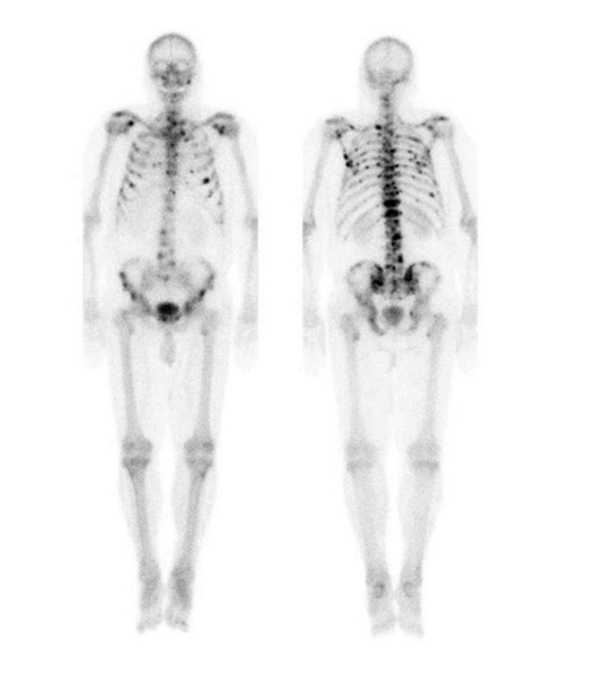

<ul><li><p>collected w/ CT data → images co-register together = more accurately localize site of uptake of injected radioactive tracer</p></li><li><p>radioactive tracer injected → taken up into targeted tissues</p><ul><li><p>emission of gamma rays detected</p></li><li><p>data reconstructed to form image</p></li></ul></li><li><p>advantage</p><ul><li><p>provides functional info, ex. nutrient + blood flow</p></li></ul></li><li><p>disadvantage</p><ul><li><p>difficult to determine exact anatomical location</p></li></ul></li></ul><p></p>
40
New cards

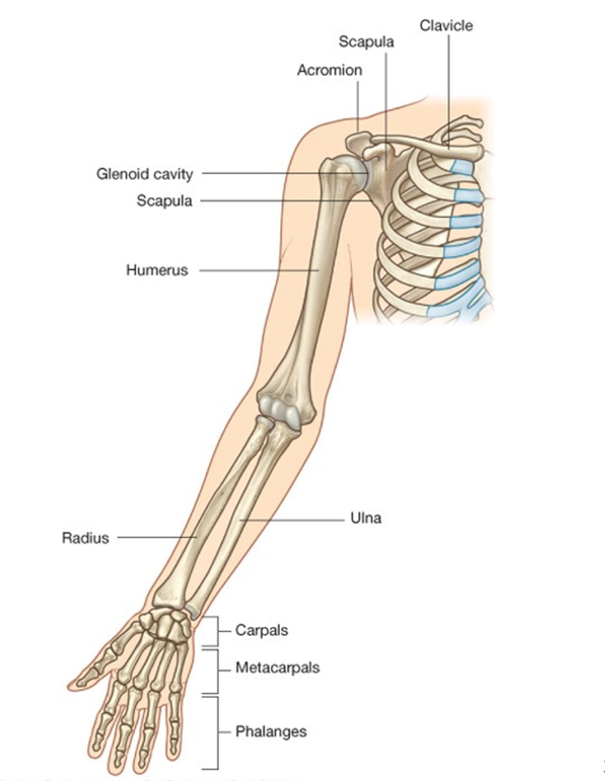
upper extremity bones

  • pectoral (shoulder) girdle

    • clavicle

    • scapula

      • sternal end = medial

      • acromial end = lateral

  • arm

    • humerus

  • forearm

    • radius

    • ulna

  • wrist + hand

    • carpals

    • metacarpals

    • phalanges


<ul><li><p>pectoral (shoulder) girdle</p><ul><li><p>clavicle </p></li><li><p>scapula</p><ul><li><p>sternal end = medial</p></li><li><p>acromial end = lateral</p></li></ul></li></ul></li><li><p>arm </p><ul><li><p>humerus </p></li></ul></li><li><p>forearm </p><ul><li><p>radius</p></li><li><p>ulna</p></li></ul></li><li><p>wrist + hand</p><ul><li><p>carpals</p></li><li><p>metacarpals</p></li><li><p>phalanges </p></li></ul></li></ul><p></p>
41
New cards

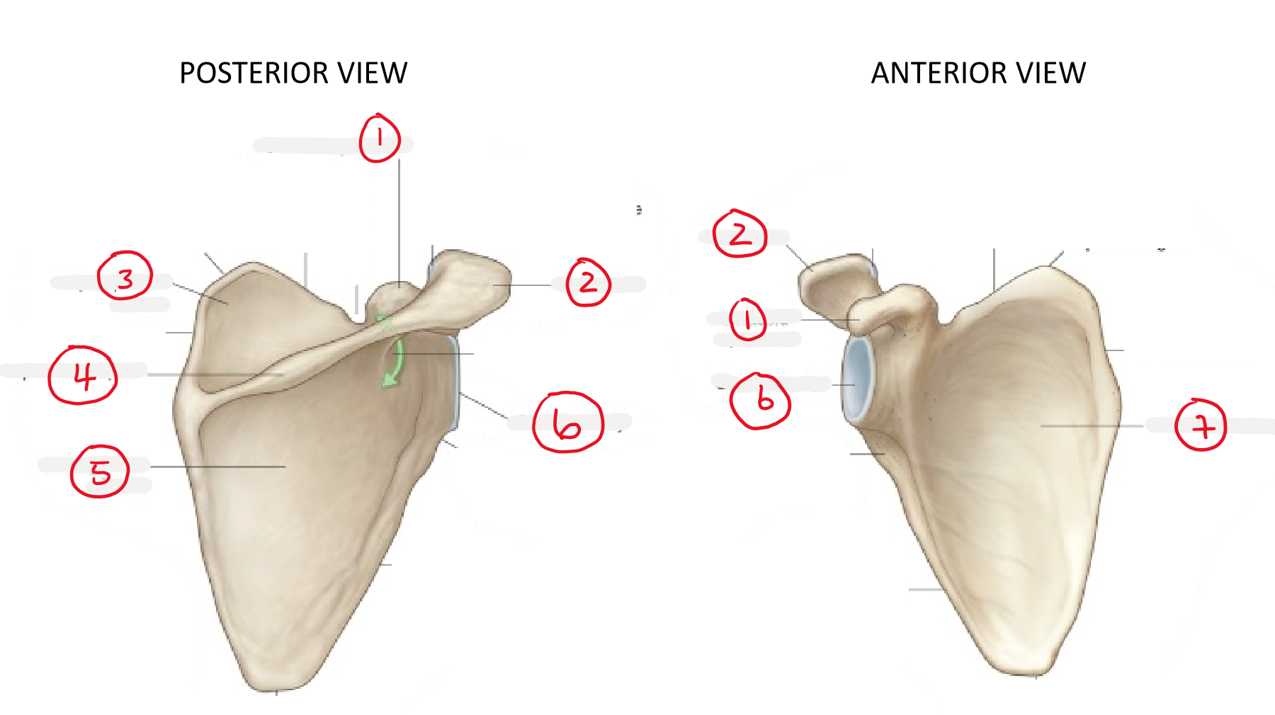
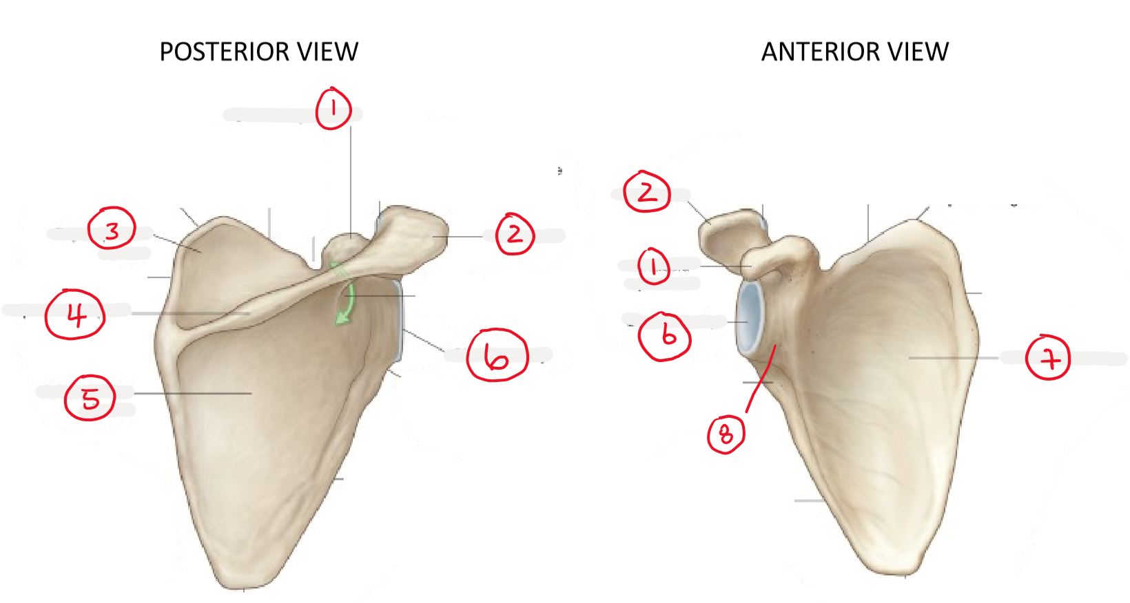
coracoid process

(1)

<p>(1)</p>
42
New cards

acromion

(2)

<p>(2)</p>
43
New cards

supraspinous fossa

(3) above spine + shallow depression

<p>(3) above spine + shallow depression</p>
44
New cards

spine of scapula

(4)

<p>(4) </p>
45
New cards

infraspinous fossa

(5) below spine + shallow depression

<p>(5) below spine + shallow depression</p>
46
New cards

glenoid cavity (glenoid fossa)

(6) articulates w/ head of humerus

<p>(6) articulates w/ head of humerus</p>
47
New cards

subscapular fossa

(7)

<p>(7)</p>
48
New cards

glenoid process

(8)

<p>(8)</p>
49
New cards

shoulder joints

  • sternoclavicular = plane

    • sternum + medial clavicle

  • acromioclavicular = plane

    • acromion + lateral clavicle

  • glenohumeral = ball + socket

    • glenoid fossa/cavity + humerus


<ul><li><p>sternoclavicular = plane</p><ul><li><p>sternum + medial clavicle </p></li></ul></li><li><p>acromioclavicular = plane</p><ul><li><p>acromion + lateral clavicle </p></li></ul></li><li><p>glenohumeral = ball + socket</p><ul><li><p>glenoid fossa/cavity + humerus</p></li></ul></li></ul><p></p>
50
New cards

pectoral girdle relational anatomy

  • acromion + acromial end of clavicle

    • superior to

      • humerus head

      • glenoid process

      • coracoid process

      • sternal end of clavicle

  • coracoid process

    • anterior to

      • humerus head

  • humerus head

    • lateral to

      • glenoid process


<ul><li><p>acromion + acromial end of clavicle </p><ul><li><p>superior to </p><ul><li><p>humerus head</p></li><li><p>glenoid process</p></li><li><p>coracoid process</p></li><li><p>sternal end of clavicle </p></li></ul></li></ul></li><li><p>coracoid process</p><ul><li><p>anterior to </p><ul><li><p>humerus head</p></li></ul></li></ul></li><li><p>humerus head</p><ul><li><p>lateral to </p><ul><li><p>glenoid process </p></li></ul></li></ul></li></ul><p></p>
51
New cards

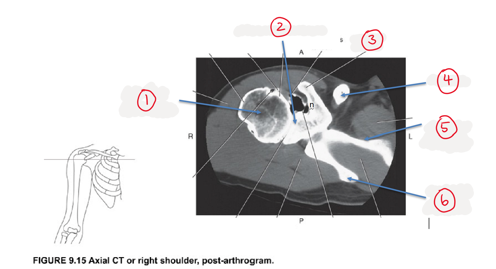
humerus

(1)

<p>(1)</p>
52
New cards

glenoid process

(2)

<p>(2)</p>
53
New cards

coracoid process

(3)

<p>(3)</p>
54
New cards

clavicle

(4)

<p>(4)</p>
55
New cards

body of scapula

(5)

<p>(5)</p>
56
New cards

spine of scapula

(6)

<p>(6)</p>
57
New cards

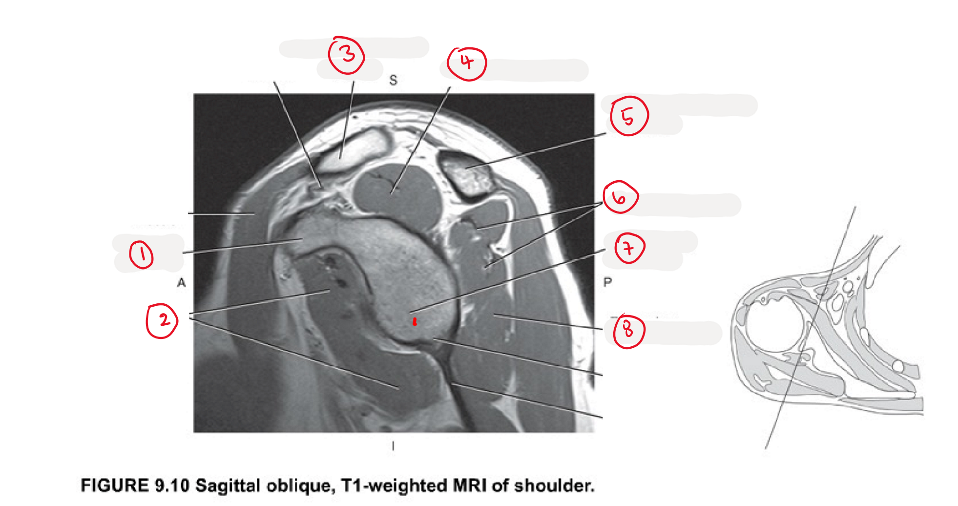
coracoid process

(1)

<p>(1)</p>
58
New cards

subscapularis

(2)

<p>(2) </p>
59
New cards

clavicle

(3)

<p>(3)</p>
60
New cards

supraspinatus

(4)

<p>(4)</p>
61
New cards

acromion

(5)

<p>(5)</p>
62
New cards

infraspinatus

(6)

<p>(6)</p>
63
New cards

glenoid fossa

(7)

<p>(7)</p>
64
New cards

teres minor

(8)

<p>(8)</p>
65
New cards

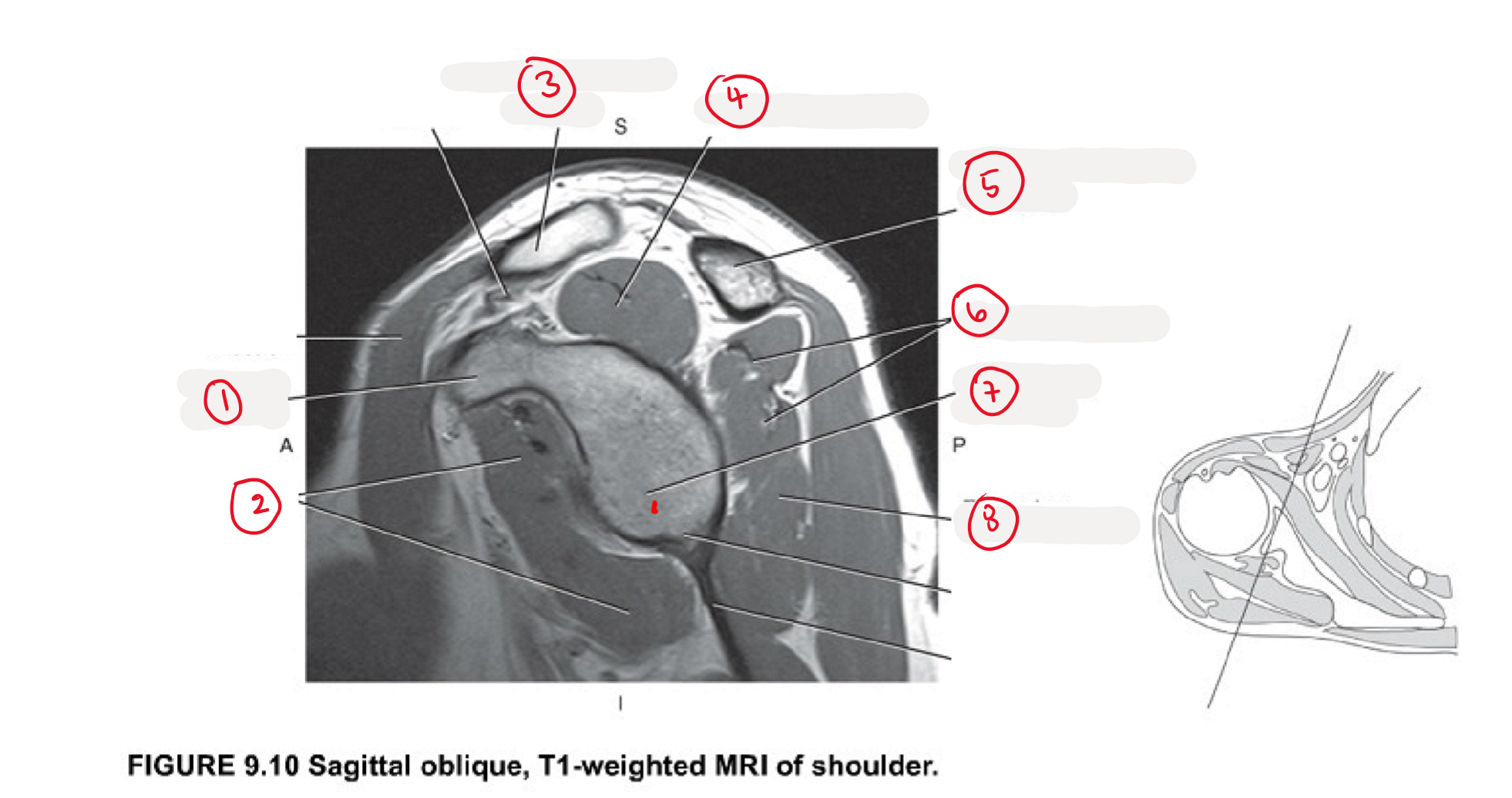
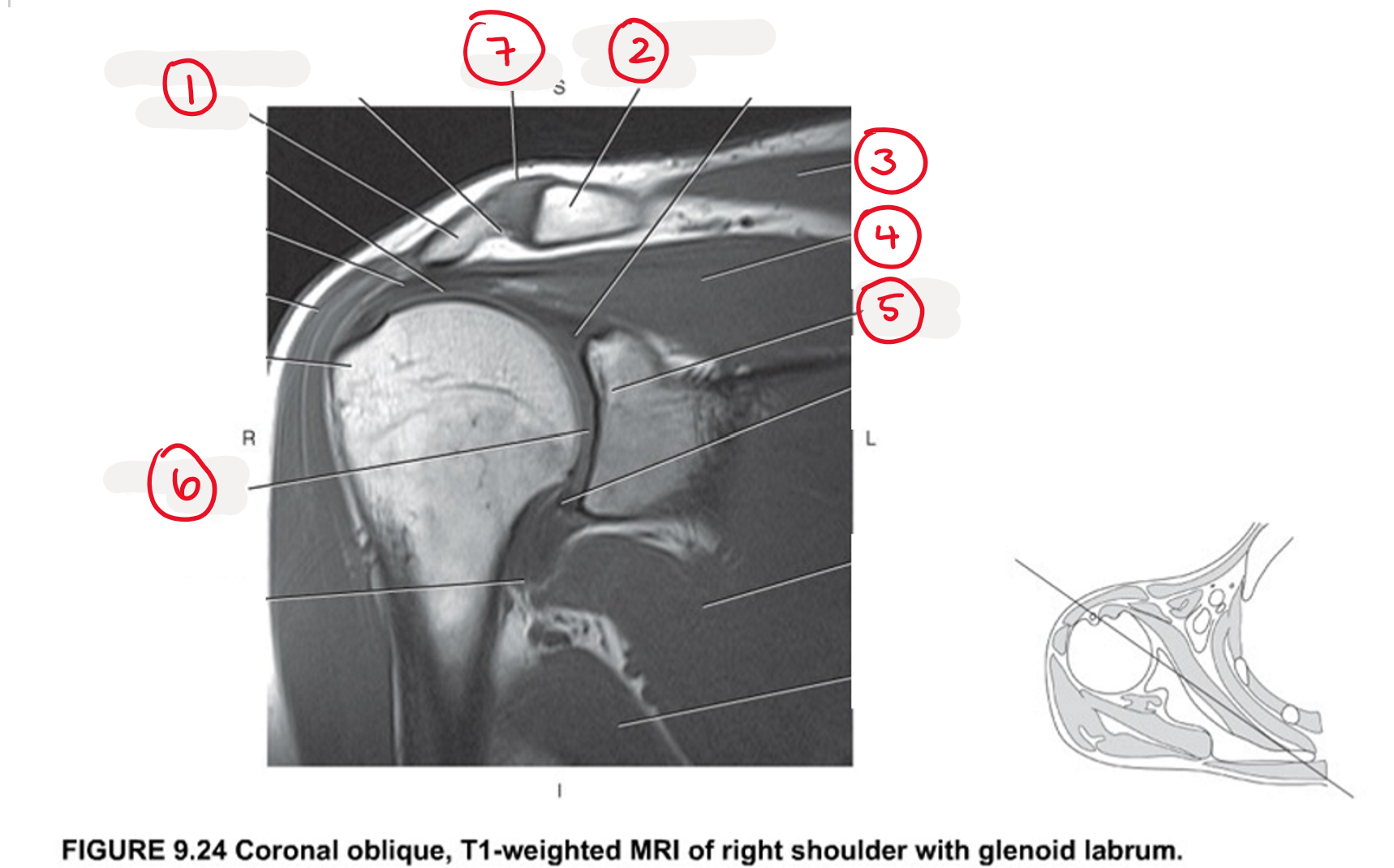
acromion

(1)

<p>(1)</p>
66
New cards

clavicle

(2)

<p>(2)</p>
67
New cards

trapezius

(3)

<p>(3)</p>
68
New cards

supraspinatus

(4)

<p>(4)</p>
69
New cards

glenoid process

(5)

<p>(5)</p>
70
New cards

glenohumeral joint

(6)

<p>(6)</p>
71
New cards

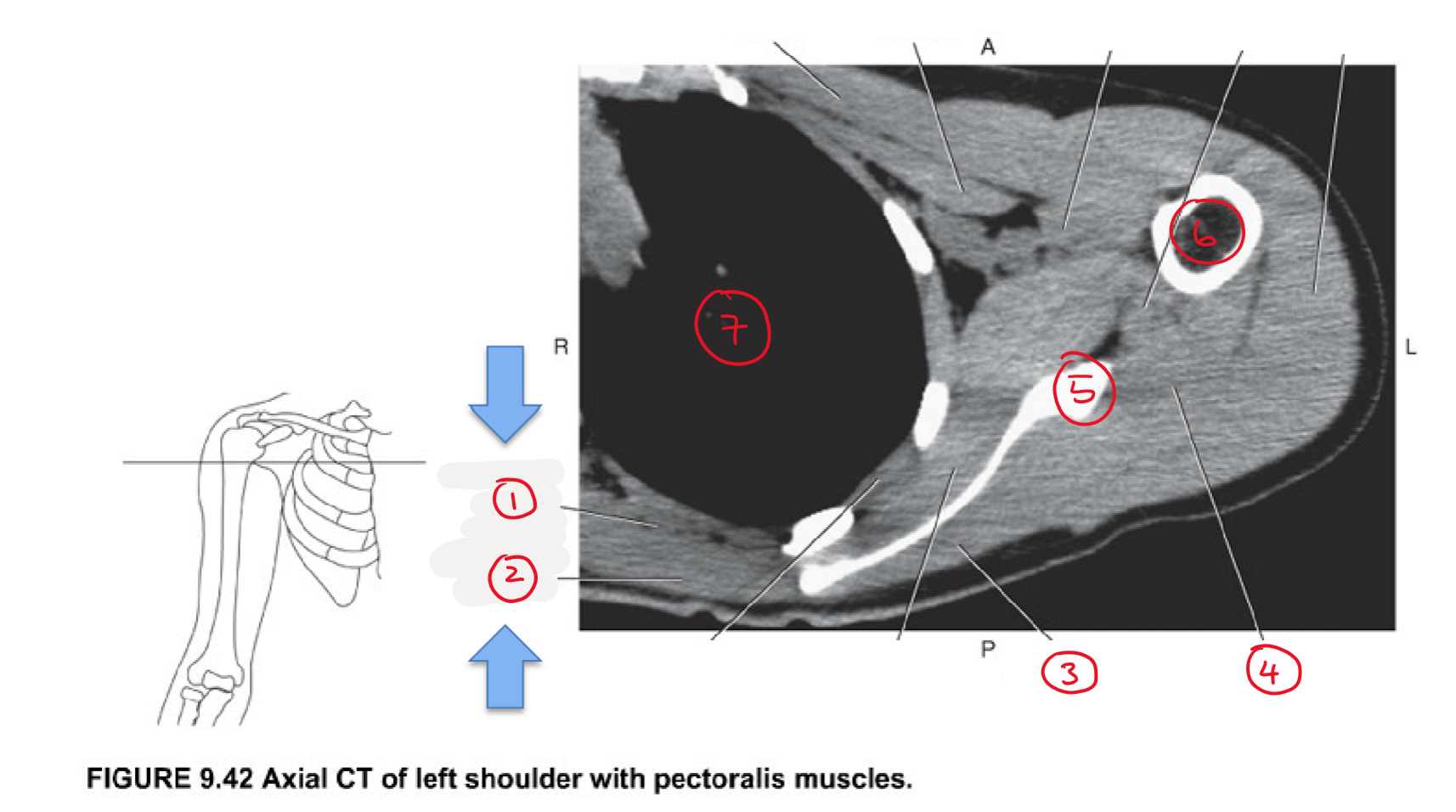
scapula position

  • posterior to ribs II-VII

  • anterior surface closely associated w/ ribs

    • subscapular fossa in contact w/ ribs

  • shoulder girdle (scapula + clavicle) coordinates shoulder movements

    • humeral head stays in glenoid fossa during arm movement


72
New cards

scapula movements

muscles insert onto scapula + work tgt to adjust scapula position = large range of motion

  • elevation + depression = shrug up + down

  • protraction (abduction) + retraction (adduction) = arm in + out front

  • upward rotation + downward rotation = raise arm up + down at side


73
New cards

scapula muscles

  • trapezius

  • levator scapulae

  • rhomboid minor

  • rhomboid major

  • pectoralis minor

  • serratus anterior


<ul><li><p>trapezius</p></li><li><p>levator scapulae</p></li><li><p>rhomboid minor</p></li><li><p>rhomboid major</p></li><li><p>pectoralis minor </p></li></ul><ul><li><p>serratus anterior</p></li></ul><p></p>
74
New cards

trapezius

muscle acting on scapula

  • origin:

    • vertebrae column

  • insertion:

    • scapula

  • superficial

  • movement:

    • elevate → upper fibers

    • retract → middle fibers

    • depress → lower fibers


<p>muscle acting on scapula</p><ul><li><p>origin: </p><ul><li><p>vertebrae column  </p></li></ul></li><li><p>insertion: </p><ul><li><p>scapula</p></li></ul></li><li><p>superficial</p></li><li><p>movement:</p><ul><li><p>elevate → upper fibers</p></li><li><p>retract → middle fibers</p></li><li><p>depress → lower fibers</p></li></ul></li></ul><p></p>
75
New cards

levator scapulae

muscle acting on scapula

  • origin:

    • vertebrae column

  • insertion:

    • scapula

  • deep

  • movement:

    • elevate


<p>muscle acting on scapula</p><ul><li><p>origin: </p><ul><li><p>vertebrae column </p></li></ul></li><li><p>insertion: </p><ul><li><p>scapula</p></li></ul></li><li><p>deep</p></li><li><p>movement:</p><ul><li><p>elevate</p></li></ul></li></ul><p></p>
76
New cards

rhomboid minor

muscle acting on scapula

  • origin:

    • vertebrae column

  • insertion:

    • scapula

  • deep

  • movement:

    • retract


<p>muscle acting on scapula</p><ul><li><p>origin: </p><ul><li><p>vertebrae column </p></li></ul></li><li><p>insertion:</p><ul><li><p>scapula</p></li></ul></li><li><p>deep</p></li><li><p>movement:</p><ul><li><p>retract</p></li></ul></li></ul><p></p>
77
New cards

rhomboid major

muscle acting on scapula

  • origin:

    • vertebrae column

  • insertion:

    • scapula

  • deep

  • movement:

    • retract


<p>muscle acting on scapula</p><ul><li><p>origin: </p><ul><li><p>vertebrae column </p></li></ul></li><li><p>insertion: </p><ul><li><p>scapula</p></li></ul></li><li><p>deep</p></li><li><p>movement:</p><ul><li><p>retract</p></li></ul></li></ul><p></p>
78
New cards

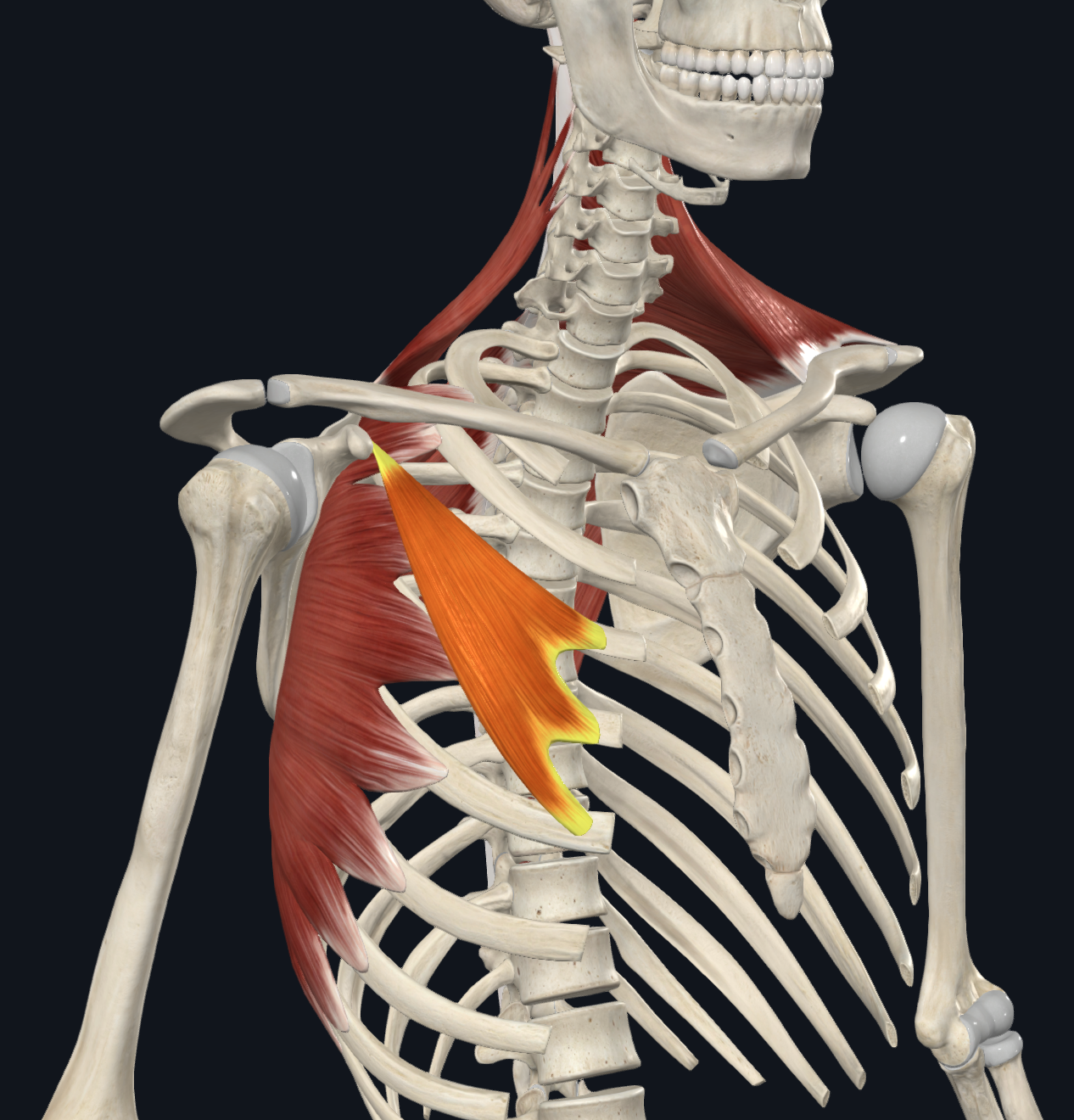
pectoralis minor

muscle acting on scapula

  • origin:

    • ribs 3-5

  • insertion:

    • coracoid process

  • movement:

    • depresses scapula


<p>muscle acting on scapula</p><ul><li><p>origin: </p><ul><li><p>ribs 3-5 </p></li></ul></li><li><p>insertion: </p><ul><li><p>coracoid process</p></li></ul></li><li><p>movement:</p><ul><li><p>depresses scapula</p></li></ul></li></ul><p></p>
79
New cards

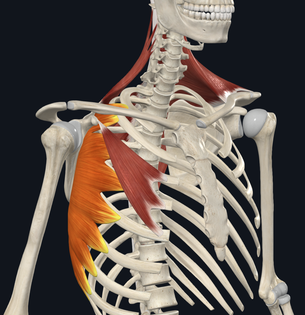
serratus anterior

muscle acting on scapula

  • origin:

    • ribs 1-8

  • insertion:

    • medial border of scapula

  • movement:

    • protracts

    • keeps scapula against thoracic wall


<p>muscle acting on scapula</p><ul><li><p>origin: </p><ul><li><p>ribs 1-8 </p></li></ul></li><li><p>insertion: </p><ul><li><p>medial border of scapula</p></li></ul></li><li><p>movement:</p><ul><li><p>protracts</p></li><li><p>keeps scapula against thoracic wall</p></li></ul></li></ul><p></p>
80
New cards

acromioclavicular joint

(7)

<p>(7)</p>
81
New cards

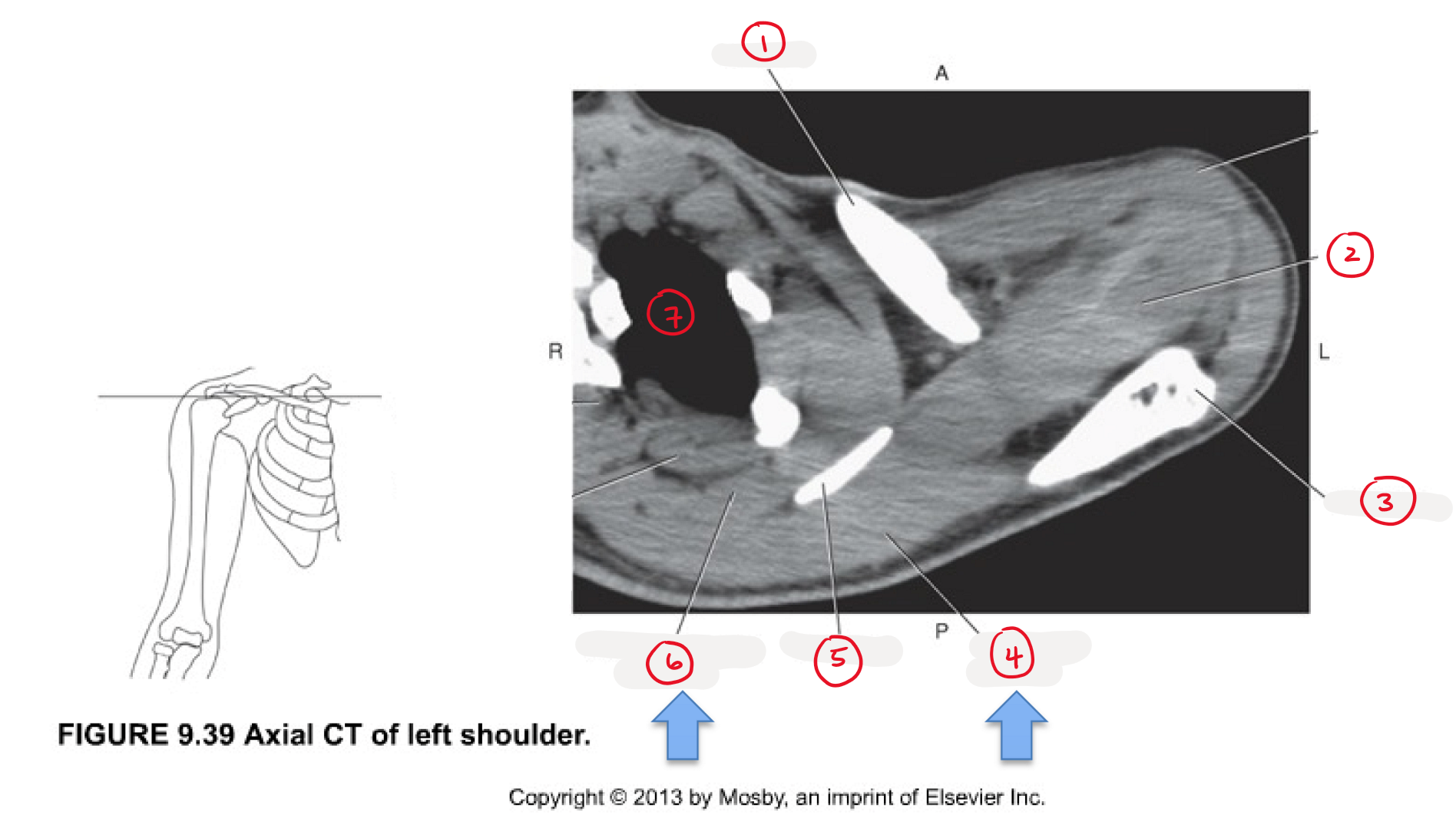
clavicle

(1)

<p>(1)</p>
82
New cards

supraspinatus

(2)

<p>(2)</p>
83
New cards

acromion

(3)

<p>(3)</p>
84
New cards

trapezius

(4)

<p>(4)</p>
85
New cards

scapula

(5)

<p>(5)</p>
86
New cards

levator scapulae

(6)

<p>(6)</p>
87
New cards

lung

(7)

<p>(7)</p>
88
New cards

rhomboid minor

(1)

<p>(1)</p>
89
New cards

rhomboid major

(2)

<p>(2)</p>
90
New cards

infraspinatus

(3)

<p>(3)</p>
91
New cards

teres minor

(4)

<p>(4)</p>
92
New cards

body of scapula

(5)

<p>(5)</p>
93
New cards

humeral shaft

(6)

<p>(6)</p>
94
New cards

lung

(7)

<p>(7)</p>
95
New cards

rotator cuff muscles

3 originate on posterior scapula + insert on greater tubercle of humerus

  • supraspinatus

  • infraspinatus

  • teres minor

  • function:

    • dynamic stabilization of GH joint

      • keep humeral head in glenoid fossa during shoulder motion

      • each rotator cuff has own additional function


1 originates on anterior scapula + insert on less tubercle of humerus

  • subscapularis

  • function:

    • dynamic stabilization of GH joint


<p>3 originate on <strong>posterior</strong> scapula + insert on <em>greater tubercle </em>of humerus</p><ul><li><p>supraspinatus </p></li><li><p>infraspinatus </p></li><li><p>teres minor </p></li></ul><ul><li><p>function: </p><ul><li><p>dynamic stabilization of GH joint </p><ul><li><p>keep humeral head in glenoid fossa during shoulder motion</p></li><li><p>each rotator cuff has own additional function</p></li></ul></li></ul></li></ul><p></p><p>1 originates on <strong>anterior</strong> scapula + insert on <em>less tubercle </em>of humerus </p><ul><li><p>subscapularis</p></li></ul><ul><li><p>function:</p><ul><li><p>dynamic stabilization of GH joint</p></li></ul></li></ul><p></p>
96
New cards

supraspinatus

  • origin

    • supraspinous fossa

  • insertion

    • greater tubercle of humerus

      • outer, lateral side of upper humerus


<ul><li><p>origin</p><ul><li><p><em>supraspinous </em>fossa </p></li></ul></li><li><p>insertion</p><ul><li><p><em>greater </em>tubercle of humerus </p><ul><li><p>outer, lateral side of upper humerus </p></li></ul></li></ul></li></ul><p></p>
97
New cards

infraspinatus

  • origin

    • infraspinous fossa

  • insertion

    • greater tubercle of humerus

      • outer, lateral side of upper humerus


<ul><li><p>origin </p><ul><li><p><em>infraspinous </em>fossa</p></li></ul></li><li><p>insertion</p><ul><li><p><em>greater </em>tubercle of humerus</p><ul><li><p>outer, lateral side of upper humerus </p></li></ul></li></ul></li></ul><p></p>
98
New cards

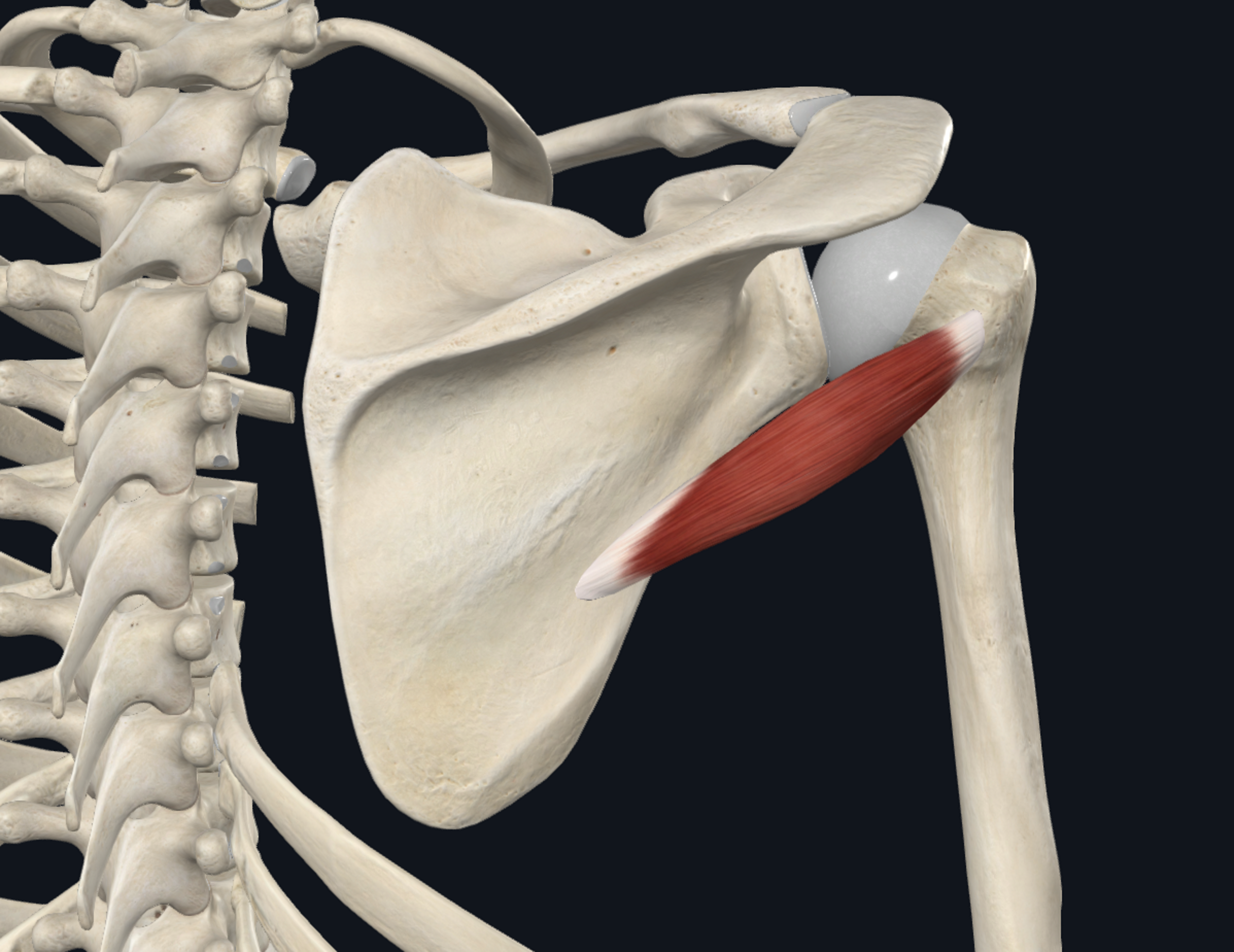
teres minor

  • origin

    • lateral border of scapula

  • insertion

    • greater tubercle of humerus

      • outer, lateral side of upper humerus


<ul><li><p>origin </p><ul><li><p><em>lateral border</em> of scapula</p></li></ul></li><li><p>insertion</p><ul><li><p><em>greater</em> tubercle of humerus </p><ul><li><p>outer, lateral side of upper humerus </p></li></ul></li></ul></li></ul><p></p>
99
New cards

subscapularis

  • origin

    • subscapular fossa

  • insertion

    • lesser tubercle of humerus

      • front, inner/medial side of upper humerus


<ul><li><p>origin </p><ul><li><p><em>subscapular </em>fossa</p></li></ul></li><li><p>insertion</p><ul><li><p><em>lesser</em> tubercle of humerus </p><ul><li><p>front, inner/medial side of upper humerus </p></li></ul></li></ul></li></ul><p></p>
100
New cards

humeral head

(1)

<p>(1)</p>