MCB 2000 Chapter 13
 Call Kai
 Learn
 Practice Test
 Spaced Repetition
 Match
 Flashcards
 Knowt Play
1/32

There's no tags or description
Looks like no tags are added yet.
33 Terms
why cells need signaling
the membrane is a barrier. cells still have to respond and coordinate
the membrane is selective
most signaling molecules can’t simply cross the PM. the message has to be received at the surface and relayed inward
the environment changes
glucose rises, glucose falls, a predator appears. cells need to detect those changes and adjust their internal chemistry
the body must coordinate
liver, muscle, brain. hormones let one tissue tell another what to do, so the whole organism acts in concert
the five-step framework
signal → reception → transduction → response → termination
hormone arrives
receptor binds it
message relayed
cell changes
signal off
without termination, the signal runs forever. cholera is exactly this failure modes.

amplification
one hormone molecule, three enzymatic steps, a thousandfold response
each step is catalytic, the receptor, the G- protein-activated enzyme, and pKa all keep working until something turns them off

why amplification matters
three reasons it shows up everywhere in physiology
Speed
economy
sensitivity
that’s why “all of the above” is the right answer to the flight-or-flight question. speed, economy and sensitivity all matter
speed
a flight-or-flight response can’t wait minutes for the cell to make enough downstream protein
amplification lets tiny signal produce a massive response in seconds
economy
hormones are expensive for the body to synthesize
amplification means you can release them at a very low concentrations and still hit every target cell
sensitivity
cells need to detect signals that are present at nanomolar or even picomolar concentrations
without amplification, those signals would be lost in the noise
GPCR
G-protein coupled receptors
Glucagon & epinephrine
glucagon released when blood glucose is low
epinephrine for fight or flight
fast, seconds
“release glucose, NOW”
follows 5-step framework
RTK
receptor tyrosine kinase
Insulin
slower, minutes
“store that glucose”
follow 5-step framework
what a GPCR looks like
7 transmembrane helices and a heterotrimeric G-protein on the inside
7 α-helices weave back and forth across the membrane
the hormone-binding site sits among them on the extracellular side
on the cytoplasmic side, the heterotrimer αβγ is tethered to the receptor.
its α subunit carries the GDP/GTP switch — binds GTP/GDP and that nucleotide shape determines whether the cascade is on or off
when epinephrine binds, the receptor changes shape and tells α to swap its GDP for GTP

GDP vs GTP
GDP = off
GTP = on
the alpha subunit is a molecular switch with two states
every G-protein in the cell uses this same trick. GDP is the resting nucleotide, GTP is the signal
when epinephrine binds the receptor, the receptor changes shape and acts like a GEF (guanine nucleotide exchange factor)
Helps GDP leave the alpha subunit so that GTP can bind
the extra phosphate on GTP changes the shape of the G-alpha subunit

the full β-adrenergic cascade
epinephrine is released from the adrenal medulla. arrives to the liver cell and binds beta-adrenergic receptor
receptor acts as a GEF so Gαs swaps GDP for GTP. active Gαs protein dissociates from the beta-gamma subunits
Gαs activates adenylate cyclase at the interface of the plasma membrane
adenylate cyclase converts ATP into cAMP (secondary messenger)
cAMP activates PKA
PKA phosphorylates downstream target proteins. In the liver, that cascade activates glycogen breakdown to release glucose into the blood

how cAMP activates PKA
resting PKA is a R2C2 tetramer, cAMP pulls it apart
R holds C off
in the resting tetramer, R subunits are inhibitors, they bind and block the catalytic Cs
cAMP binds R
2 cAMP per R subunit, four total. the Rs change shape and release the Cs
free C is active kinase
it diffuses through the cytoplasm phosphorylating serine and threonine residues on target proteins

4 ways tun the signal off
GTPase activity
phosphodiesterase
protein phosphatases
ligand dissociation
GTPase activity
Gα has slow intrinsic GTPase activity
it hydrolyzes its own bound GTP back to GDP, returning to the off state and releasing adenylate cyclase
phosphodiesterase
PDEs hydrolyze cAMP to plain AMP
with cAMP gone, R rebinds C and PKA shuts off
protein phosphatases
phosphatases remove the phosphate groups that PKA installed
targets return to their unphosphorylated, baseline state
ligand dissociation
eventually, epinephrine unbinds from the receptor
without ligand, the receptor stops acting as a GEF, and Gα isn’t being reactivated
why termination matters
a signal that won’t shut off is a disease in waiting
Normal: pulse, then silence
epinephrine arrives. PKA fires, glucose pours out
seconds late, all four shutoff mechanisms kick in, the signal decays and the cell returns to rest
response is matched to need
broken: pulse, then stuck on
same hormone arrives, PKA fires.
but one of the shutoff mechanisms is broken, Gα can’t hydrolyze its GTP, or cAMP can’t be degraded
PKA keeps firing. the cell keeps responding. this is exactly the cholera mechanism
caffeine
caffeine inhibits phosphodiesterase
phosphodiesterase is one of the four shutoff mechanisms. it hydrolyzes cAMP to AMP. block it, and cAMP isn’t being degraded
result: cAMP accumulates. PKA stays active longer and downstream phosphorylation increases
pathway logic: blocking cAMP breakdown prolongs PKA signaling. Note: caffeine’s alertness mostly comes from adenosine receptor blockade

cholera: termination broken
choleragen locks Gαs in the active GTP-bound form
what happens in the gut:
Vibrio cholerae releases a toxin, choleragen, that chemically modifies Gαs (by ADP ribosylating an arginine) so it cannot hydrolyze GTP
adenylyl cyclase keeps making cAMP so,
PKA fires continuously in intestinal epithelial cells. it phosphorylates and opens a chlordie channel (CFTR)
Cl- pours into the gut lumen, Na+ follows for electroneutrality, water follows by osmosis
Result: watery diarrhea up to a liter per hour. untreated cholera can kill in 24 hours from dehydration

not every GPCR uses cAMP
a second second-messenger system, same 5-step framework
The Gαq/ PLC pathway
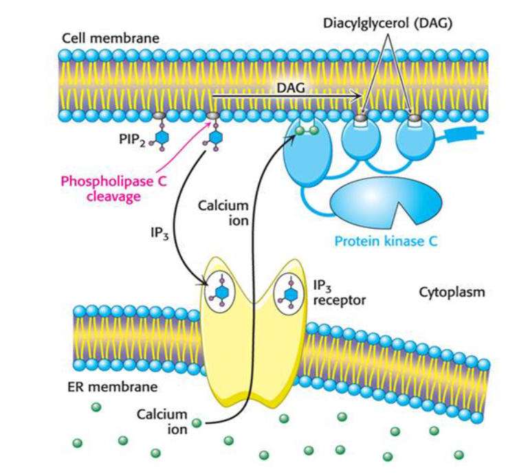
some GPCRs don’t use Gαs or cAMP. they use Gαq, which activates phospholipase C (PLC) at the inner membrane
PLC cleaves PIP2 into two second messengers: IP3 and DAG. IP3 diffuses to the ER and releases Ca 2+. DAG stays in the membrane. together they activate PKC
the point: second messengers are a strategy, not a single molecule. cAMP, IP3/DAG, Ca2+. the receptor decides which one fires
the insulin receptor: an α₂β₂ tyrosine kinase
two α subunits outside. the two β subunits crossing the membrane, with kinase domains built in
the receptor is its own kinase. pre-built as a dimer, with tow ligand-binding domains outside and two kinase domains inside
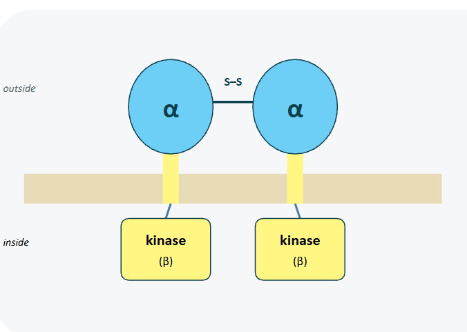
a pre-formed dimer, activated by rearrangement
insulin doesn’t assemble the receptor. it changes the shape of one already there
the dimer is already built. insulin binding repositions the kinase domains so they can activate each other

autophosphorylation
trans-autophosphorylation creates docking sites
what P-TYR does
each phosphorylation tyrosine is a docking site. cytoplasmic proteins with the right biding pocket recognize phospho-tyrosine and sticks to it
this is how the receptor recruits its downstream cascade. no diffusible messenger like cAMP is needed; the receptor displays docking sites
its a totally different signal-transduction logic from GPCRs

adapter protein dock and propagate
phospho-Tyr is the start of a cascade, not the end
the insulin cascade
phospho-Tyr sites recruit IRS-1, which itself gets phosphorylates and recruits PI3K
PI3K coverts PIP2 to PIP3 in the membrane, PIP3 recruits PDK1 and Akt. PDK1 phosphorylates and activates Akt
activated Akt drives GLUT4 translocation to the cell surface. RTKs use docking sites plus membrane lipids such as PIP3, rather than cAMP-style diffusion

the end of the cascade: GLUT4 at the surface
glucose finally enters the cell
insulin’s job: store glucose . the Atk cascade moves GLUT4 from inside the cell to the surface

termination, RTK style
phosphatases and dissociation, plus internalization
protein phosphatases: tyrosine phosphatases remove the phospho-Tyr marks on the receptor. adapter protein falls off. the cascade quiets
lipid phosphatases: PTEN converts PIP3 back to PIP2. Akt loses its docking sits at the membrane and goes idle
ligand dissociation: insulin unbinds from the receptor. without insulin, the dimer relaxes back to its inactive conformation and the kinase domains stop trans-phosphorylating
receptor internalization: the receptor itself gets endocytosed and either recycled or degraded. the cell can also down-regulate how many receptors are at the surface
signaling breakpoints

type 2 diabetes: receptor binds, signal stalls
what insulin resistance means:
hormone is fine. receptor binds
something in the IRS-1 → PI3K→ Akt chain is impaired
Phospho-Tyr signals arrive but don’t propagate
GLUT4 stays in vesicles. without GLUT4 at the surface, muscle and fat cells can’t take up glucose efficiently and blood glucose stays high
what contributes:
excess intracellular lipids and inflammatory signals can activate stress kinases that interfere with IRS1 function
chronic high insulin contributes to receptor downregulation and feedback inhibition
result= blunted signaling through the IRS1 pathway because AKT signaling is what drives GLUT 4 translocation, fewer GLUT 4 transporters reach the surface of muscle and fat cells so glucose uptake is impaired and glucose stays elevated

the compensation spiral
the pancreas tries harder. eventually it burns out

two diseases, two break points
break at transduction:
receptor binds insulin normally, but IRS/ Akt signaling is impaired. GLUT4 doesn’t reach the surface. glucose stays high
chronic, slow onset. treated with sensitizers or exogenous insulin
Break at termination
cascade fires correctly, but Gαs can no longer hydrolyze GTP. PKA stays on, chloride flows out, water follows
acute, can kill in hours. treated with aggressive rehydration

the whole story in one picture
