lec 7: bone tissue
 Call Kai
 Learn
 Practice Test
 Spaced Repetition
 Match
 Flashcards
 Knowt Play
1/89

There's no tags or description
Looks like no tags are added yet.
Name | Mastery | Learn | Test | Matching | Spaced | Call with Kai | Chat |
|---|
No analytics yet
Send a link to your students to track their progress
90 Terms
cartilage in external ear
.

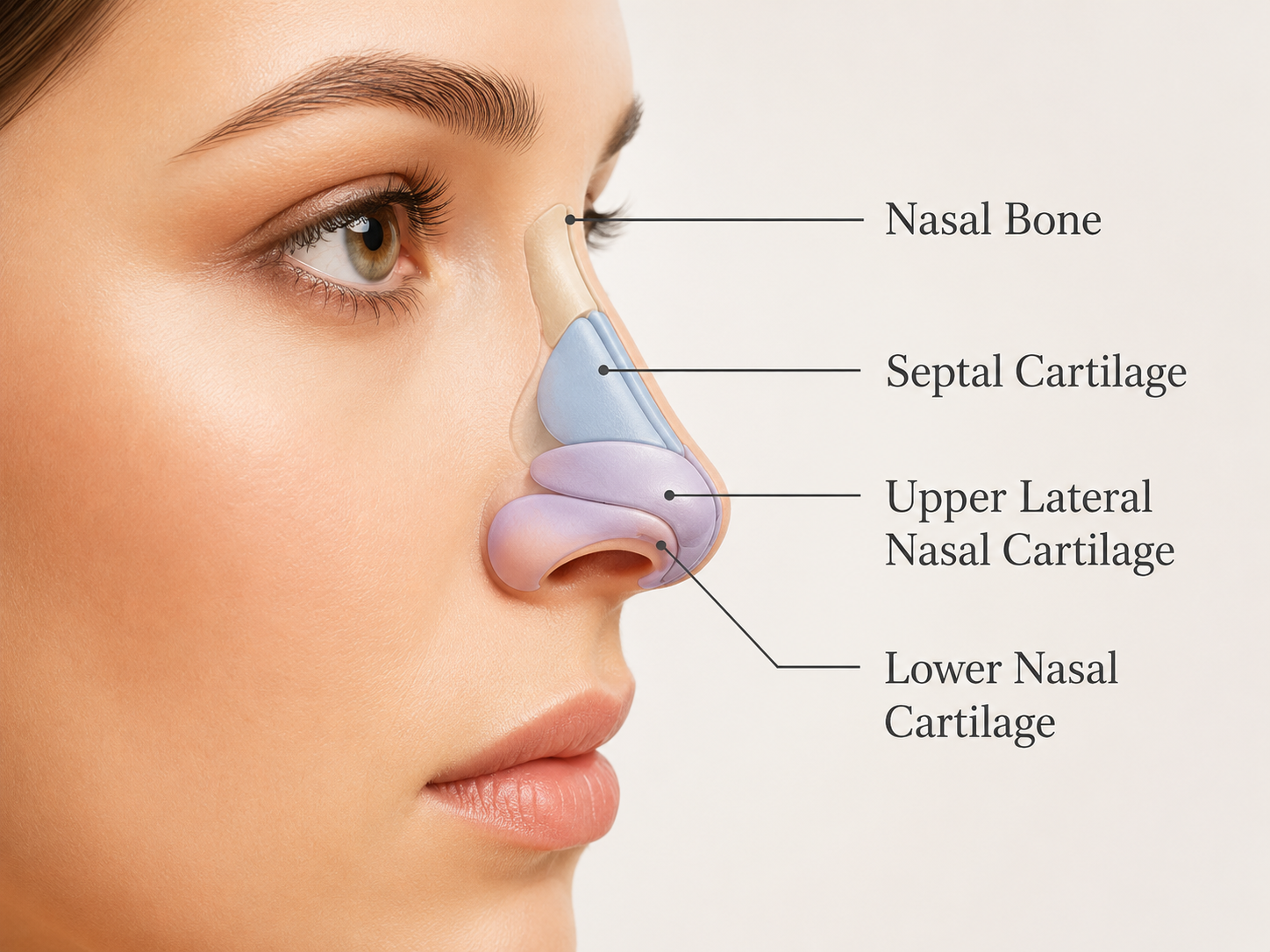
cartilages in nose
.
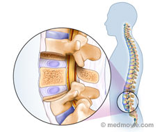
cartilage in intervertebral disc
.
articular cartilage of a joint
.

costal cartilage
.

pubic symphysis meniscus
padlike cartilage in knee joint

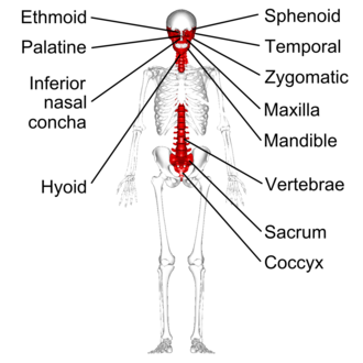
bones of a skeleton:
axial skeleton
appendicular skeleton
axial skeleton
80 bones
protects the VITAL ORGANS
skull (cranial/facial bones)
vertebral column
thoracic cage (sternum, ribs)
appendicular skeleton
126 bones
enable movement
petoral girdle/shoulders
arms
legs
pelvic girdle/hips
cartilages
hyaline cartilages
elastic cartilages
fibrocartilages

hyaline cartilages
MOST COMMON type of cartilage
contains type II collagen fibers
reduces friction in joints, absorbs shock and flexible structural support
@: ends of long bones, ribcage, nose, nasal septum, trachea and larynx
elastic cartilage
high concenrtation of elastic fibers alongside type II collagen
maintains the shape of structures while allowing a high degree of flexibility and recoil
@: external ear, epiglottis and eustachian tube
fibrocartilage
toughest and strongest type of cartilage
packed with type I and type II collagen fibers
SHOCK ABOSPORTION
@: intervertebral discs, pubic symphysis
roles of skeleton
support
protection
movement
electrolyte balance
acid-base balance
blood formation
hormone secretion
electrolyte balance
calcium and phosphate levels
HARD SALT — inorganic material
makes up the bone (reduced calcium is bad)
acid-base balance
buffers blood against large pH changes by altering phosphate and carbonate salt levels
blood formation
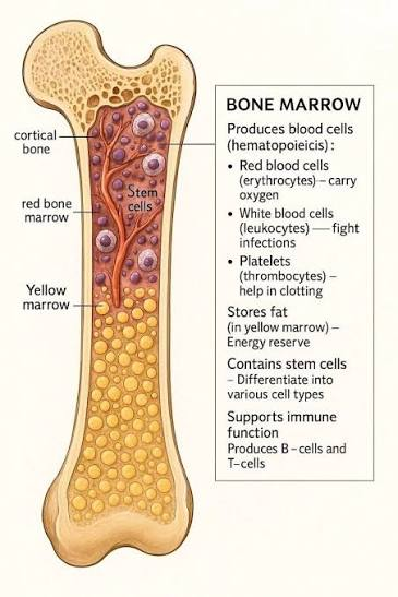
hematopoisesis tissue (red marrow) in the bone marrow cavities
found skull, vertebrae, ribsm sternum, part of pevlic girdle and proximal heads of humerus and femur
hormone secretion
bone cells secrete hormones that affect action of insulin and moderate the stress response
long bones
longer than they are wide
@: arms, legs, fingers, humerus, radius, ulna

short bones
cube shaped bones (wrist and ankle)
sesamoid bones (tendons and patella)

flat bones
thin, flat, slightly curved
@: face, skull, cranium, stermum, shoulders

irregular bones
complicated shapes (vertebrae, pelvis)
bone markings:
projections
articulations
depressions
passages
projections
tubercle
tuberosity
trochanter
crest
process
spine
tubercle
small, rounded projection
@: greater/lesser tubercles of the humerus, adductor tubercle of the femur

tuberosity
rough projection
@: ischial tuberosity (your booty when ure sitting)

trochanter
larger, rough projection in the femur

crest
prominent and rougher than a ‘line’, making it a major anchoring point
@: iliac crest (hip bone, ridge of the pelvis)

process
prominent outgrowth, bump, ridge or a projection/extension sticking out from the bone

spine
sharp, slender and often pointed projection

articulations (joints)
head
facet
condyle
head
expanded articular end often separated from the shaft by the neck—-fitting into a shallow socket

facet
small, flat articular surface

condyle
smooth, rounded articular knob that fits into a matching depression on another bone

passages
holes → needs to get through
canal (meatus)
fissure
foramen
canal
large passageway through the substance of a bone
@: internal auditory meatus/nasal cavity

fissure
elongated cleft of gap
@: superior orbital fissue

foramen
small, rounded passaageway through which blood vessels or nerves penetrate the bone

depressions:
fossa
sulcus (groove)
fossa
broad, shallow depression, pit or hollow surface
@: shoulder blades

sulcus
shallow groove, furrow or linear depression on the surface of an organ, tissue or bone

compact bone
10 years
dense outer layer

spongy (cancellous) bone
3-4 years
honeycomb of trabeculae
more porous like a sponge

membranes of a bone
periosteum
endosteum
periosteum
new bones are being formed — covered comapct bone on the outside
outer fibrous layer
inner osteogenic layer:
osteoblasts
osteoclasts
osteogenic cells

endosteum
covered spongy bone within
delicate membrane on internal surfaces of bone
contains osteoblasts and osteoclasts
osteoblasts
bone forming cells —- creates NEW bone
osteoclasts
bone destorying cells — greatest number of lysosomes
recycle bones
osteogenic cells
stem cells in periosteum and endosteum— turned into osteoblasts/osteoclasts
osteocytes
mature bone cells
microscopic anatomy of compact bone
haversian system or osteon
lamellae
central/haversian canal
perforating/volkmanns canals
lacunae
canaliculi
lamellae
concentric rings — made with a lot of collegen fibers
gives the bone strength

central/haversian canal
contains blood vessels and nerves
blue = vein
red = arteries
yelow

volksmann’s canals
at right angels to the central canal
connects blood vessels and nervse of the periosteum and central canal

lacunae
small cavities that contain osteocytes

canaliculi
hair-like canals that connect lacunae to each other and the central canal

microscopic anatomy of spongy bone
trabeculae

trabeculae
no osteons
contain lamellae, osteocytes and canaliculi

osteogenesis (ossification)
bone tissue formation — usually during puberty, resistance training, breaking/repairing a bone and baby bones
stages:
initial formation (embryo and fetus)
growth (infancy—adolescence)
remodeling (bone throughout l
intramembranous ossification
direct bone formation
membrane bone develops from fibrous membrane
forms flat bones and cranial bones
endochondral ossification
replaces bone
cartilage bone forms by replacing hyaline cartilage
forms most of the skeleton
cartilage is there FIRST—it is then broken down and bone forms
forms ALL bones bnelow skull except for clavicles
“build a temporary cartilage model, then replace it”
ossification centers appear in fibrous membrane…
produces connective tissue
osteoblasts secrete osteoid, which calcifies…
hardens—calcification of osteoid tissue and entrapment of osteocytes
interstital growth
length of long bones — epiphyseal plate
zones:
proliferation zone
hypertrophic zone
calcification zone
ossification zone

appositional growth
thickness and remodeling of all bones by osteoblasts and osteoblasts on bone surfaces
bone matrix secreted under periosteum

proliferation zone
chondroblasts quickly divide and push the epiphysis away from the diaphysis—lengthening the bone
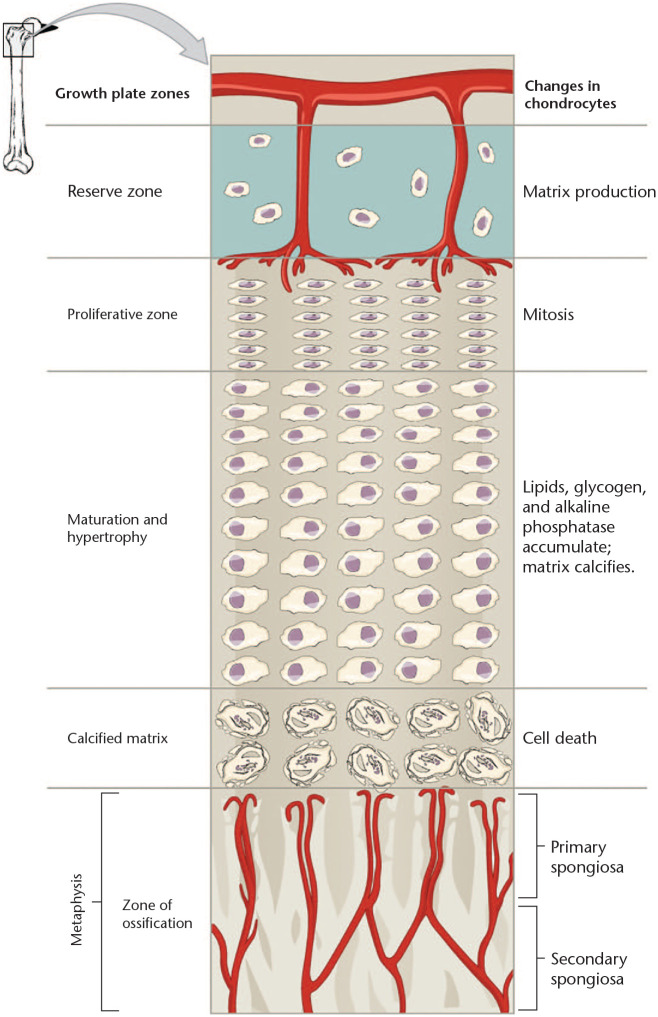
hypertrophic zone
older chondrocytes enlarge and signal the surrounding matrix to calcify
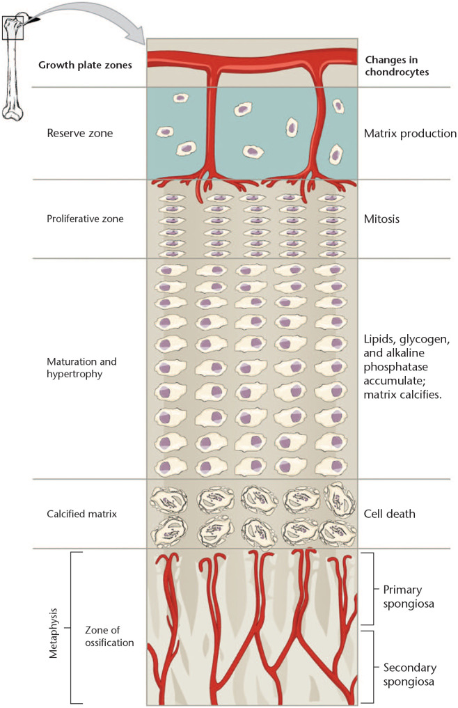
calcification zone
matrix becomes calcified; chondrocytes die

ossification zone
osteoclasts digest calcified cartilage
osteoblasts replace actual bone tissue—resullting in bone trabeculae (spongy bone)

bone growth
& bone remodelng
growth hormone
stimulates epiphyseal plate activity from the pituitary gland
thyroid hormone
modulates activity of growth hormone
testosterone and estrogens
@ puberty
promotes adolscent growth spurts
end growth by inducing epiphyseal plate closure (18 yr females; 21 yr males)
calcium and phosphate regulation
intestines: calcium and phosphate ions are absorbed from the diet
skeleton; osteoblasts are depositing new bone at the same time osteoclast release existing calcium and phosphate ions that enter the circulation—the balance between osteoblast and clasts is hormonally regulated
kidneys: levels of calcium and phosphate lost in the urine is hormonally regulated
parathyroid hormone (PTH)
blood Ca2+ levels → parathyroid glands release PTH → PTH stimulates osteoclasts to degrade bone matrix and release Ca2+ → blood Ca2+ levels
wolffs law
living bone continuously adapts and remodels itsself in response to the mechanical loads and physical stress placed upon it

fracture classification
displaced or non-displaced
open or closed
comminuted — bone fragments
spiral — excessive twisting force
compression — common in osteoporosis
depressed
greenstick
traverse
.

linear
.
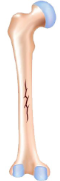
oblique nondisplaced
.
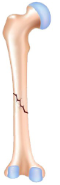
oblique displaced
.

spiral
.

greenstick
.

comminuted
.
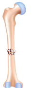
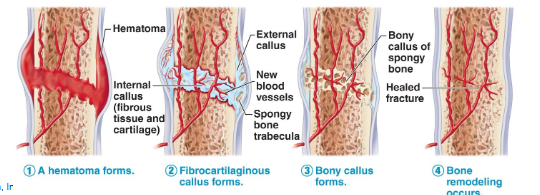
stages of healing a bone fracture
hematoma
fibrocartilaginous callus
bony callus formation
bone remodeling
hemtatoma forms
mass of clotted blood forms
site becomes swollen, painful and inflamed

fibrocartilaginous callus (soft callus)
fibroblasts secrete collagen to connect bone ends
osteoblasts begin forming spongy bone in 1 week

bony callus formation (hard callus)
osteoblasts create a bony collarto unite broken ends firm union in 6-8 weeks to heal

bone remodeling
response to mechanical stresses, fragments removed by osteoclasts, osteoblasts lay down spongy bone and then converted to compact
osteoporosis/osteopenia
loss of bone mass—bone resoprtion outspaces
treatments center around drugs that stimulate bone deposition or slow rate or resportion