Midterm 2 Proteins
 Call Kai
 Learn
Learn Practice Test
 Spaced Repetition
 Match
Match Flashcards
 Knowt Play
1/20

Earn XP
Description and Tags
Name | Mastery | Learn | Test | Matching | Spaced | Call with Kai | Chat |
|---|
No analytics yet
Send a link to your students to track their progress
21 Terms
MNase
Micrococcal nuclease (MNase)
Digestion by digestion defines protected nucleosomal DNA (defined length of DNA wrapped around histones as 146 base pairs)
preferentially cuts exposed linker DNA
light digestion releases fragments containing 1, 2, 3 or more nucleosomes
followed by heavy, more extensive digestion which trims accessible DNA until only left is DNA histone core protects
gel electrophoresis to find DNA fragment length (remove histone proteins 1st)

Histone basics
Small, positively charged proteins
abundant, among most conserved eukaryotic proteins
Basic histones (+ charge) associate highly & non-specifically with DNA (- charge)
lysine & Argentine (basic amino acids)… contain many residues
Histones octamer (8) or “core”… 2 layers of “4-slice pies”
have two copies of each (or variants of): H2A, H2B, H3, & H4
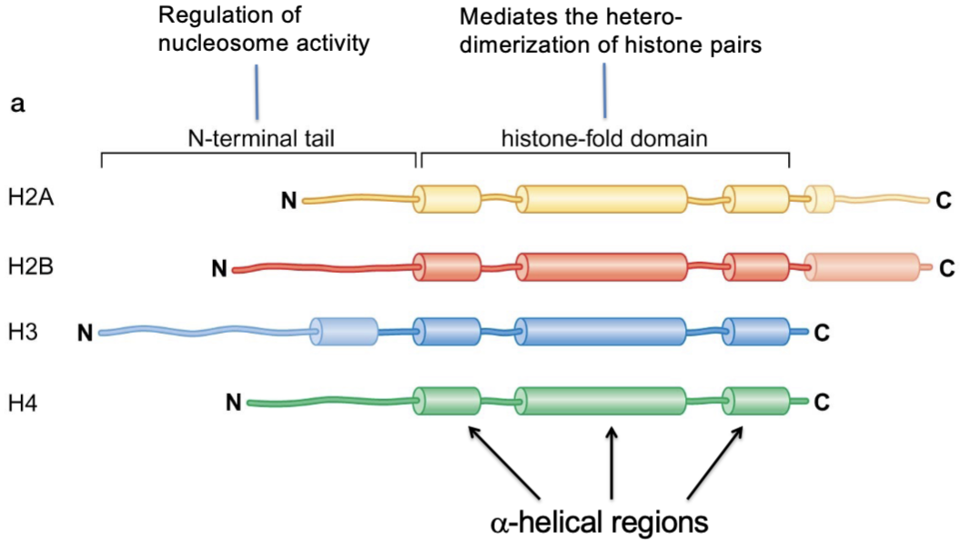
these ‘core histones; differ in size & sequence but share general organization:
N-terminal tail (regulation of nucleosome activity)
histone-fold domain (alpha-helical regions… mediates histone pairing and assembly of octamer)
H2B
Histone
The tail of H2B direct DNA wrapping in a left-handed manner (along with H3)

H3
Histone
The tail of H3 directs DNA wrapping in a left-handed manner (along with H2B)

H2AX
Variant H2A histone involved in DNA damaging signaling
phosphorylated at sites of DNA damage
involved in the DNA-damage response
CENP-A
AKA CEN-H3
Variant H3 histone found at centromeres… help specify kinetochore assembly
defines centromere chromatin
Specialized histone variant that is conserved
distinctive N-terminal region & associated proteins help interact with the kinetochore to assemble it at the correct chromosomal location
centromere identity depends o chromatin composition rather than on DNA sequence… epigenetically

H1 (linker histone)
Compacts the chromatin, forming a 30-nm fiber
additional folding increases DNA compaction to about 40-fold!
Linker histone H1 binds near DNA entry & exit (‘base’) sites of a nucleosome and promote interactions between neighboring nucleosomes
nucleosomes get closer together

CEN-H3
aka CENP-A
Specialized histone variant that is conserved
distinctive N-terminal region & associated proteins help interact with the kinetochore to assemble it at the correct chromosomal location
centromere identity depends o chromatin composition rather than on DNA sequence… epigenetically

HP1
“Heterochromatin Protein 1”
Chromodomain protein
Silences pericentromeric heterochromatin
HP1-associated histone methyltransferase Su(var)3-9 deposits H3K9me2/3, which recruits HP1 through its chromodomain
self-reinforcing domain… “spreading” of heterochromatin silencing

Caf1
Caf-1 associates with PCNA (DNA sliding clamp in eukaryotes) at the replication fork
deposits H3-H4 tetramers onto newly synthesized DNA
based on H3-H4 post-translational modifications, histone marks are redeposited on H2A-H2B and on neighboring histones
“recycles” nucleosomes from “old” DNA

Cohesin
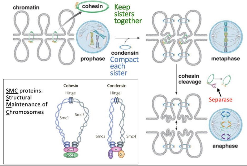
A “Structural Maintenance of Chromosomes” (SMC) protein complex
Related to condensing… has distinct yet coordinated function
Cohesins loaded onto chromatin in telophase of previous M phase and G 1
buuuut… actually cohesion becomes established as DNA replicated in S phase… as DNA replicates in S phase 2 sisters remain inside ring, establishing cohesion
Separase is the enzyme that cleaves cohesin at anaphase… allowing sisters to move toward opposite spindle poles… this happens after spindle assembly checkpoint ensures correct kinetochore attachment to microtubules
ADDITIONAL FUNCTION
cohesin also organizes interphase chromosomes by extruding DNA into progressively larger loops… “loop extrusion”
loop grows until cohesin encounters approrpiraietly oriented CTCF proteins
this mechanism brings selected genomic regions into proximity while limiting interactions across boundaries

Condesin
A “Structural Maintenance of Chromosomes” (SMC) protein
coordinated with cohesin but has distinct function
Condesin compacts each chromatid into an organized mitotic chromosome
Is loaded before S phase
condensation vs decondenstaion
chromosome compaction changes as cell progresses through cell cycle…
chromatin remains relatively decondensed during interphase (when DNA must support transcription and replication) … but becomes highly condensed during mitosis (condensation produces discrete sister chromatids that the spindle can segregate without extensive tangling)
CTCF
Protein that helps define the boundary of loops created by cohesins… helps define where loop extrusion stops
CTCF proteins must be properly oriented to do this function… CONVERGENT orientation
cohesin will then stop when it encounters these appropriate oriented CTCF proteins
helps define what genomic regions are being brought into proximity while limiting interactions across boundaries

MutS
Recognizes mismatch as a “distortion” in DNA molecule
Involved in the pathway MMR… “mismatch repair'“
Mismatch is detected by MutS protein… MutS binds mismatch
(MMR steps in bacteria slide.. for eukaryotes has homolog MSH2/6)

MutL
Binds NutS, mediated binding to MutH which is bound to farther site
(MMR steps in bacteria slide… for eukaryotes has homologs MLH + PMS where PMS has nicking activity?)

DNA Glycosylase (general)
Base excision repair (BER) reies on specific DNA glycolyses that remove abnormal bases
Is an enzyme
A specialized DNA glycosylase removes the abnormal bases… leaves an AP site
11 different DNA glycosylases found in humans
“scanning mechanism”… how enzyme locates damaged bases… unknown
but do know that it “flips out” suspicious base

oxoG DNA glycosylase
A DNA glycosylase (enzyme plays role in Base Excision Repair… BER)
This DNA glycolylase surrounds the “flipped out” damaged base
crystal structure shows that the base is “flipped out” when the glycosylase is bound… “sits in” enzymatic pocket
if the base fits into the enzyme’s recognition pocket, the enzyme cleaves it away through its active site
oxoG DNA glycosylase recognizes oxidized guanine (hence name “oxo” “G”)

O6-methylguanine methyltransferase (MGMT)… O6-methylG Methyltransferase
Catalyzes damage reversal of O6-methylG (methyl group added to O coming off top 6)
Repairs it in 1 step by transferring the methyl group to itself…
once MGMT accepts the methyl group, it becomes inactivated and is degraded…
a “suicide” enzyme
is efficient but costly… since each repair consumes one MGMT molecule

O6-methylG DNA Glycosilase
O6-methyl G… methylation of guanine (by chemicals like nitrosamines)… can be inspired to T
Involved in BER (base excision repair)… 1 of two ways to fix O6-methyl G is BER by O6-methylGDNA glycosilase
DNA glycosylases remove abnormal bases, leaving an AP site
‘flips out’ damaged base from backbone into catalytic active site

AP endonuclease
Cuts DNA backbone at AP site (AP sites= abasic site, apurinic, apyrimidinic)
Endonuclease (nickase) = cleaves DNA bonds within the DNA molecule = cuts inside DNA = makes a nick
Involved in Base Excision Repair (BER)
cuts DNA backbone at site once DNA glycosylase has removed the abnormal base
and then an exonuclease cleans out sugar + phosphate
exonuclease = cleaves nucleotides at end of DNA molecule… requires free 3’ or 5’ -OH … works with a helicase

“Mut” vs. “Uvr” vs. “Rad”