osteology of the skull
 Call Kai
 Learn
 Practice Test
 Spaced Repetition
 Match
 Flashcards
 Knowt Play
1/133

There's no tags or description
Looks like no tags are added yet.
134 Terms
how many bones is the skull made up of
22
how are the bones in the skull divided
into facial bones and cranial bones
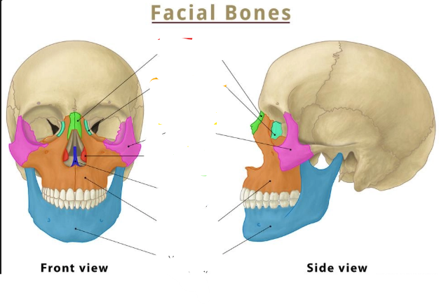
what bone is shown in green
the nasal bone
what fetures are found on the nasal bone
the nasal foreman

what bone is shows in light blue
the lacrimal bone
is the lacrimal bone a paired bone
yes
what anatomical structures are found on the lacrimal bone
has voth lateral and orbital prosseses
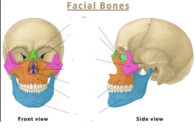
what bone is in pink
the zygomatic bone
is the zygomatic bone a paired bone
yes
what are the 3 bones the zygomatic bone articulates with
temporal bone, frontal bone and maxillary bone

what bone is this
the zygomatic bone
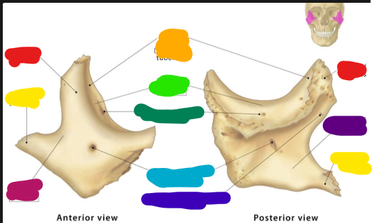
what prosses of the zygomatic bone is shown in red
the frontal prosses
what occours at the frontal prsses
articulation with the frontal bone

what is shown in orange
the orbital turburcule

what is shown in light green
the orbital surface

what is shown in dark green
the zygomaticoorbital foreman

what is shown in light blue
the zygomaticofacial foreman
what passes through the zygomaticofacial foreman
the zygomatic facial nerve

what is shown in dark blue
the zygomaticotemporal foreman
what does thezygomaticotemporal foeman carry
the zugomaticotemporal nerve

what bone is shown in red
the inferior nasal conchae

what bone is shown in dark blue
the vomer
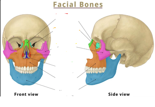
what bone is shown in orange
the maxilla

what component of the maxilla is shown in red
the frontal prosses
what are the connectins od the frontal prosses
the frontal and nasal bone

what component of the maxilla is shown in yellow
the infraorbital margin
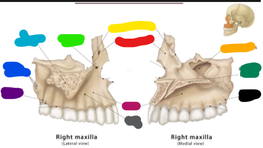
what component of the maxilla is shown in orange
the maxillary sinus
what does the maxillary sinus connect to
the nasal cavity

what component of the maxilla is shown in light green
the infraorbital foreman
what passes through the infraorbital foreman
infraorbital nerves and blood vessles

what component of the maxilla is shown in light blue
the zygomatic prosses
what does the zygomatic prosses connect with
the zygomatic bone

what component of the maxilla is shown in dark green
the paletine prosses
what does the palatine prosses form
the fron of the hard plate - roof of mouth

what component of the maxilla is shown in dark blue
the maxillary tuberosity
what is the maxillary tuberosity
a boney prominance

what component of the maxilla is shown in purple
the alveolar prosses
whats the alviolar prosses
located on the canine ridge, holds the teeth in place

what component of the maxilla is shown in pink
the body

what component of the maxilla is shown in grey
the canine fossa

what component of the aviolar prosses is shown in black
the aveolar prosses

what bone is shown in blue
the mandible

what component of the mandibule is shown in red
the condylar prosses

what component of the mandibule is shown in orange
the the coronoid prosses

what component of the mandibule is shown in yellow
the mandibular foreman
what vessles are carried in the mandibular foreman
the inferior avleolar nerve and vessles

what component of the mandibule is shown in green
the submandibular fossa

what component of the mabdibule is shown in light blue
the sublingual fossa

what component of the mandibule is shown in purple
the alveolar prosses

what component of the mandibule is shown in pink
the mental foreman
what exits from the mental foreman
themental nerve (a terminal branch of the inferior alveolar nerve)

what component of the mandible is shown in dark blue
the mental pertubance

what Vcomponent of the mandibule is shown in grey
the body

what component of the mandibule is shown in black
the ramus
what are the cranial bones seperated into
anterior middle and posterior sec

what cranial bone is shown in blue
the frontal bone

what is is the red component of the frontal bone show
the squamous part

what componenty of the frontal bone is shown in orange
the nasal component

what component of the frontal bone is shown in yellow
the trochlea part

what component of the frontal bone is shown in light green
the orbital component

what component of the frontal bone is shown in dark green
the supraorbital margin
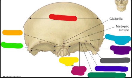
what component is shown in light blue
the supraorbital foreman
what travels through the supraorbital foreman
the opthalmic division of the trigeminal nerve

what componen is shown in darl blue
the supraorbital ridge
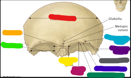
what component is shown in purple
the zygomatic prosses

what component is shown in pink
the frontal notch

what component is shown in grey
the supercillery notch
what foremen are present in the frontal bone
the frontal foreman, the froeman cecum, the supraorbital foreman and the supratrochlea foreman

what bone is shown in purple
the ethmoid bone

what part of the ethmoid bone is shown in red
the crista galli
whats the attaches to the crista galli
the faux ceribri

what component is shown in oragne
the orbital plate

what component is shown in yellow
the cribiform plate
what does the cribiform plate form
the roof of the nasal cavity
what nerve trvels over the top of the cribiform plate
the olfactory nerve

what component is shown in green
the ethmoid cells

what component is shown in blue
the superior nasl chochnea

what component is shown in pink
the ethmoid labrynths

what component is shown in grey
the purpendicular plate

what bone is shown in yellow
the sphenoid bone

what component of the sphenoid bone is highlighted red
the greater wing

what part of the sphenoid bone is highlighted in geen
the lesser wing

what component id the sphenoid bone is shown in blue
the body

what component of the sphenoid bone is shown in yellow
the pterygoid prosses

what is the lable shown in red
the optic canal
what travels through the opotic canal
the optic nerve

what label is shown in orange
the supraorbital fissure
what cranial nerves pass through the supraorbitl fissure
II,IV, V1 and VI
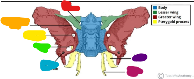
what label is shown in yellow
the foreman rotundum
what passes through the foreman rotundum
the maxiallary nerve

what lable is shown in green
the foreman ovale
what nerve passes through the foreman ovale
the mandibulr nerve

what is the lable in blue
the foreman spinosum
what artery passes through the foreman spinosum
the middle meningeal
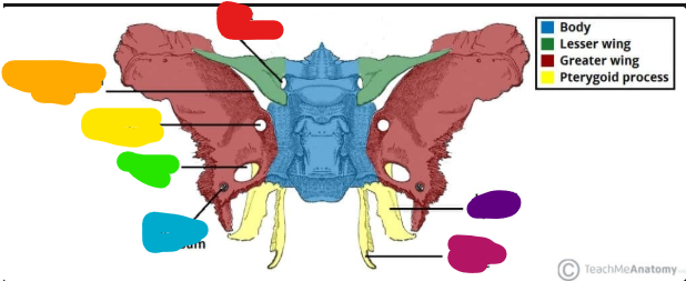
what lable is shown in purple
the lateral plate

what lable is shown in pink
the medial plate
what does the sphenoid bone look like in the cranial cavity


what bone is shown in red
the temporal bone
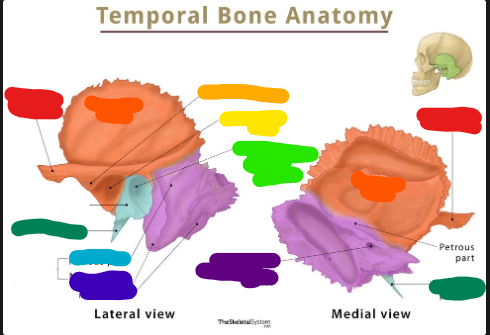
what component of the temporal bone is in orange
the squamous component

what component of the bone is shown in blue
the tympanic part