cardiac: peds
 Call Kai
 Learn
 Practice Test
 Spaced Repetition
 Match
 Flashcards
 Knowt Play
1/54

There's no tags or description
Looks like no tags are added yet.
55 Terms
Congenital heart defects are ?
severity
can be found ?
most develop when
A defect in the structure of the heart or great vessels that is present at birth
can be small or severe and not compatible with life
can be found in utero
most develop in the first 8 weeks of gestation
what drug exposures can lead to congenital defects
phenytoin - seizure med
ACE inhibitors
lithium
warfarin
valproic acid
alcohol)
Other exposures that can lead to congenital defects
viruses ?
metabolic disorders ?
age
what abnormalities
Maternal viral infections such as rubella or coxsackievirus B5
Maternal metabolic disorders such as phenylketonuria, diabetes, and hypercalcemia
Increased maternal age
Chromosomal abnormalities
how are CHD categorized
increased pulmonary blood flow
decreased pulmonary blood flow
mixed
obstructive systemic blood flow
what is usually the first sign of CHD
shunt ?
murmur
Blood flowing with a high pressure through a narrowed valve, vessel, or shunt
Shunt - an abnormal anatomic opening in the septum between systemic and pulmonary circulation
symptoms of CHD
newborns may have symptoms as soon as umbilical cord is cut
older kids - excerisce intolerance, chest pain with activity , dysrhythmias/ palpitations, syncope , sudden death
dx procedures for CHD
Cardiac Cath
Chest x-ray
Echo
EKG
CT scan
MRI
lab test to identify CHD
CBC
ABG’s - shows oxygenation levels
Serum drug tests
ESR, C-reactive protein - shows any systemic inflammation
Increased Pulmonary Blood Flow Congenital Heart Defects
how common
results from what 2 connections ?
how is blood flow affected
most common kind
Results from:
connection between the left and right side of the heart (septal defect)
Connection between the great arteries (Patent ductus arteriosus)
Increased blood volume on right side of heart
Increased pulmonary blood flow
Decreased systemic blood flow
s/s of Increased Pulmonary Blood Flow Congenital Heart Defects
Tachycardia
tachypnea
murmur
CHF
recurring Respiratory infections - kids who are frequently in hospital for this may need a cardiac workup
ASD - atrial septal defects
where is blood shunted ?
R side of heart?
pulmonary ?
leads to?
oxygenated blood is shunted from left to right side of the heart
right side of heart works harder then normal bc more blood is going there → hypertrophy
Extra blood then passes through the pulmonary artery into the lungs, causing higher pressure than normal in the blood vessels in the lungs & increased fluid in lungs
leads to signs of congestive heart failure ( crackles SOB)

s/s of ASD
Easily fatigued (in infant - not feeding, lethargic)
Poor growth bc not getting adequate nutrition
Freq. respiratory infections
Murmur from shunting of blood
Possible right side enlargement - happens over time
Or may be asymptomatic-depends on size of shunt opening
tx of ASD
small ASD?
moderate - large ?
meds ?
when do we resort to surgery
Spontaneous closure of small ASD’s occurs in the 1st 4 years of life
Mod-large ASD are closed by a septal occlude via cardiac cath (device that plugs the shunt)
ASA 81 mg is prescribed for 6 months after the procedure
Surgery to close or patch the ASD is performed when increased pulmonary blood flow causes HF.
prognosis of ASD
risk for
how
Middle-aged adults with uncorrected small-mod size ASDs may have no symptoms but are at risk for stroke.
Small clots that develop in the atrium can pass through the ASD into the left atrium and systemic circulation
Ventral Septal Defect (VSD)
similar to ASD, but happens in the ventricles
Oxygenated blood is shunted from left to right side of the heart via defect in the ventricles
•A larger volume of blood than normal must be handled by the right side of the heart ⇨hypertrophy
Extra blood then passes through the pulmonary artery into the lungs, causing higher pressure than normal in the blood vessels in the lungs ⇨ congestive heart failure

VSD s/s
Associated with CHF
Poor growth - associated w poor appetite
Dyspnea
Tachycardia
Decreased exercise tolerance
Increased number of pulmonary infections
Pulmonary hypertension
Systolic murmur and or thrill may be present
Failure to thrive
tx of VSD
closure @
if not managed well?
response to sugery
prognosis ?
non surgical or surgical closure at 1 yrs old
if HF symptoms cant be manage w meds , closure within first 6 months
children respond well to surgery and catch up with peers
prognosis- r/o arryhthmias, and heart blocks, some children may need pace maker
non-surgical closure for VSD
cardiac catheterization to patch the opening.
Umbrella-shaped patch is closed and inserted into the damaged area through a small tube, then opened to cover the defect
requires a small incision, avoiding the need for open-heart surgery
surgical closure for VSD
large mm?
if child doesn’t have surgery ?
Large VSD 6-10 mm require surgery or child will develop CHF and Failure to thrive by age 3-6 month
open heart surgery to patch the hole
surgeons stitch or patch the defect
Patent ductus arteriosus
Blood shunts from aorta to the pulmonary artery (right)
Returns to the lungs causing increase pressure in the lung - fluid in lungs
Congestive heart failure

s/s of PDA
resp
cardiac
on ascultatation
Gi
Dyspnea on exertion
Tachycardia, tachypnea
Forceful, bounding pulse
Murmur over pulmonary artery; wooshing Machine like murmur
Feeding difficulties and slow weight gain
Possible heart enlargement and left sided heart failure
closure of PDA
may ?
2 meds
what are we checking on the meds ?
May close spontaneously during the first year, usually during neonatal period
Intravenous Ibuprofen
Indomethacin (Prostaglandin inhibitor): good success rate
Prostaglandin inhibitors cause vasoconstriction and closure of DA
Must Check kidney function - BUN, creatintie , I and O
Check bleeding - easy bruising
tx of PDA
Cardiac Catheterization repair with Coil Technique
Surgical Repair-Ligation
Prophylactic Antibiotics
Prevent Congestive Heart Failure
Types of CHD that Decrease Pulmonary Blood Flow
Pulmonic Stenosis (PS)
Tetrology of Fallot (TOF)
Pulmonic stenosis (PS)
what is increased?
R V?
Narrowing of entrance to pulmonary valve that decreases blood flow
Increases preload causes right ventricular hypertrophy
Afterload is increased

s/s of pulmonic stenosis
Mild cyanosis
Dyspnea and fatigue with exertion
Heart failure: SOB; edema
Murmur
complications of pulmonic stenosis
Endocarditis - inflammation of endocardium
Cardiomegaly - enlarged heart
heart failure
Pulmonary regurgitation may result but is not significant
tx of PS
Balloon dilation of the valve to push open mechanically
Surgical Valvotomy - surgically opening valve
Replacement of valve if needed

tetralogy of fallot
what defects are included
what happens to blood flow
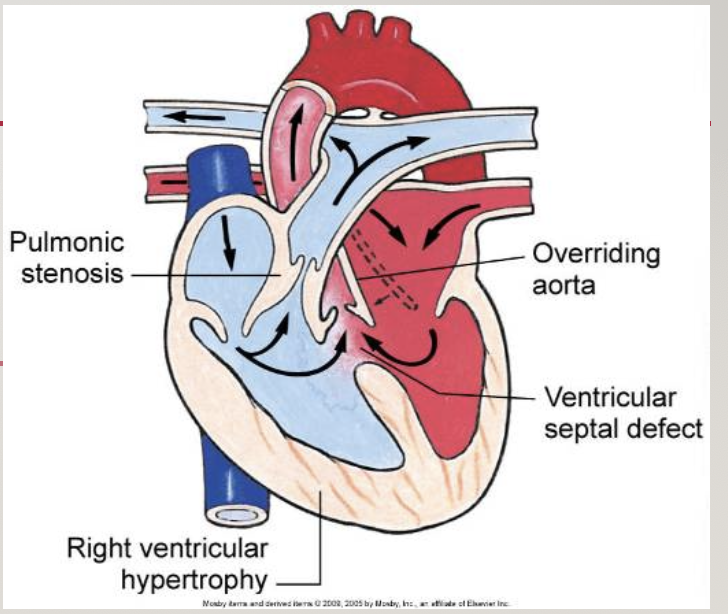
4 different defects:
A large ventricular septal defect (VSD)
Pulmonary stenosis
Right ventricular hypertrophy
An overriding aorta - wall is shifted and opening is more narrow
Both oxygenated and deoxygenated blood enter aorta and is delivered to rest of body
s/s of TEF
as PDA closes ?
cardiac ?2
resp?
growth?
position?
blood?
cyanosis (lips fingers and toes ) as PDA closes - may develop cyanosis in the first year of life.
Tachycardia
Systolic murmur at left sternal border
Severe dyspnea on exertion
Slowed growth and development
Squatting (less surface area for blood to reach in this position, child will feel better in this position )
Polycythemia - increased RBCs
Tet spells

tet spells
most often occurs when
starts at age /
s/s ?
Most often occurs in morning after feedings, defecation, or crying bc it causes lots of exhaustion for the child
most commonly start around 4 to 6 months of age
Acute cyanosis (quick)
Hypercapnia (high CO2)
Inconsolable crying, makes it worse
Hypoxia which leads to acidosis
SOB
tx of TEF
Most infants have corrective surgery by 1-2 yrs of age
Symptomatic children have corrective surgery 3-4 months of age
Not all children are cured by surgery but quality of life is improved
TEF later in life 2
may need
Pulmonary regurgitation may occur requiring a valve replacement 10-20 yrs later
Ventricular arrythmias may occur years later causing Sudden Death
Must be followed throughout lifetime and may need a pacemaker later on in life
Nursing Care for TEF
Decrease Respiratory Distress , keep as calm as possible
Decrease Cardiac Workload
Administration of Cardiac Medications
Maintain Nutrition and fluids (carefully, small feedings with breaks in between and oxygenation status checked)
what do you do during a tet spell
position ? parent ?
administer ?
meds ?
prophylactic?
Knee to chest position, or fetal position against parents Chest
Administer 100% O2 via tent or face mask
IV Morphine sulfate
IV propranolol and Oral propranolol used to prevent TET spells
IV fluids as ordered
Have parents hold to decrease anxiety and try to calm

Defects that Obstruct Blood Flow
Aortic Stenosis (AS)
Coarctation of the Aorta (COA)
Hypoplastic Left Heart Syndrome (HLHS)
Aortic stenosis
blood flow?
narrowing of entrance of aorta
increase in afterload when blood is trying to be pumped In through the aorta

s/s of aortic stenosis
Decrease CO/ hypotension → dizziness
Weak peripheral pulses
Tachycardia
Poor feeding
chest Pain
Surgeyy in utero for AS
done under ultrasound
ballon catheter to open aortic valve
catheter is small enough for heart to usually heal on its own, but baby is always monitored after for bleeding

ROSS procedure
grafts?
replaces ?
To correct aortic stenosis
grafts the pulmonary artery to the aorta. (Autograft)
Replacement of the aortic valve with synthetic/metal valve
coarctation of the aorta (COA)
what is it? causes ?
BP?
pulses ?
other s/s?
•Narrowing of Aorta causing obstruction of left ventricular blood flow
⇧ B/P in upper extremities
⇩B/P in lower extremities
Radial pulses full/bounding and femoral or popliteal pulses weak or absent
Leg pains, fatigue
Epistaxis - nose bleeds

Compliactions of COA
from high BP
LV?
failures ?
Risk of Intracranial bleeds from high BP and ruptures
Ruptured Aorta
Left Ventricular hypertrophy
Heart failure
Kidney failure (decrease blood volume to kidneys, monitor I & Os)
tx for COA
meds ?
to prevent HF?
procedures ?
lifetime ?
A continuous intravenous medication, prostaglandin (PGE-1), is used to open the ductus arteriosus (and maintain it in an open state) allowing blood flow to areas beyond the coarctation.
Treatment to prevent HF inotropic meds (increases contractility), diuretics
Balloon valvuloplasty to open narrowed area
Surgery and balloon dilation are palliative. COA may reoccur. Lifetime follow-up needed.
s/s of congestive HF in children
Infant tires easily (especially during feeding)
Failure to thrive
Diaphoresis
Irritability betong consolidation
Frequent respiratory infections
Tachy (heart and resp)
Nasal flaring/grunting
Cough/crackles
nursing management of children with CHF
Assess vitals
Obtain a Health history
I+O’s - weigh infant diapers
Assess for Periorbital edema
Assess for Peripheral edema
promote rest - Cluster care, Encourage older children to watch videos or games
Family assessment - Encourage parents to play with child, teach not to be fearful
Developmental Assessment
meds for CHF
position ?
Lasix,
ACE inhibitors
IV inotropic
Digoxin - lowers HR and O2 demand of heart
give humidified O2
Semi-fowlers position an infant car seat
nutrition of CHG
Encourage Breast feeding
Feedings should last no longer than 30-40 mins, if dyspnea No longer than 20 min
Frequent small feedings with breaks in between
Angle child at 45 degrees for feeding
High calorie formulas or food
Acute Rheumatic Fever is ?
caused by ?
main concern ?
when will s/s occur
Inflammatory disorder
Caused by Group A beta-hemolytic Streptococci (GAS)
Big concern: May cause long term damage to heart valves
1- 3 weeks after an untreated strep infection s/s may occur
Manifestations of Rheumatic Heart Disease
cardiac ? 3
hx of ?
joints ? skin? 2
movement ?
labs?
New murmur
Chest pain/ carditis (swelling, primarily of mitral valve)
Prolonged PR interval
fever w/ hx of sore throat
Inflamed joints (polyathristis)
Subcutaneous nodules (often over joints)
Erythema marginatum - rash
Chorea (St. Vitus dance) - spastic movement, twitch like
Elevated C-reactive protein (shows inflammation)

tx of RHD
3 meds
what test
Antibiotics (penicillin or erythromycin)
Aspirin (for fever joint pain)
Corticosteroids (Heart inflammation)
Routine Echos to check valve function
Kawasaki disease is?
what kind of response
most cases occur in what age
what 3 things happen to the vessels? causes ?
risk of
Acute systemic vasculitis of unknown cause
Autoimmune response, common after covid
75% of cases in children <5 years old
Inflammation , edema and necrosis occurs in vessels
causes Structural integrity of vessel impaired
Risk of aneurysm and coronary artery dilation

s/s of Kawasakis
fever for more than 5 days
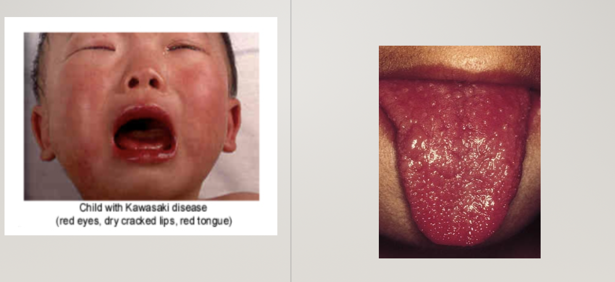
red, dry cracked lips and strawberry tounge (hallmark sign)
swollen lymph nodes
widespread rash
red eyes
swelling and redness in hands and feet
peeling of skin around nails and toenails after 1-2 weeks
3 phases of Kawasaki disease
Acute: sudden high fever, unresponsive to antipyretics and antibiotics (lasts 1-2 weeks)
Sub acute: end of fever through end of all KD clinical signs (lasts 2-4 weeks)
Convalescent: clinical signs resolved, but laboratory values not returned to normal; completed with normal values (6-8 weeks)
tx of Kawasaki
IV med? should be administered within ? to avoid ?
for fever? once afebrile ?
hospitalization if ?
if coronary involvement ?
Single high dose of 2g/kg IV immune globulin (IVIG) over 10 -12 hours
If administered within 7-10 days of onset reduces risk of Coronary artery aneurysms
High doses of Aspirin are given for fever
Low dose ASA once child is afebrile 2-3 days
Hospitalization depends on presence of lesions and if fever persists
Children with coronary artery involvement may have an MI within 1st year of onset
nursing management
check what ? how often ?
asses
s/e of meds ?
mobility ?
teach parents ?
follow up?
Temps every 4 hours
Assess extremities for Edema, redness, swelling, pain
Medication side effects - Bleeding and GI upset
PROM exercises
Parental teaching - Avoid contact sports
follow up with cardiologist