Bones (Ch 6)
 Call Kai
 Learn
 Practice Test
 Spaced Repetition
 Match
 Flashcards
 Knowt Play
1/16

There's no tags or description
Looks like no tags are added yet.
17 Terms
The skeletal system
Bones of the skeleton
Cartilages, ligaments, and other connective tissues
Functions:
movement and locomotion
Structural support and attachment
Protection: thoracic cage, vertebrae, skull
Storage of minerals and lipids (calcium and yellow bone marrow)
Blood cell production
Bone Structure: Long Bone
Diaphysis:
Wall of compact bone
Inside: medullary cavity with yellow bone marrow
Metaphysis: narrow zone between diaphysis and epiphysis
Epiphysis:
Spongy (trabecular) bone filled with red bone marrow
Cortex: thin covering of compact bone

Bone Structure (Flat bone)
Core of spongy bone with red bone marrow
Covered by compact bone
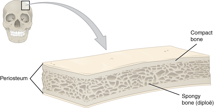
Features of bone
Osteocytes are the main cell type
Depend on diffusion through canaliculi for nutrient and waste removal
Little ground substance with dense mineralized matrix
Surrounded by periosteum (dense irregular connective tissue)
Good blood supply: high regeneration and repair
Bone matrix (identical in compact and spongy bone)
Composed of:
Hydroxyapatite crystals: Ca3(PO4)2 and Ca(OH)2
Calcium carbonate: CaCO3
Minerals for compressive strength
Collagen fibers: flexible and tensile strength
Composite materia
Osteoprogenitor cells
Mesenchymal stem cells produce osteoblasts
Located in inner cellular layer of periosteum/endosteum
Assist in fracture repair

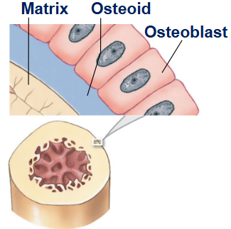
Osteoblast cell
immature bone cells
Secrete matrix compounds to produce new bone (osteogenesis)
Osteoid: matrix produced by osteoblasts, not yet calcified
Secrete calcium phosphate
Surrounded by bone become osteocytes
Osteocytes
Mature bone cells: maintain bone matrix
Live in lacunae (1 cell each)
Between lamellae (layers) of matrix
Connect by cytoplasmic extensions through canaliculi
Do not divide but may produce osteoblasts that repair the matrix
Osteoclast
Derived from same stem cells as monocytes
Giant, multinucleate cells
Secrete acids and proteases (protein digesting enzymes) to dissolve matrix (osteolysis)
Release stored minerals, making bones weaker
Osteoblast=add matrix vs Osteoclast=destroy matrix
Structure of Compound Bone

Structure of Spongy Bone
Matrix formed by a network of trabeculae (no osteons)
Produces red blood cells
No blood vessels in trabeculae
Some yellow bone marrow stores fat

Periosteum
Covers the exterior of all compact bones except at joint capsules
Contiguous with articular connective tissue and synovial membranes
Made of an outer, fibrous layer and an inner cellular layer
Perforating fibers: collagen fibers of periosteum entering bones
Also ligaments and tendons
Endosteum
Lines the medullary cavity
lines central canals
Covers trabeculae of spongy bone
Contains osteoblasts, osteoprogenitor, and osteoclasts
Active in bone growth and repair

Endochondral Ossification
Hyaline cartilage grows (interstitial)
Chondrocytes get bigger (so do the lacunae)
Matrix gets calcified and chondrocytes die
Blood vessels grow
Cells from perichondrium become osteoblast
Compact bone is produced through appositional growth
Capillaries and fibroblast invade cartilage
Fibroblast take space left by chondrocytes
Fibroblasts become osteoblasts
Primary ossification center forms (spongy bone)
Osteoclast form medullary cavity
Shaft becomes thicker
Cartilage at the edge lifts
Capillaries and osteoblast create secondary ossification center
Epiphyses fill with spongy bone
Some stays as articular cartilage
Epiphyseal cartilage separates
New bone grows
Dermal Ossification (intramembranous)
Mesenchymal cells and differentiate as osteoblasts
Bone expands as spicules
Spicule interconnect, trapping blood vessels
Bone remodels into spongy bone
Repair of a Bone Fracture
Hematoma production
An external callus (cartilage and bone) and internal callus (bone) stabilizes the broken bone
Effects of Parathyroid Hormone
Decreases Ca2+ storage in bones; stimulates osteoclast activity and Ca2+ release from bone into plasma
Decreases Ca+ excretion in kidneys and induces production of calcitriol Vit D
Indirectly, increases intestinal absorption of Ca+