Types of tissues
 Call Kai
 Learn
Learn Practice Test
 Spaced Repetition
 Match
Match Flashcards
 Knowt Play
1/36

There's no tags or description
Looks like no tags are added yet.
Name | Mastery | Learn | Test | Matching | Spaced | Call with Kai | Chat |
|---|
No analytics yet
Send a link to your students to track their progress
37 Terms
Major Tissue Types: Muscle Tissue
Contracts and produces force
Major Tissue Types: Epithetal tissue
Protects surfaces, and facilitates gas and nutrient exchange
Major Tissue Types: Connective tissue
Supports and transports
Major Tissue Types: Nervous Tissue
Transmits information
Connective Tissue Components
Consists of cells
extracellular fibers
and matrix
Connective Tissues Classifications
Connective Tissue Proper
Fluid connective tissue
Supporting connective tissue
Connective Tissue Proper (Viscous matrix)
Loose: Fibers create a loose, open framework (e.g., reticular, adipose, and areolar tissue).
Dense: Fibers are densely packed (e.g., dense regular, elastic, and dense irregular tissue).
Fluid Connective Tissues (Watery matrix)
Blood: contained in circulatory system
Lymph: contained in lymphatic system
Supporting Connective Tissues (Dense matrix)
Cartilage: Features a solid, rubbery matrix (includes hyaline, elastic, and fibrocartilage).
Bone: Features a solid, crystalline matrix.

Areolar Tissue Locations (Loose connective tissue)
Found within and deep to the dermis of the skin
covered by epithelial lining of the digestive, respiratory, and urinary tracts
between muscles; and around blood vessels, nerves, and joints

Areolar Tissue Functions (Loose connective tissue)
Cushions organs
provides support while permitting independent movement,
contains phagocytic cells that defend against pathogens

Adipose Tissue Locations (Loose connective tissue)
Located beneath the skin (especially at the sides, buttocks, and breasts), posterior to the eyeballs, and around the kidneys

Adipose Tissue Functions (Loose connective tissue)
Provides padding, cushions shocks, insulates against heat loss, and stores energy
Dominated by adipocytes (fat cells)

Reticular Tissue Locations (Loose connective tissue)
found in the liver
kidney
spleen
lymph nodes
and bone marrow
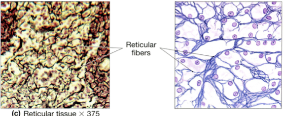
Reticular Tissue Functions (Loose connective tissue)
Provides a supportive framework for organs

Dense Regular Connective Tissue Locations (Dense connective tissues)
Between skeletal muscles and skeleton (tendons and aponeuroses)
between bones (ligaments)
covering skeletal muscles, and in deep fasciae
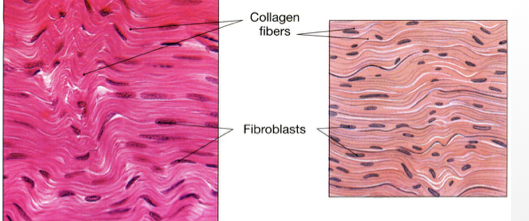
Dense Regular Connective Tissue Function (Dense connective tissues)
Provides firm attachment
conducts pull of muscles
reduces friction between muscles
and stabilizes relative positions of bones

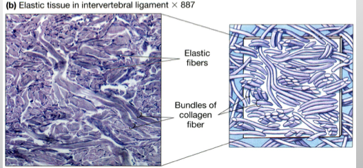
Elastic Tissue Locations (Dense Connective Tissues)
Between vertebrae of the spinal column (ligamentum flavum and ligamentum nuchae)
ligaments supporting the penis
underlying transitional epithelia, and in the walls of blood vessels

Elastic Tissue Function (Dense Connective Tissues)
Stabilizes positions of vertebrae and penis
cushions shocks,
allows expansion and recoil of organs
Dense Irregular Connective Tissue Locations (Dense connective tissue)
Capsules of visceral organs
dermis of skin, periostea and perichondria
and nerve and muscle sheaths

Dense Irregular Connective Tissue Function (Dense connective tissue)
Provides strength to resist forces applied from many directions
helps prevent overexpansion of organs like the urinary bladder

Fluid Connective Tissue
Red blood cells, white blood cells, Platelets
Supporting Connective Tissue
Cartilage
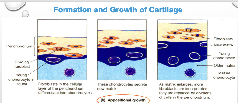
Appositional Growth (Growth from the outside edge)
Fibroblasts in the perichondrium's cellular layer divide and differentiate into chondrocytes.
Newly formed chondrocytes begin secreting fresh cartilage matrix onto the surface.
As the matrix thickens, new fibroblasts are continuously incorporated and replaced via divisions within the perichondrium.

Interstitial Growth (Expands Cartilage from Within)
An existing chondrocyte inside a lacuna divides
The two daughter cells secrete additional matrix while enclosed in the matrix.
The accumulation of new matrix pushes the daughter cells apart, enlarging the cartilage structure from the inside out

Skeletal Muscle Tissue Locations:
Cells are long, cylindrical, striated, and multinucleate
Location: Combined with connective tissue and nervous tissue in skeletal muscles (such as arm or leg muscles)

Skeletal Muscle Tissue Function:
Moves or stabilizes the skeleton
guards entrances and exits to the digestive, respiratory, and urinary tracts
generates heat
protects internal organs

Cardiac Muscle Tissue Locations
Cells are short, branched, and striated, usually with a single nucleus; interconnected by intercalated discs
Location: Found only in the heart

Cardiac Muscle Tissue Functions
Circulates blood and maintains hydrostatic blood pressure

Smooth Muscle Tissue Locations:
Cells are short, spindle-shaped, nonstriated, and have a single central nucleus.
Locations: Encircles blood vessels and is found in the walls of digestive, respiratory, urinary, and reproductive organs.

Smooth Muscle Tissue Functions:
Moves food, urine, and reproductive secretions
controls diameters of respiratory passageways
regulates blood vessel diameter and tissue blood flow.
Neural Tissue Locations
Cells have dendrites, body, and axon(s)
Location: Combined with connective tissue, in brain, spinal cord, and throughout bod
Neural Tissue Function:
Conduction of electrical impulse
Fasciae
connective tissue framework of the body
Superficial fasciae, deep fascia, subserous fascia
Superficial Fascia
Subcutaneous Layer / Hypodermis
Location: Found between the skin and underlying organs.
Composition: Composed of areolar tissue and fat.
Deep Fascia
Location: Bound to organ capsules, tendons, and ligaments.
Composition: Composed of dense connective tissue.
Function: Forms a strong, fibrous internal framework throughout the body
Subserous Fascia
Location: Positioned between serous membranes (such as the parietal peritoneum) and deep fascia
Composition: Composed of areolar tissue