Found 4.1 Brain
 Call Kai
 Learn
 Practice Test
 Spaced Repetition
 Match
 Flashcards
 Knowt Play
1/77

There's no tags or description
Looks like no tags are added yet.
78 Terms
Reviewing The Action Potential - review
how many phases?
MP vs TP
Na enter/exit?
K enter/exit?
A vs R refractory period

Action Potential Types
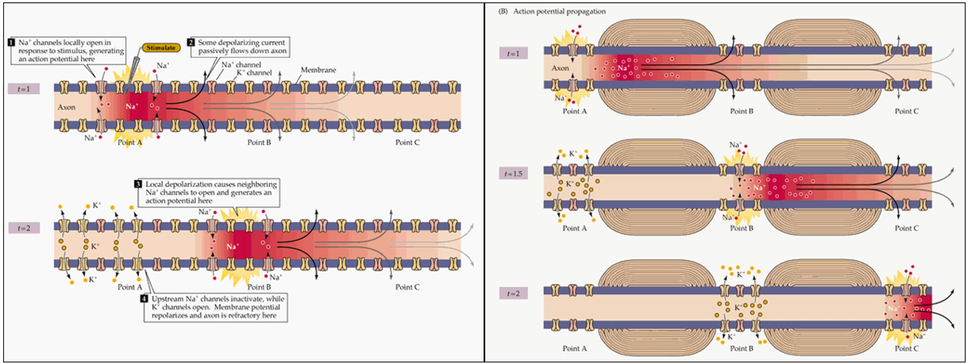
Continuous vs Saltatory Conduction
Continuous vs Saltatory Conduction
Jumping over the node of Ranvier over allows the impulse to move faster.
And that's because the cell is myelinated.
white matter is myelinated
gray matter is unmyelinated
So impulses move more slowly through gray matter.
The Neuron
dendrites vs axon hillock

Basic Parts of Neuron:
Dentrite → carry graded signals that spread towards and collect at axon hillock
Cell Body = Soma
Nucleus
Axon hillock → once combined impulse reaches threshold, it will send the AP down the axon
Axon → conduct impulse away from soma
The Presynaptic Terminal
what e-?
Voltage-gated Ca2+ is the start of everything here


Neurotransmitters
dopamine
GABA
serotonin aka
ACh
nor/epi
gycline
glutamate
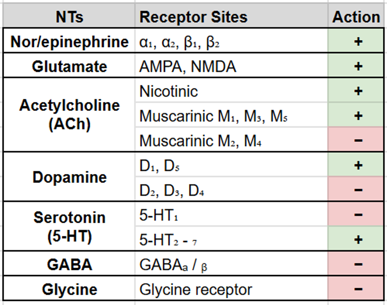
NOT inherently excitatory or inhibitory by nature, but they do have a tendency based on the receptor they bind to
It's what receptor site it's acting on that determines what its property is.
CNS Cells
Functional vs Glial
3 vs 4
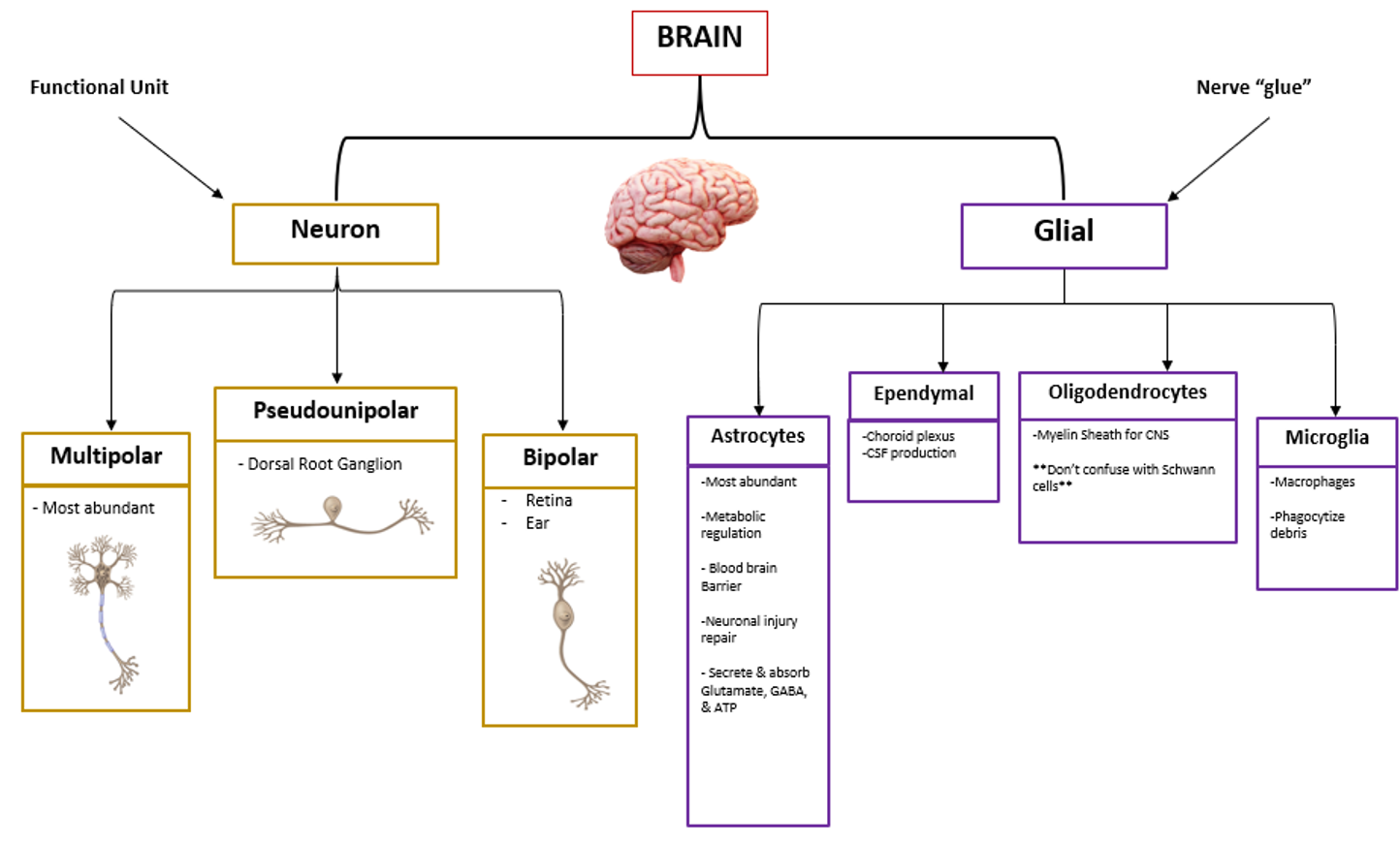
Neurons are more functional.
Glial are more like structural. They say glue here. They're kind of holding things together, if you will.
Neuron - Multipolar - 1
axon vs dendrites
ex? 1
it's got one axon vs many dendrites. So it's accepting and receiving a lot of information.
This is the most abundant neuron in the body.
Ex. a lot of motor neurons are multipolar.

Neuron - Pseudounipolar - fx
axon vs dendrite
(where body relative to both)
where?
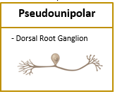
And it's gonna be primarily carrying sensory information from the periphery to your spinal cord to generate a response.
And it's found in the dorsal root ganglion outside SC (PNS)
Neuron - Bipolar
axon vs dendrite
(where body relative to both)
ex? 2
But the bipolar is going to be your specialized sensory pathways, specifically on your retina and in your ear.
So there's two sides = you have two eyes and you have two ears.

Glial Cells - Astrocytes - 1
fx 2
secretes/absorbs 3
forms/maintains x
Astrocytes are your most abundant glial cell
It's going to be responsible for metabolic regulation like glucose delivery.
And it's going to secrete and absorb glutamate, GABA, and ATP.
It's going to be responsible for helping to repair neurons that have been injured.
It's going to create or form the blood brain barrier, which we will talk about towards the end of lecture today.
Glial Cells - Oligodendrocytes - fx
oligo vs schwan
they are the myelinating cells.
So they're going to form the myelin sheath for your CNS, but don't confuse that with Schwann cells, which are in the PNS.
Oligodendrocyte (CNS): one cell myelinates segments of multiple axons
many cells myelinates many segments of an axon
Schwann cell (PNS): one cell myelinates a segment of one axon
So oligodendrocytes is a lot vs Schwann is a 1:1
Glial Cells:
Ependymal - fx, where? 2
Microglia - fx
Ependymal glial cells are what line the ventricles in your brain and the central canal of your spinal cord because they form the choroid plexus, which are what produce your CSF or your cerebrospinal fluid is what CSF stands for.
Microglia, which are like your immune cells in your CNS. They're the macrophages of the CNS, and they eat the debris, which is phagocytosis
Surface Anatomy
gyri
sulci 2
fissure 2
Gyri → elevated convolutions
Sulci → shallow grooves
Central Sulcus → divides frontal and parietal lobes
Parieto-Occipital Sulcus → separates occipital lobe
Fissure → deep grooves
Medial Longitudinal Fissure → divides cerebral hemispheres
Sylvian (Lateral) Fissure → divides temporal lobe from frontal/parietal

Brain Overview - tentorium compartments?
5
3
3 aka
Enclosed in cranium
Compartments divided by tentorium cerebelli
Supratentorium
Frontal - responsible for executive function
Parietal - parietal lobes are gonna be responsible for things like processing, detecting like sensory information, spatial awareness
Temporal - primarily hearing and language
Occipital - responsible for your vision
Diencephalon, which includes your thalamus and your hypothalamus
Infratentorium aka posterior fossa
Brainstem - vital functions: breathing, heart rate, consciousness, your reflexes like cough gag reflex, corneal reflexes
Cerebellum - coordination and balance

Brodmann Areas - NTK

Frontal Lobe - % cortex
Fx 2
Prefrontal cortex Fx 2
Broca’s Fx 2, where?
1/3 of cortex: which is just that outer gray matter of cerebrum. It's that wrinkled surface that you see is also called cortex.
Fx: Premotor & supplementary motor
Initiation of voluntary movement, which just means it basically plans the movements before the movements are fired.
So it does some of the organization of the movements also, but it's primarily responsible for just your voluntary movements
Prefrontal Cortex
Intellectual traits
Executive function
Organization, insight, judgement, reasoning
hence why immaturity can be attributed to an under developed frontal lobe.
Males not developed til >20 yr old
Broca’s Area found in the dominant hemisphere
Motor Speech
Language
Frontal Lobe - which side usually?
dominant - 5
non-dominant - 4
Dominant hemisphere fx [usually left side]
Language
Skilled motor formulation
Math & analytics
Music
Direction (written instructions)
Nondominant hemisphere fx
Visual-spatial analysis
Math & organization
Music (untrained people)
Direction (spatial orientation)
Parietal Lobes - fx 2
blue vs red zones, name & where
which body part of homunculus is largest 2
Posterior to central sulcus
Primary sensory areas for auditory, visual, and somatosensory
Helps you plan motor movements
So your blue somatosensory cortex is what receives the information.
So your red motor strip is gonna send movement commands
So it's receiving that something is hot and your motor strip is going to say, what do I do about it?
I need to take my hand off of it.
You got your processing center and your action center.

homunculus. That is just Latin for little man.
And it is a scaled map of sensory sensitivity, not like size of things.
So it's just showing you that like how sensitive your fingertips are compared to like your knees.
Temporal Lobes - Fx 5
Wernicke’s Area 1
bridge
Basic auditory cortex (Detect sound quality) like pitch and volume.
Complex Auditory association area of speech, music
Basic olfactory cortex (Interpret smell)
Complex Visual association to interpret visual info.
Information retrieval (memory)
Wernicke’s Area
Sensory speech (comprehension)
Arcuate fasciculus → a white matter bridge connecting Broca’s & Wernicke’s
Broca’s & Wernicke’s - where
expressive vs receptive aphasia
Broca’s and Wernicke’s areas are typically in the same language-dominant hemisphere
Damage to Broca’s area — expressive aphasia
- Speech is difficult and effortful; the person may know what they want to say but struggle to get the words out.
- Comprehension is relatively preserved, though it can be affected for complex language.
- They’re often aware of the difficulty and frustrated.
Damage to Wernicke’s area — receptive aphasia
- Speech is fluent but may not make sense (“word salad”).
- Understanding spoken language is impaired, so following commands can be difficult.
- The person may be less aware that their speech is confusing.
Occipital Lobes - where?
Primary Visual Cortex 2
Visual Association Area 1
2 pathways, where does it go?
Posterior brain
Receives/processes visual info
Step 1: Primary Visual Cortex
Detection of visual stimuli - raw input stage.
It sees lights, edges and movements but it doesn't understand what it's looking at.
Step 2: Visual Association Area
Complex processing of visual stimuli processed down 2 pathways
Dorsal pathway → “where” travels toward the parietal lobe.
“Where is it/how do I interact with it?” Helps track direction, location and guide actions.
Ventral pathway → “what” travels toward the temporal lobe.
“What is it?” Helps verify objects and faces.
So dorsal means direction, if that helps you remember.
Ventral, I remember ventral is verify.
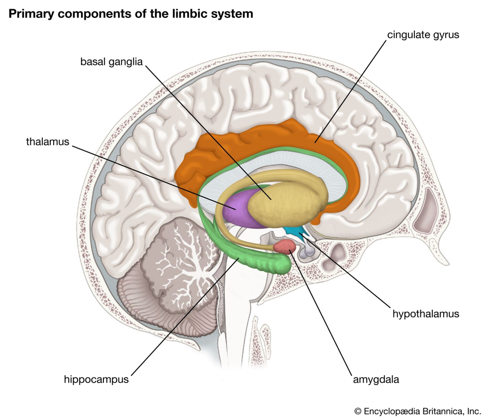
The Limbic System
4 parts and fx
The limbic system is not one single structure.
It's a group of interconnected structures that handle Cognitive function, memory consolidation, & emotional response
Hippocampus - memory consolidation
amygdala - emotional processing
part of hypothalamus - emotional state in response to a physical state
Insular Cortex → “The 5th Lobe”
Part of limbic system → processing emotion
Higher level cognitive information
helps your brain process pain: Influences afferent nociceptive input & efferent response

Diencephalon - where? connects what?
3 parts
2
3 anes?
1
Fx: Connects the midbrain to forebrain that sit deep in the center part of the brain.
Thalamus
Relays sensory and motor impulses
Regulates consciousness (sleep & alertness)
Hypothalamus
Homeostasis: Synthesizes and secretes neurohormones, communicating with pituitary gland
Controls temperature, hunger, thirst, fatigue, sleep, & Circadian rhythm
So another thing to remember is the hypothalamus is going to be one of our targets for general anesthesia
Which makes sense because it's regulating our consciousness, right?
So it's going to interrupt this thalamocortical pathway. And that's that relay station, how the thalamus talks to the cortex. So our drugs are going to interrupt that pathway so that we can't know we're awake, basically.
Pineal gland. Melatonin.

Pituitary Gland - aka, where?
A/P review 5/2
AKA “Master Endocrine Gland”
Housed in Sella Turcica
Anterior (adenohypophysis)
HGH, TSH, ACTH, beta-endorphin, prolactin, LH, FSH, & melanocyte-stimulating hormones
Posterior (neurohypophysis)
Oxytocin & ADH

Basal Ganglia - fx 2
parts 5
outcome ex
4
1
1
Plays a role in cognitive, limbic & motor function
It is, so your cortex is going to send ideas for a lot of movements all at once.
And the basal ganglia is going to pick the one that you want and help suppress the rest.
So it's taking that information and deciding what we want to do and what we don't want to do and making it smooth so that we don't have jerky movements or unrefined movements.
Paired Components located on each side of the brain, mainly lateral to and surrounding the thalamus
Putamen
Caudate nucleus
Globus pallidus (externa/interna)
Substantia nigra
Subthalamic nucleus
One of the principal FX of the basal ganglia in motor control is to function in association with the corticospinal system to control complex patterns of motor activity
Writing alphabet
Cutting with scissors
Throwing a baseball
Shoveling dirt
Many aspects of vocalization
Controlled eye movements
Caudate Nucleus & Putamen: aka 2
2 circuits
receives from? passes what? excludes? 1
receives from? passes what? excludes? 3
Striatum is Caudate Nucleus & Putamen aka internal capsule
Input comes from many areas from the cerebral cortex
Output → other parts of BG via 2 circuits
Putamen circuit usually receives input from motor cortex and then its output is primarily to the motor cortex.
Passes through putamen → GPe/GPi → substantia nigra/subthalamus → VA/VL thalamus
Skips caudate
Caudate circuit plays a role in the cognitive control of motor activity and receives input from association areas of the cortex as opposed to like the primary motor cortex.
Passes through caudate → putamen → GPe/GPi → VA thalamus
Skips substantia nigra/subthalamus and VL thalamus

Globus Pallidus - receives from?
2 zones inside, stimulation of each?
output to?
Input comes from striatum basically a giant input hub.
Striatum inhibits Globus pallidus interna (GPi) (the “Int” segment) with GABA → GPi inhibits the thalamus less → the thalamus can excite the cortex more → movement is facilitated.
Striatum inhibits Globus pallidus externa (GPe) (the “Ext” segment) with GABA → the subthalamic nucleus is less inhibited → it excites GPi with glutamate → GPi inhibits the thalamus more → movement is suppressed.
Output → primarily thalamus & secondary to other areas via 2 pathways
Substantia Nigra - supplies what? where? fx?
patho: too much vs too little
It's going to function similarly to the pallidum that it's got a lot of output.
Supplies dopamine to the striatum, which is going to fine tune how the whole circuit behaves.
Too little = Parkinson’s Disease
Too much = Schizophrenia
Subthalamic Nucleus - fx?
input from? output where?
with what? fx? 2
which is like the modulator
Input comes from striatum & cortex
Output → striatum
Produces glutamate and it's primarily excitatory and it's going to act as like the brake and balance on the entire circuit.
Basal Ganglia Overview - fx 2
excitatory NT 1
inhibitory NT 2
Think kinetic modulation or moving modulation. It's not initiating these movements, it's fine tuning the movements
Inhibitory action counterbalances the excitatory action from the cerebellum is what's driving and coordinating the movement and your basal ganglia is going to kind of tap the brakes on that system.
Coordinates with corticospinal system to control complex patterns of skilled motor activity
Input to BG → Striatum
Output from BG → Globus Pallidus
Primary excitatory NT = Glutamate
Primary inhibitory NTs = DA & GABA
DA = dopamine
Basal Ganglia NTs - visual
inhibition of GPi
inhibition of GPe

The key is that GABA inhibits, glutamate excites, and dopamine adjusts the balance
hence the double arrow heads
BG Visual -

Brainstem - fx 2 3
CN 2, relevant why?
4 parts
2
1
1
3
CV/resp control
Cough, gag, & pupil reflexes
Nuclei for CN III and XII - brain death exam
Midbrain (has two i’s)
Auditory & visual tracts
Pons
Autonomic integration
Medulla
Autonomic integration
Reticular Activating System
kind of the on-off switch because it controls consciousness, arousal, and sleep
Cerebellum - fx 3
how many lobes? fx?
decussation?
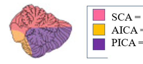
blood supply 3
Processes proprioceptive input (where you are in space is proprioception), axial posture, & gait (coordinating movement and balance)
3 Lobes
anterior lobe is responsible for regulating muscle tone with skilled motor movement.
Posterior lobe plans your voluntary activity.
Flocculonodular lobe is your eye movements and balance is primarily housed here.
Ipsilateral effect via double decussation - So the right side controls the right side and that's because of this double crossing over.
So you have your superior cerebellar artery, your anterior inferior cerebellar artery, and your posterior inferior cerebellar artery are the blood supply to the cerebellum.

Other Important Brain Structures
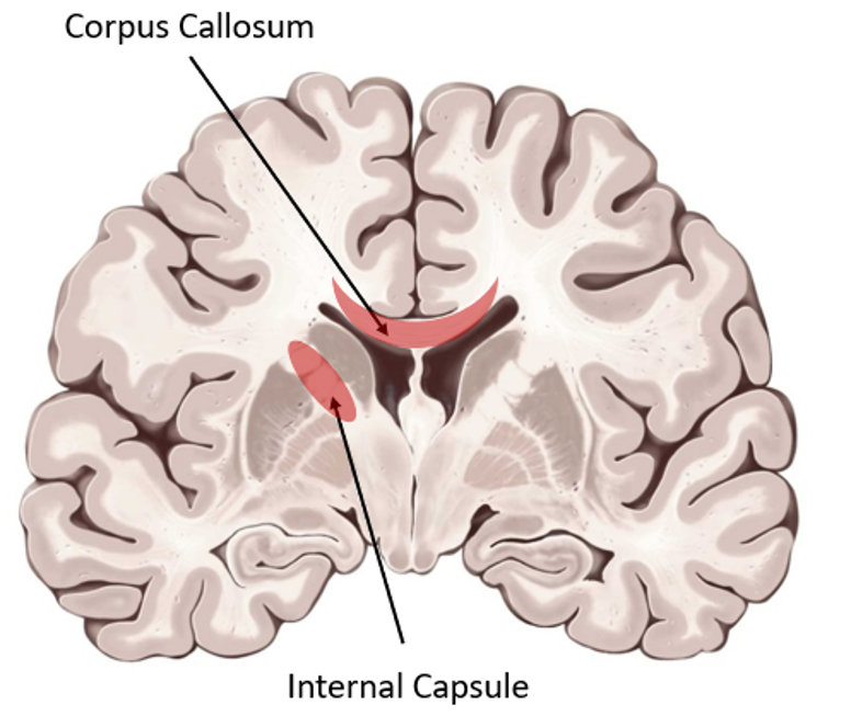
Corpus Callosum 2
Internal Capsule 2
Hippocampus 2
Amygdala 3, where?
Corpus Callosum
Interconnects brain hemispheres
actually white matter
Internal Capsule
“Interconnects” brain to body
also white matter and it connects cortex information to the brainstem and spinal cord.
Hippocampus
Memory & Learning
Amygdala
a little almond shaped cluster of nuclei that are housed like medial temporal lobe
Emotion, appetite, pain/stressor response (fear)
Reticular Activating System - fx 3
zones? 3
TMN: where? fx
VTA: fx
TILN: fx
anes
tip: thalamic tubero tegmental
Diffuse collection of brainstem neurons
Mediates awareness, consciousness, arousal, & sleep
Contents
Reticular Formation - neuron network itself
Tuberomammillary nucleus - which is what is mainly in the posterior hypothalamus, and it promotes wakefulness
Ventral tegmental area - which is the reward system: dopamine releasing area
Thalamic intralaminar nucleus - relays the RAS signals up to the cortex broadly.
Target of many anesthetic agents
A lot of our anesthetic agents work here to suppress the reticular activating system. Like we want to put it to sleep because it's promoting wakefulness.
Extrapyramidal System - Not part of 2, fx? 2
components 3
psych meds?
Not part of corticospinal tract & primary motor
Going to refine and Modify motor function
Components
Basal Ganglia
Cerebellum
Auditory/Vestibular pathways
psych medications have extra pyramidal side effects
So if you've ever heard of somebody with like twitching or uncontrolled motor movements after being on some of these psychiatric medications, it's because you are affecting the function of these components.
Meninges - 3 layers
meninges, which are the covering in the nervous system.
Fx: They offer support and protection.
You have dura mater, arachnoid mater, pia mater, and that's from superficial to deep.
Dura Mater - describe it 3
forms what system?
Innervation 2
Blood supply 1 patho?
EPH vs SDH
Thick, tough, leathery
Adheres to inner skull
Support, forms sinus system - So it helps drain blood from the brain
Innervation → C2 & C3
Blood supply
Middle meningeal artery source of epidural hematoma which is b/w skull and dura
SDH: is from venous bridging injuries and is b/w dura and arachnoid layer
Arachnoid Mater - describe it? fx?
contains? where? exits where?
patho? 2
b/w dura & pia
Spongy/fibrous layer
Keeps brain suspended
Subarachnoid space → contains CSF
Arachnoid granulations
where CSF exits into venous system
CSF turns over a few times per day as it's produced and taken away to house a certain amount at a time.
So this is what's responsible for maintaining that CSF balance, if you will.
subarachnoid hemorrhage, but this is where those hemorrhages occur.
And one reason these patients get Hydrocephalus is when they have bleeds here from aneurysms rupturing that actually blocks the output of the CSF sometimes.
So they will sometimes have big ventricles.
Because if you block these granulations and you can't get CSF out, it's just going to continue to build up.
Plus, if you have blood in the area, it's going to cause expansion and brain compression.
Pia Mater - 2
Innermost layer
Adheres to cortical surface or the cortex of the brain itself
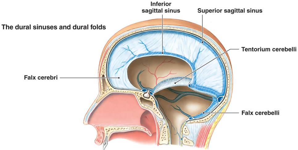
Dural Reflections (Septa)
3 components
2
3
1
tip: falx
Falx Cerebri
Down longitudinal fissure, b/w hemispheres
Encloses superior and inferior sagittal sinuses
Tentorium Cerebelli
Extends inward b/w cerebellum & occipital lobe which is like your supra and your infratentorial divider.
Encloses straight, transverse, & petrosal sinuses
Brainstem passes through tentorial notch
Falx Cerebelli
which is the cerebellar hemisphere divider
Cerebrospinal Fluid - fx 3? where 2?
production by?
ml/hr? ml at one time? replaced qxhr?
reabsorption 2
Cushions brain, provides buoyancy (helps to reduce the actual pressure on the brain), & provides ideal conditions for neurologic function
Locations
4 Ventricles (L/R lateral, 3rd, 4th)
SA space in brain/SC
Production
by choroid plexi in ventricles @ 20 mL/hr
Constant volume ~ 150 mL at any given time
Volume replaced every 3-4 hours
Reabsorption
Arachnoid villi at superior sagittal sinus into venous circulation via pressure gradient
which is one reason they may have you raise the head of the bed because they want to force drainage out of the head and neck to help reduce ICP sometimes because it's gonna follow that gradient down.
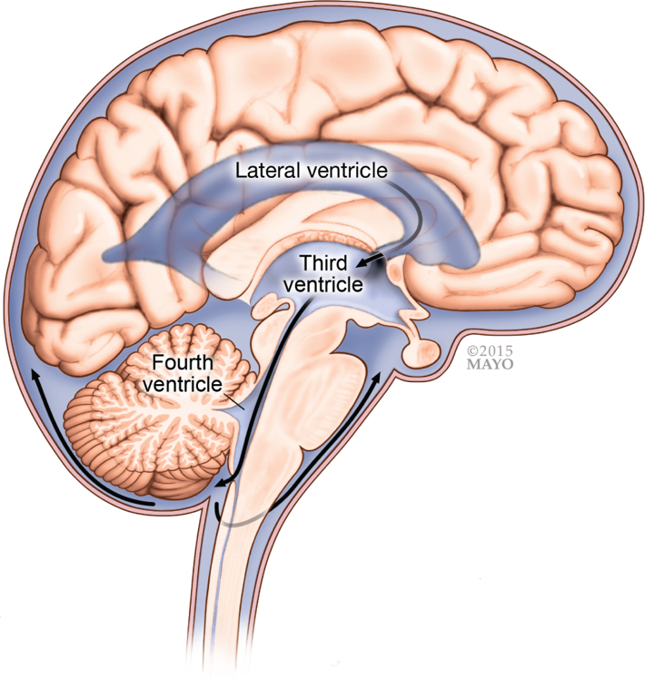
CSF Pathway - 8 layers

This is how the CSF is gonna travel through your body, through your left and right ventricles to the foramen of Monroe, which is an opening that's going to travel to the third ventricle through the aqueduct of Silvius and then to the 4th ventricle. And then it's going to travel to the foramen of Magendi and Luschka and they're paired on each side to that subarachnoid space out the superior sinus. And that's how it's going to travel all the way to being drained out so that it's not overproduced or accumulating in your central nervous system. CSF in the space around the spinal cord is continuous with the CSF space around the brain. It circulates and is eventually reabsorbed
Cerebrospinal Fluid - how many exits?
how many openings per exit?
patho?
2 exits that let CSF leave the fourth ventricle and enter the SA space to be reabsorbed:
Magendie: one opening in the middle → cisterna magna
Luschka: two openings on the sides → spaces near the cerebellum and brainstem
They matter because CSF needs these openings to circulate around the brain and spinal cord. If they’re blocked, CSF can back up in the ventricles and cause obstructive hydrocephalus

Cerebrospinal Fluid Table
osmo
Na
Cl
K
pH
PaCO2
HCO3
Glucose
Protein
fx of mannitol and 3% NS
Protein (x2 and 2 zeroes)

High ICP: Mannitol or hypertonic saline (e.g., 3%) raises plasma osmolality, shifting water out of swollen brain tissue; mannitol also promotes diuresis.
Intact BBB: Keeps most large proteins out of CSF, so CSF normally has much less protein than plasma.
Lumbar puncture: CSF WBCs, protein & glucose can help identify infection or inflammation. High CSF protein may suggest BBB disruption, but isn’t proof by itself.
The brain tightly regulates extracellular K⁺ because when neurons fire, they release K⁺ outside themselves. If that outside K⁺ builds up, neurons become more excitable and may fire abnormally.
Blood Brain Barrier - fx 2
restricts? 4
permeable to? 3 2
Separates CSF from plasma
Unlike many body capillaries, BBB capillaries have tight junctions, not open pores. Substances cross selectively—through specific transporters or directly across the lipid membranes
Another way of saying this is that it effectively isolates the extracellular compartment from the intravascular compartment in the brain/SC
Endothelial cell tight junctions restrict passage of large molecules & ions
Substances with molecular weight > 500 daltons
Polar molecules (amino acids, glucose, mannitol)
hydrophilic drugs
Proteins
Permeable to lipid-soluble molecules
VA, O2, CO2, ETOH, nicotine
BBB not present where 6?
CRTZ = chemo receptor trigger zone
triggers your vomiting reflex = zofran inhibits this
Area Prostrema
Choroid plexus
Hypothalamus
Posterior pituitary
Pineal gland
Cerebral Blood Flow: Arterial
Anterior %, pathway 3
Posterior %, pathway 4
patho?
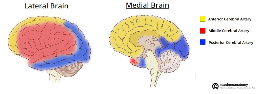
Anterior
Internal carotid arteries → Circle of Willis → hemispheres
80% of CBF
Posterior
subclavian arteries → vertebral arteries → Basilar artery → Posterior brain and cervical spinal cord
20% of CBF - 10% on each side because you have a vertebral artery on each side
Both anterior/posterior is an interconnected arterial loop that can provide collateral blood flow around some blockages.
How well it compensates depends on the blockage’s size and location and the person’s anatomy. An aneurysm is a vessel bulge; it doesn’t automatically block flow.

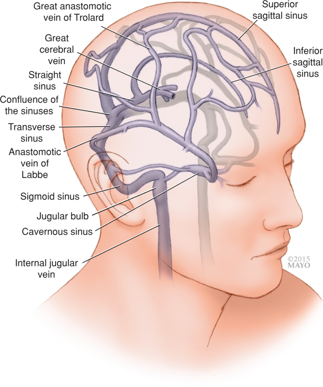
Cerebral Blood Flow: Venous
superior 2 vs inferior 3 aka
Cortex/cerebellum
Superior sagittal sinus
Dural sinus
Basal brain
Inferior sagittal sinus
Vein of Galen (great cerebral vein)
Straight sinuses
All blood exits brain into jugular veins
Major Cerebral Arteries - visual
Cerebral Blood Flow - formula?
regulated by?
normal 2? CO%?
ischemia vs cell death @?
penumbra
CBF = CPP/CVR
Regulated by “flow-metabolism coupling”
Normal = 50 mL/100g tissue/min (15% CO)
750 mL/min (15% CO)
Ischemia <20 mL/100g tissue/min
Cell death <10 mL/100g tissue/min
“Time is brain”: In stroke, rapid treatment can limit brain injury.
Penumbra: Ischemic brain tissue around the infarct core that is impaired but still viable. Restoring blood flow—by clot-busting medication or thrombectomy—may save it.
Cerebral Blood Flow
Five Key Determinants
Cerebral Metabolic Rate (CMRO2) - cerebral metabolic rate. That's the demand component here is your metabolic rate
CPP
↑ PaCO2 = vasodilation and an ↑ CBF
hyperventilation to ↓ CBF = ↓ ICP
PaO2 - doesn’t matter til <60. Hypoxia will trigger cerebral vasodilation
Venous Pressure = keep >HOB
CMRO2 - r/ts CBF
total O2 consumption?
%/% for electrical vs cell activity
Don’t go lower than %?
↑ CMRO2 4
↓ CMRO2 3*2
Monitoring?
CBF has direct correlation with CMRO2 (↑ demand = ↑ supply)
↑ CMRO2 = ↑ CBF
Oxygen consumption
20% of total body oxygen consumption
60% for electrical activity; 40% for cellular integrity
Takeaway: can only ↓ CMRO2 up to 60%. Like that's the max that you can decrease it with like your anesthetics
↑ CMRO2
Seizures
Ketamine
N2O
Hyperthermia
↓ CMRO2
Halogenated anesthetics
IV anesthetics (Propofol, Etomidate, Barbiturates)
Hypothermia
↓ 7% per 1℃ body temperature (suppression @18-20℃)
Use EEG to monitor pt activity because these drugs are going to cause burst suppression and reduce the metabolic demand on their brain
Cerebral Perfusion Pressure - formula?
autoregulation range? what happens at both ends?
autoregulation impaired by 4*
CPP = MAP – ICP (or CVP); whichever is higher
CBF autoregulation
< 50 mmHg = max vasodilation
50-150 mmHg = constant/consistent CBF
> 150 mmHg = max vasoconstriction
When is autoregulation impaired?
Intracranial Tumor
Head Trauma
VA
Chronic HTN (right shift in CPP curve)
And chronic hypertension patients have a shift to the right in their CPP curve because they require higher perfusion rates because their body is used to regulating at higher pressures.
So some of these patients are going to be more sensitive to being HPT, because their brain is going to be used to being perfused at a higher nap.
So you may notice that people get a little loopy when their blood pressure drops even if it is at normal ranges
Venous Pressure - r/ts intracranial drainage & volume
causes 4
↑ CVP = ↓ cerebral drainage = ↑ cerebral volume
Causes of impairing venous drainage
Jugular compression (positioning)
↑ intrathoracic pressure (cough, PEEP)
Thrombosis
Vena cava syndrome which is like a compression or an obstruction of the superior vena cava, they're not going to have as much flow either.
PaCO2 - r/ts CBF
1 mmHg ↑ = ?
range at both ends & outcome?
metabolic acidosis? why?
Direct correlation with CBF
If you remember, CO2 is one of those things that freely crosses BBB
↑ PaCO2 = ↑ CBF
CSF pH alters cerebral arteriolar diameter
↑ 1 mmHg PaCO2 = ↑ 1-2 mL/100g brain tissue/min
Max vasodilation @ >80 mmHg PaCO2
Max vasoconstriction @ <25 mmHg PaCO2
Think about how respiratory acidosis/alkalosis affect CBF
Metabolic acidosis does NOT affect CBF → H+ can’t cross BBB
So metabolic acidosis isn't going to have the same effect.
Remember, respiratory acidosis is going to have the same effect because CO2 can cross.
And the CO2 is what's converted into carbonic acid to change the pH level.
So that's how it's going to be different and how it's going to change the arterial tone.
PaO2 - 2
PaO2 >60 mmHg does NOT affect CBF
PaO2 <50-60 mmHg = cerebral vasodilation = ↑CBF
Hypoxia will trigger cerebral vasodilation
Intracranial Pressure - normal?
initial s/s? 6
body’s compensatory?
Cushing’s triad
Normal = 5 – 15 mmHg
Intracranial HTN
ICP > 20 mmHg
Initial S/S = HA, N/V, papilledema, ↓LOC, seizure, coma
Initial rise in volume is compensated by shunting CSF into spinal cord, then shoved into the subarachnoid space to be reabsorbed.
Cushing’s Triad
HTN (body’s attempt to maintain perfusion)
Bradycardia (baroreceptor reflex) - Their heart rate is going to plummet as a baroreceptor reflex. Don't give these people beta blockers. It's not going to be good.
Irregular respirations (medullary compression) - should already be intubated and ventilated at this point
Monro-Kellie Hypothesis - 3 volume %
examples that can ↑ partition
2
3
4
point graph system
Cranium = rigid box with 3 volumes:
Brain = 80% → swelling, tumor, etc.
Blood = 12% → increased CBF (volatile anesthetics, vasodilators, etc) and intracranial bleeding
CSF = 8% → increased CSF production, decreased CSF reabsorption, impaired BBB, CSF flow obstruction
↑ in one requires a ↓ in others to maintain the same pressure

Points 1 – 2 are compensated
Points 2 – 3 is where compensation begins to fail and you start having s/s of intracranial HTN and focal ischemia
Points 3-4 is where you start having risk of global ischemia & intracranial herniation
Ways to Lower ICP
least invasive 2
more invasive 3*
meds
dose, fx
dose, fx
fx
Restrict Fluids
>HOB
Hyperventilate (effect lasts 4-6 hours)
Hypothermia (not recommended for CHI - closed head injuries only because that can cause other things like coagulopathies and infections)
Normotensive (slightly on hypertensive side)
Mannitol 0.25-1 g/kg - the osmotic diuretic
Furosemide 0.5-1 mg/kg
Corticosteroids - steroids to reduce vasogenic edema
Spinal Cord Physiology - formula
arter of x? why is it important?
Very similar to brain physiology
Autoregulation rules apply
Spinal Cord Perfusion Pressure = MAP – Spinal Subarachnoid Space Pressure
Therefore, we sometimes place a lumbar drain to decrease the SSSP when clamping the aorta is planned (increased perfusion pressure
The concern is spinal cord ischemia. Clamping the aorta can reduce blood flow through arteries that supply the spinal cord—including the artery of Adamkiewicz, a major feeder to the anterior spinal artery. If the cord doesn’t get enough blood, it can be injured, potentially causing leg weakness or paralysis.
A lumbar drain removes a small amount of CSF around the spinal cord. That lowers the pressure pushing back against incoming blood, improving the spinal cord’s perfusion pressure:
Cerebral Blood Flow Summary Graph -
MAP
CPP
PaCO2
PaO2
ICP

Below autoregulation limit → vessels are maximally dilated → pressure dependent
Above autoregulation limit → vessels are maximally constricted → pressure dependent
The vessel diameter is only blue because the graph had MAP as blue.
This picture almost captures the whole picture for cerebral blood flow. It omits “CPP” in the graph. Autoregulation for CPP is 50-150 mmHg. Note that to have a CPP of 50-150 you need a MAP of about 60-160 mmHg.
So like basically you're Cerebral blood flow is constant when your map, which is that blue line, is between 50 and 150. So it's straight across. It's just showing you how it's staying constant.
And your cerebral blood flow is going to rise as your PA CO2 rises, which is the green line, the green line, until max dilation is reached at about 80.
And then your cerebral blood flow is going to stay constant until your PaO2 drops below 60.
So it's actually kind of, it's the red line and it's kind of backwards from the other line.
And then your cerebral blood flow is going to decrease as your ICP increases, which is the purple line.
So if you take it one at a time, it kind of makes sense.
Types of Cranial Nerves *
motor
1
3
sensory
1
5
both
Cranial nerves are part of the peripheral nervous system, even though they come directly from the brain
1) Motor
Skeletal Motor
Voluntary muscle
Autonomic Motor
Smooth muscle
Cardiac muscle
Glands
2) Sensory
Somatosensory
Special Sensory
Smell
Sight
Hearing
Taste
Equilibrium
3) Both (mixed)
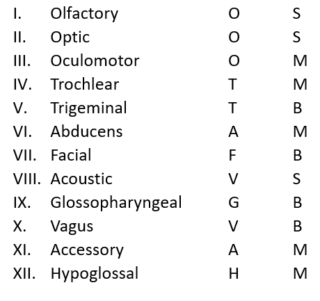
All CN’s - table
also mark if it [S - M - B]
nmemonics
some say merry money, but my brother says big boobs matter more
Olfactory Nerve (I) - 1
Special Sensory for smell
Optic Nerve (II) *
2 vision types
3
3
patho?
Only CN that is part of CNS (surrounded by dura)
Lateral vision
Travels medially via optic nerve
Decussates at optic chiasma
Travels to contralateral brain
Medial Vision
Travels laterally via optic nerve
Does NOT in cross in optic chiasma
Travels to ipsilateral brain
Pituitary gland
Note that it’s adjacent to the optic chiasma
Enlargement can compress chiasma, resulting in loss of peripheral vision (lateral vision)

Oculomotor (III)
skeletal
autonomic 2
Skeletal
Most extrinsic ocular muscles. So it moves the eyeball itself.
Autonomic
Intrinsic ocular muscles
Miosis & accommodation (lens)
And it's also your intrinsic ocular muscles, which are your meiosis or pupil constriction and accommodation, which is the lens shape for your near vision focus.

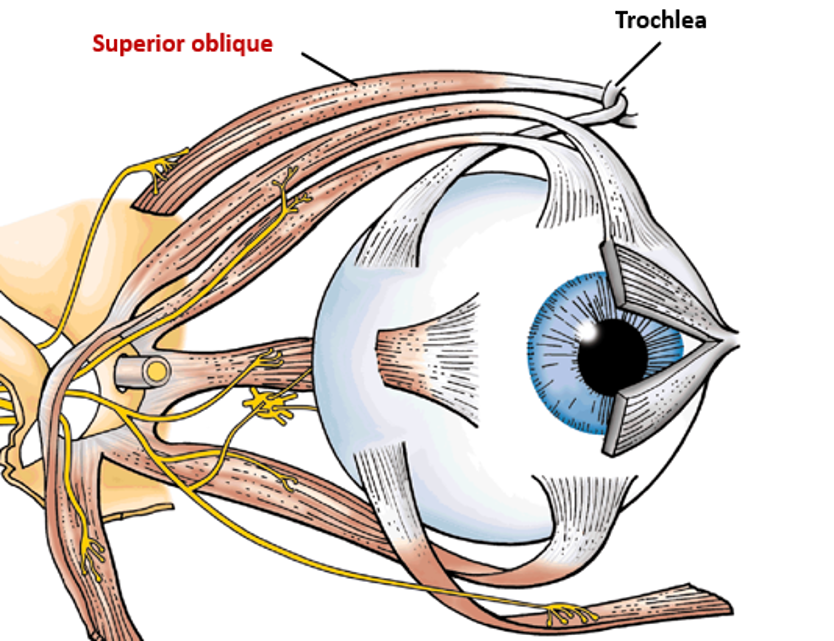
Trochlear (IV) - 1
Skeletal motor to superior oblique muscle
Moves eye down & outward
Trigeminal (V) - 2
what branches 3?
Trigeminal is going to be both motor and sensory.
And they provide sensory innervation to your face.
The motor function is mastication, so chewing.
So it's called trigeminal because it's got three branches that branch out into your face.
You have the ophthalmic, maxillary, and mandibular branches of the trigeminal nerve.

Abducens (VI) - 1
Skeletal motor to lateral rectus muscle
Abducens is LR6.

Facial (VII)
autonomic
sensory
s. sensory
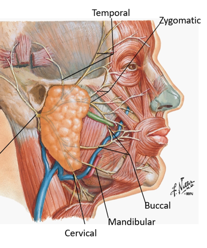
sk. motor - 5 branches
Autonomic Motor
Lacrimal, submandibular & sublingual glands
General Sensory
Skin on external ear
Special Sensory
Taster anterior 2/3 tongue
Skeletal Motor
Facial expression
Temporal
Zygomatic
Buccal
Marginal mandibular
Cervical
Vestibulocochlear (VIII) - 2, aka
aka acoustic
Special Sensory
Hearing
Equilibrium
Glossopharyngeal (IX)
sensory 4
autonomic 1
s. sensory 1
sk. motor 1
Number 9, glossopharyngeal is both as well sensory and motor.
And if you remember, it's going to help with chemoreceptors and everything in your carotid body and sinus to help with blood pressure regulation.
So autonomic control in your parotid gland
special sensory, so taste on the posterior 1/3 of your tongue.
And then it's also going to be down here controlling the stylopharyngeus muscle that is one of the muscle that assists with swallowing.

Vagus (X)
autonomic %
sensory 3
Autonomic
75% of all parasympathetic nervous system activity as it travels all throughout your body.
Sensory
does have a little bit of sensory to Larynx, thoracic & abd viscera
Spinal Accessory (XI)
convergence where?
enters/exit where?
innervates which muscles?
fx 2
fx 1
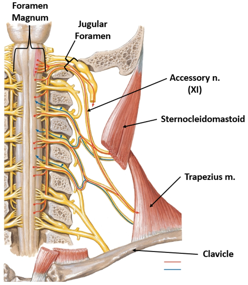
Convergence of C1-C5
Enters skull via foramen magnum
Exits skull via jugular foramen
Skeletal motor innervation of:
Sternocleidomastoid
Lateral Flexion
Rotation of head
Trapezius muscles
Shoulder shrug
Hypoglossal (XII)
innervates which muscles?
1
2
Skeletal Motor
Intrinsic tongue muscles
Tongue shape
Extrinsic tongue muscles
Genioglossus
Tongue movement - So if you go from side to side or stick it out or pull it in, which is your genioglossus muscle, which is protrusion and retraction of your tongue.

Right Eye Muscle Cranial Nerves - CN#? 3
8 directions

I always remember LR6SO4
And then LR6 is your lateral rectus abducens nerve #6
So if you can remember, SO4 is your superior oblique and that's #4
So if you can remember SO4 and LR6 for your eye nerves and muscle innervation, then every other muscle is the oculomotor nerve #3