Anatomy and Physiology - CARDIOVASCULAR COMPLETE
 Call Kai
 Learn
 Practice Test
 Spaced Repetition
 Match
 Flashcards
 Knowt Play
1/200

There's no tags or description
Looks like no tags are added yet.
201 Terms
Cardiovascular system
Consists of the heart and blood vessels
Transports blood throughout the body
Delivery of O2, nutrients; removal of CO2, wastes
Providing adequate perfusion (sufficient delivery to maintain cells’ health)
Requires continual pumping of the heart and open, healthy vessels
Perfusion
Delivery of blood per time per gram of tissue (mL/min/g)
Blood vessels
Arteries carry blood away from the heart; most (besides pulmonary arteries) carry oxygenated blood
Veins carry blood back to the heart; most (besides pulmonary veins) carry deoxygenated blood
Capillaries are sites of exchange (ex: of gases) between blood and air in lungs/body cells

Atria
Superior chambers that receive blood and send it to ventricles
Right atrium: receives deoxygenated blood from body
Left atrium: receives oxygenated blood from lungs

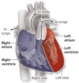
Ventricles
Inferior chambers that pump blood away
Right ventricle: pumps deoxygenated blood to the lungs
Left ventricle: pumps oxygenated blood to the body


Great vessels
Transport blood to and from heart’s chambers
Right side:
Venae cavae: drain deoxygenated blood into right atrium
Pulmonary trunk: transports blood from right ventricle; splits into pulmonary arteries
Left side:
Pulmonary veins: drain oxygenated blood into left atrium
Aorta: transports blood from left ventricle

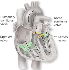
Heart valves
The heart has two sets of valves: atrioventricular (AV) and semilunar
Ensure one-way flow of blood through heart; prevents backflow
Made of endothelium-lined fibrous connective tissue cusps (flaps)
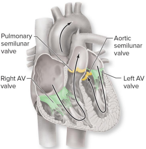

Atrioventricular (AV) valves
Sit between atrium and ventricle of each side
Right AV valve (has 3 flaps = tricuspid)
Left AV valve (has 2 flaps = bicuspid or mitral)

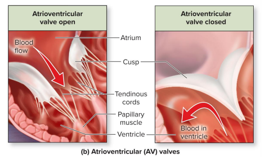
Atrioventricular (AV) valves mechanism of action
AV valves close when ventricles contract, forcing blood superiorly
The flaps are connected to papillary muscles by tendinous cords; when these muscles contract (get shorter), the tendinous cords are pulled taut, closing the flaps to prevent inverting into atria

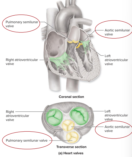
Semilunar valves
Sit at boundary of ventricle and arterial trunk; each has three cusps
Pulmonary semilunar valve: located between right ventricle and pulmonary trunk
Aortic semilunar valve: located between left ventricle and the aorta


Semilunar valves mechanism of action
Prevent backflow to ventricles
Open when ventricles contract and blood is forced into arteries (ventricular pressure > arterial pressure)
Close when ventricles relax (arterial pressure > ventricular pressure); as blood starts to slide backward, it catches and fills cusps and closes valves


Heart flow
Body
Superior/inferior vena cavae
Right atrium
Tricuspid valve
Right ventricle
Semilunar pulmonary valve
Pulmonary arteries
Lungs
Pulmonary veins
Left atrium
Mitral/biscupid valve
Left ventricle
Semilunar aortic valve
Aorta
Body

Pulmonary circulation
Between the blood and the lungs
Deoxygenated blood from right side of heart to lungs
At lungs, blood picks up oxygen and releases carbon dioxide
Blood vessels return blood to left side of heart
Systemic circulation
Between the blood and the body cells
Oxygenated blood from left side of heart to systemic cells
At systemic cells (for example, skin, muscles), blood exchanges gases, nutrients, and wastes
Blood vessels return blood to right side of heart
Congestive heart failure
Impaired ability of the heart to pump blood
Edema (swelling) is a characteristic symptom
Systemic edema (CHF)
May occur if right ventricle impaired
More blood remaining in systemic circulation
Additional fluid entering interstitial space
Pulmonary edema (CHF)
May occur if left ventricle impaired
More blood remaining in pulmonary circulation
Swelling and fluid accumulation in the lungs
Breathing difficulties and impaired gas exchange

Heart position within the thoracic cavity
Located in the mediastinum (space in the thoracic cavity between the left and right pleural cavities)
Sternum is anterior
Thoracic vertebra is posterior


Pericardium
Three layers
Fibrous pericardium: outermost covering
Parietal layer of serous pericardium
Visceral layer of serous pericardium: innermost covering
The top two layers form the pericardial sac
The two serous layers continuous and separated by pericardial cavity
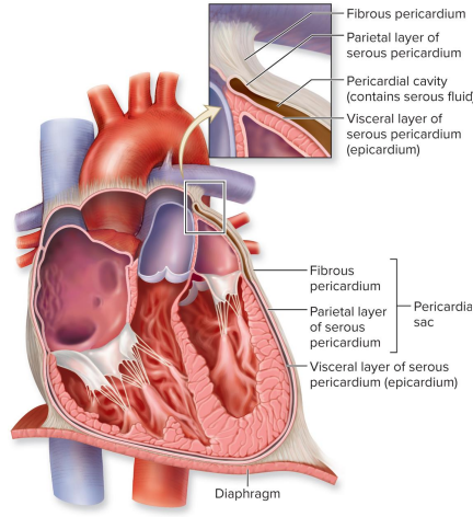

Fibrous pericardium
Dense irregular connective tissue
Attaches to diaphragm and base of aorta, pulmonary trunk
Anchors heart and prevents its overfilling, similar to brain meninges


Parietal layer of serous pericardium
Simple squamous epithelium and areolar connective tissue
Attaches to fibrous pericardium

Visceral layer of serous pericardium
AKA the epicardium
Simple squamous epithelium and areolar connective tissue
Attaches directly to heart


Pericardial cavity
The two serous layers continuous and separated by pericardial cavity, containing serous fluid (acts as lubricant)

Pericarditis
Inflammation of the pericardium
Caused by viruses, bacteria, or fungi
Increased capillary permeability
Fluid accumulation in pericardial cavity
Becomes real space as it fills with fluid, increasing pressure on the heart and pericardial sac
May restrict heart’s movement and keep chambers from filling
Results in cardiac tamponade, where the heart is unable to pump
Friction rub can be heard with stethoscope; a crackling sound as pericardial layers rub

Layers of the heart wall
Heart wall varies in thickness
Ventricles (pumping chambers) have thicker walls than atria
Left ventricle has thicker wall than right ventricle because left must generate high pressure to force blood through systemic circulation; right just pumps to nearby lungs


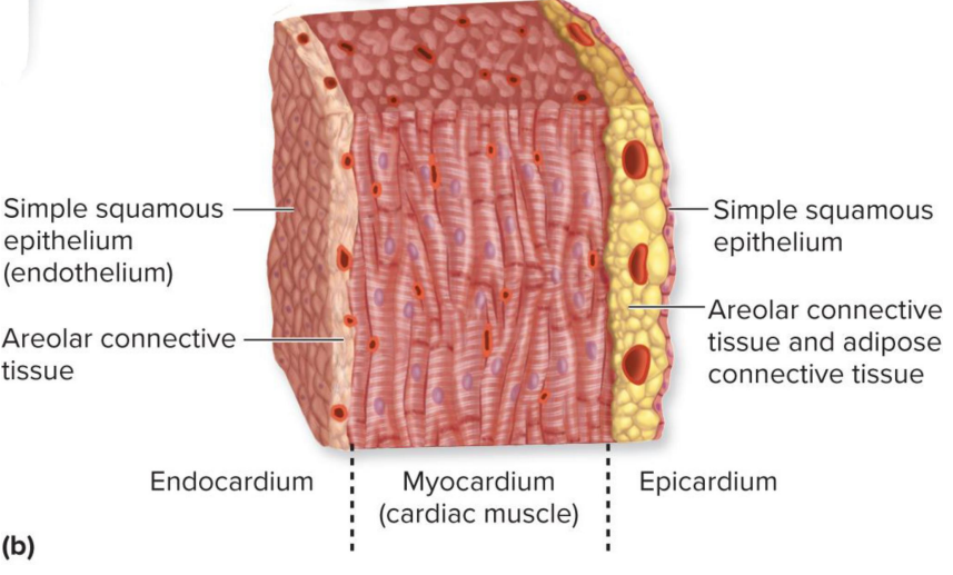
Heart wall
Inner to outer
Endocardium: inner lining of heart chambers; simple squamous epithelium (endothelium), areolar connective tissue
Myocardium: cardiac muscle
Epicardium: areolar connective tissue and adipose connective tissue, simple squamous epithelium

Fibrous skeleton of the heart
Fibrous skeleton of dense irregular connective tissue
Functions:
Structural support at boundary of atria and ventricles
Anchors the valves
Provides framework for attachment of cardiac muscle
Acts as electrical insulator preventing ventricles from contracting at same time as atria
Muscle cells are attached to fibrous skeleton and arranged in spiral bundle


Arrangement of cardiac muscle around fibrous skeleton
Atrial contraction moves walls inward to narrow the heart; bundle looks like a pretzel
Ventricular contraction resembles wringing a mop to shorten the heart; begins at apex and compresses superiorly


Coronary circulation
Heart has its own coronary arteries and cardiac veins to provide itself oxygen and nutrients
Coronary arteries: left coronary artery and branches (circumflex, anterior interventricular); right coronary artery and branches (posterior interventricular, right marginal)
Cardiac veins: coronary sinus; great, middle, small cardiac veins

Coronary heart disease
Buildup of plaque in coronary arteries (called atherosclerosis)
Reduces blood flow to heart muscle
Coronary spasm: sudden narrowing of vessels
Both of the above can lead to angina or myocardial infarction
Angina pectoris
Heart pain during strenuous activity
Usually felt on left side of chest, left arm, or jaw
Treatments induce vascular dilation
Myocardial infarction
Heart attack
Sudden and complete occlusion (blockage) of coronary artery
Myocardium deprived of oxygen, possible tissue death
Excruciating chest pain radiating down left arm
Weakness, shortness of breath, nausea, anxiety, and sweating

Structure of cardiac muscle cells (cardiomyocyte)
Covered in endomysium, inside which lie the sarcolemma, intercalated discs, T-tubule openings, nucleus, and mitochondria
Intercellular junctions between cardiomyocytes contain intercalated discs on folded sarcolemma
High number of mitochondria because of high energy consumption
Sarcoplasmic reticulum stores calcium ions required for contraction
Contains myofibrils, which are composed of myofilaments (protein filaments)

Sarcolemma
Plasma membrane of a muscle cell
Different elements of the sarcolemma have to contract at the same time
Intercalated disc
Connect cardiomyocytes mechanically and electrically
Includes gap junctions: ion channels that allow ions to move freely between the cells; crucial for heart synchronization across millions of cardiomyocytes
When intercalated discs slide, they become shorter → cardiac muscle contracts
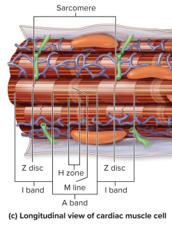
Sarcomere
Functional units of skeletal muscle and cardiac muscle
Have an H zone, M line, Z discs, I band, and A band

Innervation of the heart
Includes parasympathetic and sympathetic innervation
Together, cardioinhibitory and cardioacceleratory centers make up the cardiac center in the medulla oblongata
Reflexive modification of cardiac activity

Parasympathetic innervation
Cardioinhibitory (CI) center sends nerve signals along the vagus nerves (CN X) → decrease in heart rate, lengthens delay of signals at AV node
Nerve signals such as Ach release from the CI center → PS motor neurons (both preganglionic and ganglionic) → right and left vagus nerves → SA and AV nodes, respectively


Sympathetic innervation
Cardioacceleratory (CA) center sends nerve signals along cardiac nerves → increase in heart rate, force of contraction, stroke volume, and vasoconstriction
Nerve signals such as NE release from the CA center → preganglionic sympathetic motor neurons → spinal cord → ganglionic sympathetic neurons within the cardiac nerve → reach the SA node, AV node, coronary arteries, and myocardium

Baroreceptor and chemoreceptors (cardiac center)
Located in the aorta, arteries, and right atrium; receive sensory info and transmit it through the glossopharyngeal nerve (CN IX) and left vagus nerve to the cardiac center
Baroreflex: high blood pressure = ↑ PS stimulation, ↓ S stimulation
Chemoreflex: low O2, high CO2, or low PH = ↓ PS stimulation, ↑ S stimulation

Heart conduction system
Sinoatrial (SA) node: pacemaker in right atrium; synchronizes cell
Atrioventricular (AV) node: also in right atrium; activated by gap junctions
Atrioventricular (AV) bundle in the interventricular septum extends from AV node and splits into right and left bundle branches
Purkinje fibers branch from the bundle branches


Steps of heart conduction system
SA node generates an action potential → spreads via gap junctions between cardiac muscle cells throughout the atrial myocardium to the AV node
AP is delayed at the AV node before it passes to the AV bundle within the IV septum
AV bundle conducts the AP to the left and right BB and then to the Purkinje fibers
From the Purkinje fibers, the AP is spread via gap junctions between cardiac muscle cells throughout the ventricular myocardium


Heart contraction: cardiac muscle cells
Action potential is propagated along the sarcolemma of cardiac muscle cells.
Muscle contraction: Thin filaments slide past thick filaments and sarcomeres shorten within cardiac muscle cells.

SA node cellular activity
Relies on voltage-gated cation, Ca2+, and K+ channels on the plasma membrane
SA nodal cells’ resting membrane potential is -60 mV
Reaching threshold: voltage-gated cation channels open → Na+ enters → threshold reached (-60 mV → -40 mV) → cation channels close
Depolarization: voltage-gated Ca2+ channels open → Ca2+ enters → depolarization occurs (-40 mV → just above 0 mV) → Ca2+ channels close
Repolarization: voltage-gated K+ channels open → K+ exits → repolarization occurs (+ mV → -60 mV) → K+ channels close
Ectopic pacemaker
Pacemaker other than SA node
Conduction system cells other than SA node also have ability to spontaneously depolarize, but depolarize at slower rates than SA node
AV node is the default pacemaker if SA node impaired: inherent rhythm of 40 to 50 beats/min, fast enough to sustain life
Cardiac muscle with rate of 20 to 40 beats/min: usually too slow to sustain life
What happens in cardiac muscle cells following conduction system stimulation?
Propagation of an action potential at the surface of each of the cardiac muscle cells, specifically at the sarcolemma (plasma membrane)
Contraction of sarcomeres within each of the cardiac muscle cells.
(Remember that both of the above events occur twice per heartbeat: first in the atria, then in the ventricles)
Cardiac muscle cell electrical events
Relies on fast voltage-gated Na+ channels, voltage-gated K+ channels, and slow voltage-gated Ca2+ channels on the plasma membrane
Cardiac muscle cell’s resting membrane potential is -90 mV
Depolarization: fast voltage-gated Na+ channels open → Na+ rapidly enter → depolarization occurs (-90 mV → 30 mV) → Na+ channels close
Plateau: voltage-gated K+ channels open → K+ flows out and slow voltage-gated Ca2+ open → Ca2+ enters → no electrical change; depolarized state is maintained
Repolarization: voltage-gated K+ channels remain open → K+ exits → voltage-gated Ca2+ channels close → repolarization occurs (30 mV → -90 mV)

Skeletal muscle single stimulation
Muscle contraction and relaxation take 100 ms
Repolarization occurs immediately following depolarization → relatively short (1-2 ms) refractory period
Therefore, while sarcomeres are still contracting in skeletal muscle fibers, repolarization has already occurred to allow for new stimulation


Skeletal muscle frequent stimulation
Since it has a relatively short refractory period, it can be restimulated at a frequency that doesn’t allow skeletal muscle enough time to relax completely → condition of sustained muscle contraction is called tetany


Cardiac muscle single stimulation
Muscle contraction and relaxation takes 250 ms
Repolarization is not immediately after depolarization → relatively long (250 ms) refractory period (due to the plateau event delaying repolarization)
Therefore, while sarcomeres are still contracting and relaxing, repolarization has not occurred to allow for new stimulation

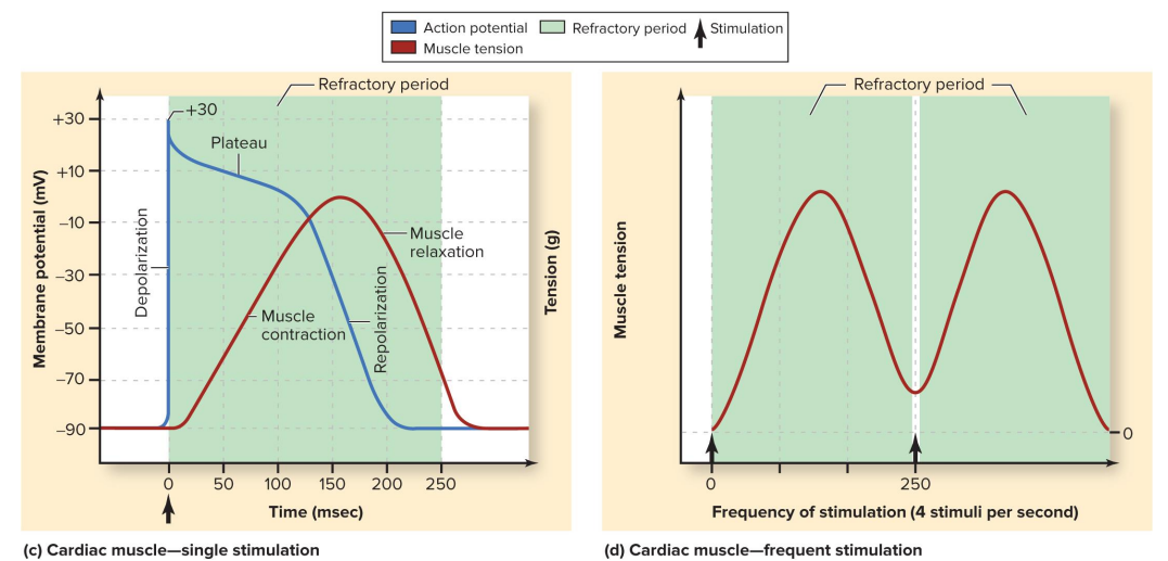
Cardiac muscle frequent stimulation
Longer refractory period → extended period of time where it cannot be restimulation → delay in restimulation allows sarcomeres to fully contract and relax before being stimulated again
Therefore, cardiac muscle doesn’t exhibit tetany, but continues to both contract and relax following each stim—essential for pumping blood


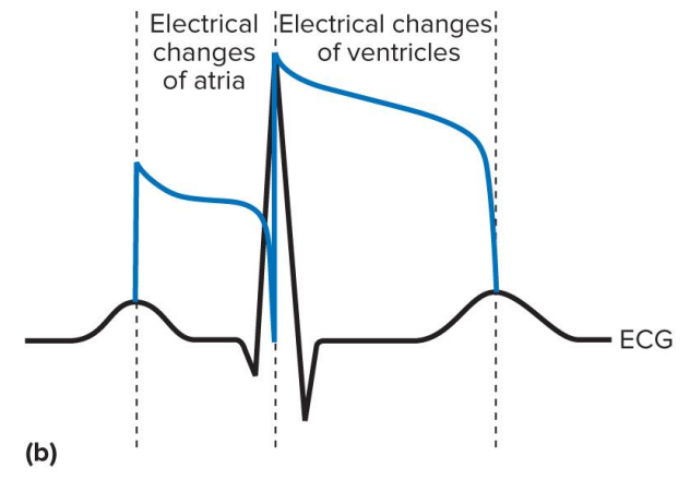
Electrical changes of atria on EKG
Peak of P-wave to peak of R-wave
P-wave: atrial depolarization
PQ segment: atrial pleateau
a. Atrial repolarization is not visible on ECG, as it is concurrent with ventricular depolarization
PR interval: represents conduction from the SA node to the AV node
Depolarization = contraction, systole, repolarization = relaxation, diastole

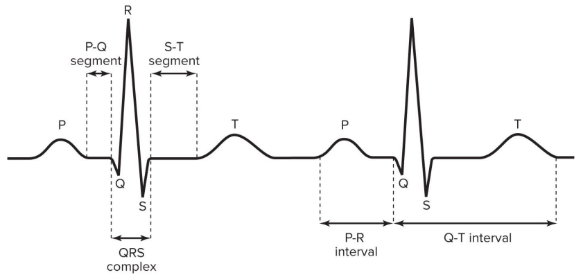
Electrical changes of ventricles on EKG
Peak of R-wave to peak of T-wave
b. R-wave/QRS complex: ventricular depolarization
ST segment: ventricular plateau
T-wave: ventricular repolarization
QT interval: represents ventricular depolarization and repolarization
Depolarization = contraction, systole, repolarization = relaxation, diastole
Cardiac arrhythmia
Any abnormality in heart’s electrical activity
Heart blocks: impaired conduction; may result in light-headedness, fainting, irregular heartbeat, chest palpitations
Premature ventricular contraction
Atrial fibrillation
Ventricular fibrillation
First-degree AV block
PR prolongation
Slow conduction between atria and ventricles
Second-degree AV block
Failure of some atrial action potentials to reach ventricles
Third-degree AV block
Complete block
Failure of all action potentials to reach ventricles
Premature ventricular contractions (PVC)
Abnormal action potential within AV node or ventricles
Result from stress, stimulants, or sleep deprivation
Not detrimental unless they occur in large numbers
Atrial fibrillation (a-fib)
Chaotic timing of atrial action potentials
Ventricular fibrillation (v-fib)
Chaotic electrical activity in ventricles
Uncoordinated contraction and pump failure
Leads to death of heart cells
Treated with paddle electrode defibrillator or automated external defibrillator (AED)
Cardiac cycle
Atrial relaxation and ventricular filling
Both atria and ventricles are in diastole, with AV valves open and semilunar valves closed
Blood goes through vena cavae to RA; simultaneously, blood from the lungs travels through the pulmonary veins to LA
Blood from both atria flows through open AV valves into ventricles
Atrial contraction and ventricular filling
Atrial systole forces any remaining blood into ventricles (still in diastole), allowing them to expand and completely fill with blood
Isovolumic contraction
Ventricular systole closes AV valves to prevent backflow while atria relax and SL valves stay closed
Ventricular ejection
Pulmonary SL opens, and RV expels blood through pulmonary artery to lungs; meanwhile, aortic SL opens, and LV expels blood through aorta to rest of body
Isovolumic relaxation
Cardiac cycle repeats as both atria and ventricles enter diastole and close valves to allow the heart to fill with blood
Sympathetic innervation of SA nodal cells
Adrenal medulla releases EPI and NE / synaptic vesicles of a sympathetic neuron axon releases NE into synaptic cleft
Beta1-adrenergic receptors on SA nodal cells absorb NE, activating G-protein-mediated signal transduction pathway
G-protein binds to adenylate cyclase to form cAMP → activates protein kinase enzyme → phosphorylates calcium ion channels, opening them
Ca2+ enters the cell through open channels → increases pacemaking speed and heart rate
__ increases release of NE
Nicotine
__ increases numbers of beta1-adrenergic receptors on SA nodal cells
Thyroid hormone
__ inhibits NE reuptake
Cocaine
__ inhibits breakdown of cAMP
Caffeine
Cardiac output
Blood pumped per minute
Chronotropic agents (factors affecting cardiac output)
Alter SA node and AV node activity
Positive agents (ex: NE) increase HR
Negative agents decrease HR
Stroke volume
Blood pumped per beat
Venous return (factors affecting cardiac output)
Volume of blood returning to heart alters stretch of heart wall; aka preload
Directly correlated with stroke volume (venous return ↑, stroke volume ↑)
Inotropic agents (factors affecting cardiac output)
Alter Ca2+ levels in sarcoplasm
Positive agents increase stroke volume
Negative agents decrease stroke volume
Afterload (factors affecting cardiac output)
Increased resistance in arteries (from HTN, stenosis, etc)
Inversely correlated with stroke volume (afterload ↑, stroke volume ↓)
Bradycardia
Persistently low resting heart rate in adults
Below 60 beats/minute
Normal change in athletes
Abnormal due to: hypothyroidism, electrolyte imbalance, and congestive heart failure
Tachycardia
Persistently high resting heart rate
Over 100 beats/minute
Caused by heart disease, fever, and anxiety
Label the arteries
https: //www.purposegames.com/game/arteries-of-the-full-body
Label the veins
https: //www.purposegames.com/game/veins-of-the-full-body
Label the arteries of the head and neck
https: //www.purposegames.com/game/arteries-of-the-head-game
Label the cranial arteries
https: //www.purposegames.com/game/circulation-cranial-arteries-quiz
Label the veins of the head and neck
https: //www.purposegames.com/game/veins-head-game
Label the dural venous sinuses
https: //www.purposegames.com/game/dba35d3d68

Circle of Willis/cerebral arterial circle
“Backup system” with circular blood flow; in the event of an occlusion on either side of the brain, it will supply blood to an adjacent artery
Due to the innervation of small capillaries, it is still able to supply that part of the brain with blood or oxygen that it would otherwise have been deprived of

Dural venous sinuses
Cranial cavities
Some blood drains to vertebral veins, but most blood drains through these large modified veins between two layers of dura mater
Also receive excess cerebrospinal fluid
Drain primarily into internal jugular veins

Vessel structure
Space inside of vessel called lumen
Walls composed of three layers called tunics:
Tunica externa: outermost layer layer composed of connective tissue with elastic and collagen fibers; helps anchor vessel to other structures
Tunica media: smooth muscle cells with elastic fibers; contraction causes vasoconstriction (narrows lumen), relaxation causes vasodilation (widens lumen) — much thicker in arteries
Tunica intima: innermost layer composed of endothelium

Capillary structure and characteristics
Small vessels connecting arterioles to venules
Contain only tunica intima (no subendothelial layer)
Composed of endothelium on a basement membrane
Thin wall and small diameter are optimal for gas and nutrient exchange between blood and tissue fluid
Average length = 1 mm; diameter = 8 to 10 micrometers
Erythrocytes travel single file (rouleau)
Types: continuous, fenestrated, sinusoid
Arteriole
The smallest type of artery
Venule
The smallest type of vein
Intercellular clefts
Gaps between endothelial cells of capillary wall

Continuous capillaries
Endothelial cells form a continuous lining; tight junctions connect cells but don’t form a complete seal
Large particles (e.g., cells, proteins) cannot pass, but smaller molecules (e.g., glucose) can pass through wall
Commonly found in muscle, skin, lungs, central nervous system


Fenestrated capillaries
Endothelial cells form a continuous lining, but the cells have fenestrations (pores) for smaller plasma proteins
Found in areas where much fluid transport happens (e.g., intestine capillaries absorbing nutrients, kidney capillaries filtering blood to form urine)


Sinusoid capillaries
Endothelial cells form an incomplete lining with large gaps; the basement membrane is incomplete or absent
Openings allow transport of large substances (formed elements, large proteins)
Found in bone marrow, spleen, and some endocrine glands

Atherosclerosis
Plaque buildup in arteries due to increased cholesterol in the blood (hypercholesterolemia)
Makes prone to disease
Males more affected than females
Smoking and hypertension increase vascular injury & overall risk
Treatment: balloon angioplasty, mesh stent insertion, coronary bypass surgery
Aneurysm
Part of arterial wall thins and balloons out
Wall more prone to rupture → can cause massive bleeding and death
With age, elastic and muscular arteries are less able to withstand forces from pulsating blood
Risk increasing with age
Most common in aorta or arteries at the base of the brain
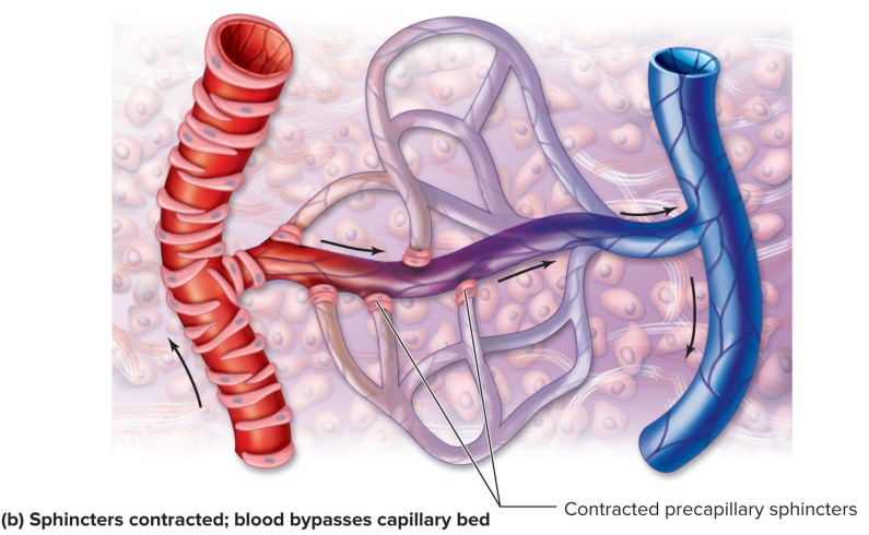
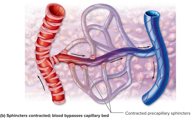
Capillary bed
Precapillary sphincters: collections of smooth muscle cells
Precapillary sphincters relaxed → blood from the arteriole flows through all capillaries in the bed and drains into postcapillary venule
Precapillary sphincters contracted (pictured) → most blood bypasses the bed on its way to the venule, reducing gas exchange
Systemic veins acting as blood reservoirs
Blood can be moved from veins into circulation via vasoconstriction of veins (e.g., when more blood is needed during exertion)
Blood can be shifted back into reservoirs via vasodilation (e.g., when less blood is needed during rest)
Percentages of total blood volumes at certain locations
Pulmonary circulation: ~18%
In the heart: 12%
In systemic circulation: ~70%
Systemic veins: 55%
Systemic capillaries: 5%
Systemic arteries: 10%
Bulk flow at capillaries
Exchange processes include diffusion (move down concentration gradient), vesicular transport (active transport up concentration gradient), and bulk flow
Net filtration pressure (NFP) = net hydrostatic pressure (HP) - net colloid osmotic pressure (COP)
Arterial end: filtration; HP > COP → net pressure out, fluid forced out of vessel
Venous end: reabsorption; COP > HP → net pressure in, fluid drawn into vessel
Blood pressure
Force of blood against vessel wall; highest in arteries and lowest in veins
Arterial blood pressure
Blood flow in arteries pulses with cardiac cycle
Systolic pressure occurs when ventricle contracts (systole)
Highest pressure generated in arteries (they are stretched)
Upper number
Diastolic pressure occurs when ventricles relax (diastole)
Lowest pressure generated in arteries (they recoil)
Lower number
Pulse pressure: pressure in arteries added by heart contraction
Systolic minus diastolic blood pressure
It reflects the elasticity and recoil of arteries and tend to decline with age and disease
Allows for palpation of a throbbing pulse in elastic and muscular arteries
Pulse
Throbbing of arterial wall
Allows determination of heartbeat
More forceful pulse associated with higher pressure
Absence indicates flow to body part is lacking
Pulse points
Where artery may be compressed against solid structure (e.g., radial, common carotid, femoral, dorsalis pedis, and others)