CLSC 131 Final Studyguide
 Call Kai
 Learn
 Practice Test
 Spaced Repetition
 Match
 Flashcards
 Knowt Play
1/135

There's no tags or description
Looks like no tags are added yet.
Name | Mastery | Learn | Test | Matching | Spaced | Call with Kai | Chat |
|---|
No analytics yet
Send a link to your students to track their progress
136 Terms
Cell that carries gasses
Red Blood Cells (Hemoglobin)
Cell responsible for fighting immunity
Lymphocytes
Cell responsible in hemostasis
Platelets
Cell involved in buffering blood and what component of that cell
Red Blood Cells (Hemoglobin)
Site(s) for extramedullary Hematopoiesis
Spleen, liver, and Lymph nodes
First stage where hgb is recognized
Polychromatophilic Normoblast (Rubricyte)
Last stage capable for mitosis
Rubricyte (Polychromatophilic Normoblast)
Polychromatophilic Normoblast is another name for what cell
Rubricyte
Name the RBCs that are normally seen in peripheral blood
Polychromatophilic Erythrocytes (Reticulocytes) and Erythrocyte (Mature Red Blood Cell)
Give examples of megaloblastic Erythropoiesis
Vitamin B12 Deficiency
Pernicious Anemia
Folic Acid Deficiency
What cell size is seen in megaloblastic Erythropoiesis
Macrocytes
What cell size do we see in IDA
Microcytes
Give two examples of Microcytic anemia and state which is seen more frequently
Iron Deficiency Anemia (IDA)- more common
Thalassemia
Name the stain used for peripheral smears
Wrights Stain
Name two stained used for Reticulocytes
New Methylene Blue
Brilliant Cresyl Blue
Normal values for reticulocytes (ages)
Adults: 0.5-2.5%
Neonates: 2.0-6.0%
What are we actually seeing stained in a retic
Residual Ribosomal RNA (Cytoplasmin RNA)
Mitochondria
Ribosomes
What is the purpose for doing a retic count
To estimate the rate at which new RBCs are entering the peripheral blood from the bone marrow
An increased count shows that there is increased bone marrow production of RBCs or rapid RBC regeneration
Define Erythropoiesis
Erythro: red
-poiesis: Formation
Erythropoiesis: Formation of Red Blood Cells
Define anisocytosis
Variation in the size of the erythrocytes
Define poikilocytosis
Variation in the shape of erythrocytes
Define asynchronism
A defect which causes the cytoplasm and the nucleus to develop at different rates
Echinocyte/Burr Cell

Acanthocyte

Codocyte (Target Cell)

Drepanocyte (Sickle Cell)

Elliptocyte

Ovalocyte

Schistocyte

Dacrocyte (Teardrop)

Stomatocyte (Mouth or cup Cells)

Keratocyte (Blister)

Spherocytes

Helmet (Bite Cell)

Microcytes

Macrocytes

Basophilic Strippling
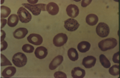
Howell-Jolly Bodies

Cabot Rings

Siderocytic Granules
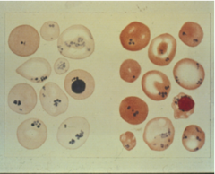
Pappenheimer Bodies

Heinz Bodies

Malaria

Hemoglobin Crystal CC

Hemoglobin Crystal SC

A decrease in the normal Globin chain synthesis of the heme part of the hgb molecule is called
Hemoglobinopathies
Calculation for a retic count
Number of Reticulocytes Counted
——————————————————————- x100
1000 (Total RBC's Counted)
(Round to the 1st Decimal place)
Recall what inclusions are seen in Wrights Stain and what inclusions need a supravital stain
a. Basophilic Strippling: Wrights Stain
b. Howell-Jolly Bodies: Wrights Stain
c. Cabot Rings: Wrights Stain
d. Siderocytic Granules: Prussian Blue Stain
e. Pappenheimer Bodies: Wrights Stain
f. Heinz Bodies: New Methylene Blue, Crystal Violet
g. Malaria: Wrights Stain
h. Hemoglobin Crystal CC: Wrights Stain
i. Hemoglobin Crystal SC: Wrights Stain
What makes up Basophilic Strippling
Made up of ribosomes, protoporphyrin, and mitochondrial remnants (RNA)
What Makes up Howell-Jolly Bodies
Inclusion is predominantly composed of DNA
What Makes up Cabot Rings
Inclusion is thought to be past of the Mitotic spindle, part of the microtubule, or fragment from the nuclear membrane (Nuclear Remnant)
What makes up Siderocytic Granules (Pappenheimer Bodies)
RBCs contain small iron particles (ferritin)
What Makes up Heinz Bodies
Denatured (precipitated) Hemoglobin
What Makes up Malaria
Parasite that lives in RBCs
What Conditions lead to Basophilic Stippling
i. Lead poisoning
ii. Megaloblastic Anemia
iii. Other Severe Anemias
iv. Normal in neonate RBCs
What conditions lead to Howell-Jolly Bodies:
i. Splenectomy
ii. Hemolytic Anemia
iii. Megaloblastic Anemia
iv. Thalassemia
v. Sickle Cell Anemia
What condition leads to Siderocytic Granules (Pappenheimer Bodies)
i. Iron- Loading Anemias
ii. Hemolytic Anemias
iii. Hyposplenism
What conditions lead to Heinz Bodies
i. Congenital Hemolytic Anemia
ii. Hemolytic Anemia secondary to oxidizing drugs
iii. Glucose-6-Phosphate Dehydrogenase Deficiency (G6PD)
iv. Splenectomy
What Conditions lead to Hemoglobin Crystal CC
i. Hemoglobin CC
ii. Patient is homozygous for Hemoglobin C instead of Hemoglobin A
iii. Crystals increase after Splenectomy
iv. Resembles a "Bar of Gold"
What conditions lead to Hemoglobin Crystal SC
i. Patient inherits abnormal Hemoglobin S and abnormal Hemoglobin C
Define Polychromatophilic RBCs and list their characteristics
When RBCs stains pale, blue-gray color on the wrights stain
These cells are usually reticulocytes
It indicates an increased number of reticulocytes
Polychromasia: Wrights Stain
Reticulocytes: New Methylene Blue or Brilliant Cresyl Blue
State the hormone involved in RBC production
Erythropoietin- a glycoprotein
- Produced primarily by the kidney
What circumstances increase Erythropoietin production
i. Hypoxia- Lack of oxygen
ii. Pituitary Hormones
iii. Adrenal Gland Hormones
iv. Testosterone (Androgenic Hormones)
v. Thyroid Hormones
Note the stages in granulocytes for non-specific (primary granules) and specific (secondary granules)
Primary Granules: Promyelocyte
Secondary Granules: Myeocyte
Stage of granulocytes where we can differentiate between neutrophil, eosinophil, or basophil
Metamyelocyte
WBC stages that can undergo mitosis
Myeloblast
Promyelocyte
Myelocyte
Tissue macrophage is associated with what cell
Monocytes
WBCs which are in the maturing/storage pool
Metamyelocytes (45%)
Banded Neutrophils (35%)
Segmented Neutrophils (20%)
WBC granulocytes NORMAL for the peripheral blood
Banded Neutrophils
Segmented Neutrophils
Basophils
Eosinophils
First Line of Defense
Skin-Epithelial Barriers
Second Line of Defense
Phagocytosis
Third Line of Defense
B-Lymphocytes
Which cell if the last to arrive at the infection site
Monocytes
Cell associated with Mast Cell
Basophils
CD4
T helper/effector cells- Release cytokines (lymphokines) to activate other immune cells (B-cells)
Decreased in AIDS
CD8
T- suppressor/cytotoxic cells- Suppress other immune cells
Organ Transplant rejection
Cell responsible for Cellular Immunity
T-Lymphocytes
Cell responsible for humoral immunity
B lymphocyte
May- Hegglin Anomaly
• 50% of these patients have bleeding disorders
• Inherited-Platelet disorders

Pelger Huet
• Inherited
o Pince-new Form
o Functionally Normal

Psudo Pelger-Huet
• Round Nucleus
• Clumped Chromatin
• Hypogranulation
• Granulocytic Leukemia

Chédiak-Higashi Syndrome
• Very rare
• Children die very young
• Albinism
• Silver hair
• Bleeding Problems

Function of Natural Killer Cells
Immunity against viral and tumor cells that do not require any tiger recognition
Function of Platelets
Important for blood coagulation:
i. Maintains blood vessel integrity- keep strong/healthy
ii. Acts as plugs
iii. Initiate the formation of fibrin
Promotes vascular healing
Receptors on platelets for fibrinogen and VonWillebrand Factor
Coagulation Factors
Role of PF3
Platelet Factor 3 (procoagulant)
- Participates in the activation on the clotting Factors to form Fibrin
Characteristics of the maturation of a platelet
a) Increase in Nuclear Lobes (Endomitosis-Genetic Material Replicating)
b) Increase in Cell size
c) Increased Cytoplasmic Grannulation
Components of alpha and dense granules
Alpha: Coagulation Factors, Platelet Factor 4, Platelet Derived Growth Factor (PDGF)
Dense: Serotonin, ADP
Glanxmann's Disease
Inherited- diseased platelet aggregation to dense granule components such as ADP; Serotonin
Defect in the Glycoprotein IIb/IIIa complex which binds fibrinogen in the presence of Calcium and initiates platelet Aggregation
VonWillebrand's Disease
Platelets fail to adhere to collagen, decrease in Factor VIII
VIII has 2 parts, vWf is just on of the two
ITP
Idiopathic Thrombocytopenia Purpura- develops an antibody against own platelets
Bernard Soulier Disease
Rare inherited disorder, Platelets fail to adhere to collagen
- Defect in the Glycoprotein Ib complex
Giant Platelet Syndrome (60%-80% of the platelets have increased Diameter (2.5-2.0 um in diameter)), Only 2-4% function normally
DIC and the results of DIC
Disseminated Intravascular Coagulation
Massive Coagulation uses up Platelets in forming Clots
Thrombocytopenia
a decease in the number of platelets
Thrombocytosis
an increase in the number of platelets >450x10^9/L
Asprin's effects on platelets
Aspirin suppresses the enzyme Cyclooxygenase which prevents the release of granules and thereby decreases platelet aggregation
Enzyme activity returns seven days or when new plates are produced
The belted ing time in normal persons are increased 1-2 minutes after a single dose of aspirin
Tests to measure platelet function
Platelet counts
Bleeding time
Clot retraction test
Semiquatitative test for hgb
Specific Gravity
Major Basic protein is affiliated with what
The granular content within Eosinophils
Mesoblastic Period
first 2-8 weeks of fetal lifedevelops in then yolk sac. Mesenchymal stem cellsfrom primitive nucleated red cells