Radiology Ch.28
 Call Kai
 Learn
Learn Practice Test
 Spaced Repetition
 Match
Match Flashcards
 Knowt Play
1/87

There's no tags or description
Looks like no tags are added yet.
Name | Mastery | Learn | Test | Matching | Spaced | Call with Kai | Chat |
|---|
No analytics yet
Send a link to your students to track their progress
88 Terms
radiolucent
The portion of the processed radiograph that is dark or black
Lacks density, permits passage of x-ray
radiopaque
The portion of the processed radiograph that appears white
Appears dense, absorbs/resists passage of x-ray
radiolucent
Caries are:
radiopaque
Enamel, dentin, and bone are:
enamel
the outermost radiopaque layer of the crown of a tooth
dentin
comprises most of the tooth structure, not as radiopaque as enamel
dentinoenamel junction
the junction between dentin and enamel
gingival tissue
the grayish invisible-looking area behind the last molar teeth
lamina dura, alveolar crest, and periodontal ligament space
Anatomic landmarks of the alveolar process include the
lamina dura
A dense radiopaque line that surrounds the root of a tooth
The wall of the tooth socket, made of dense cortical bone
alveolar crest
The most coronal portion of the alveolar bone found between teeth
Appears radiopaque, typically 1.5 to 2.0 mm. below the cementoenamel junction
periodontal ligament space
The space between the root of the tooth and the lamina dura
A thin radiolucent line around the root of a tooth
anterior regions
Normal alveolar crest appears pointed and sharp between the teeth
alveolar crest appears as a dense radiopaque line
posterior regions
Normal alveolar crest appears flat and smooth between the teeth
alveolar crest appears less dense and less radiopaque
cancellous bone
The soft, spongy bone located between two layers of dense cortical bone
Appears primarily radiolucent
cortical bone
The dense outer layer of bone
Appears radiopaque on a radiograph
process
marked prominence or projection
ridge
linear prominence or projection
spine
sharp, thornlike projection
tubercle
small bump or nodule
canal
tubelike passageway through bone that contains nerves and blood vessels
foramen
opening or hole that permits the passage of nerves and blood vessels
fossa
broad, shallow, scooped-out, or depressed area
sinus
hollow space, cavity, or recess
septum
A bony wall or partition that divides two spaces or cavities
Radiopaque
suture
An immovable joint representing a line of union between adjoining bones of the skull
Thin radiolucent line
maxilla
composed of two paired bones that meet at the midline of the face
Forms the: floor of the orbit of the eye, sides and floor of the nasal cavities, hard palate
incisive foramen
A hole in the bone located at the midline of the anterior hard palate directly posterior to the maxillary central incisors
Radiolucent
superior foramina of the incisive canal
Two small round radiolucencies located superior to the apex of the maxillary central incisors
median palatal suture
The immovable joint between the two palatine processes of the maxilla
Radiolucent
lateral fossa
A smooth, depressed area of the maxilla located between the canine and lateral incisor
Radiolucent
nasal cavity
A pear-shaped compartment of bone located superior to the maxilla
Large radiolucent area
nasal septum
A vertical bony wall or partition that divides the nasal cavity into the right and left nasal fossae, formed by Vomer and part of Ethmoid bone
radiopaque
floor of nasal cavity
A bony wall formed by the palatal processes of the maxilla and the horizontal portions of the palatine bones
Radiopaque
anterior nasal spine
A V-shaped radiopaque area located at the intersection of the floor of the nasal cavity and the nasal septum
inferior nasal conchae
Wafer-thin, curved plates of bone that extend from the lateral walls of the nasal cavity
radiopaque mass
maxillary sinus
Paired cavities or compartments of bone located within the maxilla
Located above the maxillary premolar and molar teeth
septa within maxillary sinus
Bony walls or partitions that appear to divide the maxillary sinus into compartments
Radiopaque lines
nutrient canals
Tiny, tubelike passageways through bone that contain blood vessels and nerves
Radiolucent band
inverted Y
The intersection of the maxillary sinus and the nasal cavity, located above the maxillary canine
Radiopaque
maxillary tuberosity
A rounded prominence of bone that extends posterior to the third molar region
radiopaque
hamulus
A small hooklike projection of bone extending from the medial pterygoid plate of the sphenoid bone
zygomatic process
A bony projection of the maxilla that articulates with the zygoma
J- or U-shaped radiopacity
zygoma
Articulates with the zygomatic process of the maxilla
Radiopaque
mandible
The largest and strongest bone of the face
Divided into three main parts
ramus, body, alveolar process
What are the three main parts of the mandible
ramus
Vertical portion of mandible found posterior to the third molar
body
Horizontal U-shaped portion of mandible stretching from ramus to ramus
genial tubercles
A ring-shaped radiopacity below the apex of the mandibular incisors lingual surface
lingual foramen
A small radiolucent dot inferior to the apex of the mandibular incisors lingual surface
mental ridge
A linear prominence of cortical bone of the mandible
Thick radiopaque band that extends from the premolar region to the incisor region
mental fossa
A scooped-out, depressed area of bone located on the external surface of the anterior mandible
radiolucent area
mental foramen
An opening or hole in bone located on the external surface of the mandible in the region of the mandibular premolars
Radiolucent
mylohyoid ridge
A linear prominence of bone located on the internal surface of the mandible
Dense radiopaque band
mandibular canal
A tubelike passageway through bone that travels the length of the mandible
A radiolucent band outlined by two thin radiopaque lines
internal oblique ridge
A linear prominence of bone located on the internal surface of the ramus
Radiopaque
external oblique ridge
A linear prominence of bone located on the external surface of the body of the mandible
Radiopaque
Superior to the internal
submandibular fossa
A scooped-out, depressed area of bone located on the internal surface of the mandible inferior to the mylohyoid ridge
Radiolucent
coronoid process
A marked prominence of bone on the anterior ramus of the mandible
Radiopaque
coronoid process

submandibular fossa

external oblique ridge
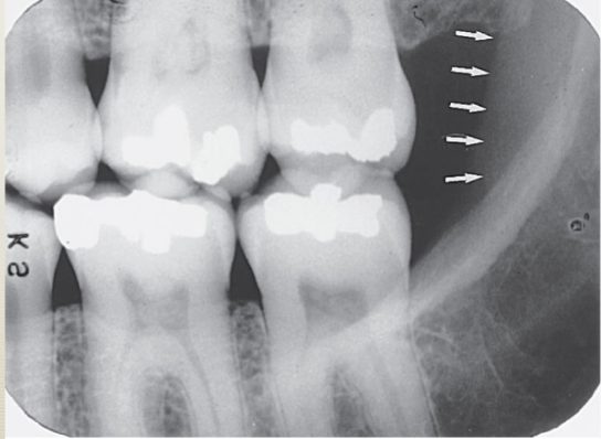
internal oblique ridge
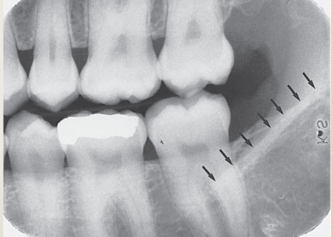
mandibular canal

mylohyoid ridge
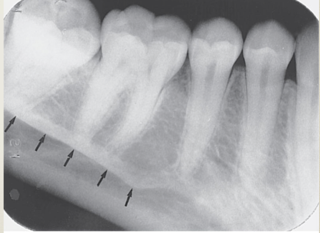
mental foramen

mental fossa

mental ridge
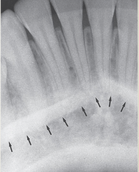
nutrient canals

lingual foramen

genial tubercles

zygoma

zygomatic process

hamulus

maxillary tuberosity

inverted Y
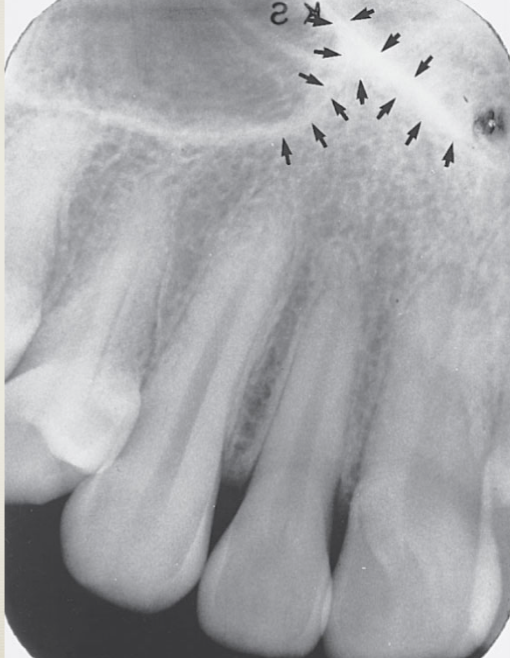
nutrient canals

septa

maxillary sinus

inferior nasal conchae

anterior nasal spine
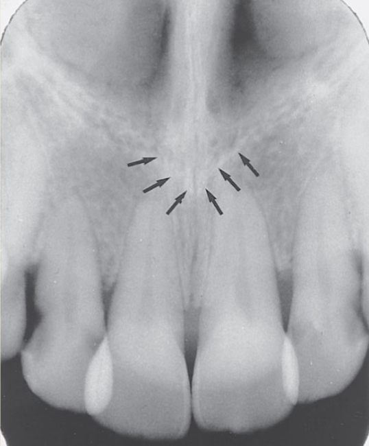
floor of nasal cavity

nasal septum

nasal cavity
The large radiolucent area

lateral fossa
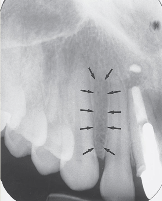
median palatal suture

superior foramina of incisve canal

incisive foramen
