Mast Cell Tumor and Transitional Cell Carcinoma
 Call Kai
 Learn
 Practice Test
 Spaced Repetition
 Match
 Flashcards
 Knowt Play
1/33

There's no tags or description
Looks like no tags are added yet.
34 Terms
What is the most commonly diagnosed skin tumour in the dog?
Mast cell tumour
At what sites can mast cell tumours be found?
Skin, subcutaneous tissue, conjunctiva, oral mucosa, GIT
(Very variable behaviour)
What breeds are predisp to MCT?
Boxer, Boston terrier (no age/sex predis)
Where do MCT commonly metastasis to?
local LNs
spleen, liver
How do mast cells tumours present clinically?
Cutaneoius mass of variable external appearance
Anywhere in body
Most solitary mass (can be mutliple in 11-14% esp in certain breeds —> pug, GR)
commonly reoccur post-tx
Intracystoplasmic granules containing histamine, heparin and proteases cause degranulation
Local effects eg erythema, oedema, pruritus, haemorrhage (Darier’s sign)
Systemic signs (rare): V+, meleana, rarely collapse
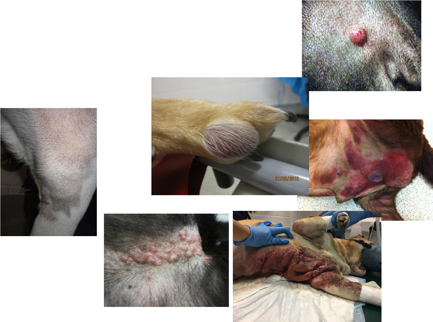
visceral form —> rare, v. aggressive

How is MCT diagnosed?
FNA (usually diagnostic)-> round cells, characteristic purple granules
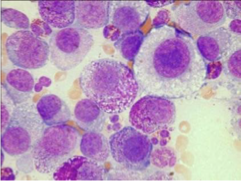
Diff quick usually fine but occ need specialist stains (e.g.- toluidine blue)
sometimes poorly stain granules —> esp in higher stage as less differentiated
(For some poorly differentiated biopsy +/- IHC)
What tests can be done to stage a MCT?
Haematology/ Biochemistry/Urine analysis
FNA or biopsy of local LN:
Abdominal ultrasound
FNA spleen and liver
Thoracic Rx
Lung metastasis uncommon
evaluate sternal LNs
Buffy coat
Bone marrow aspirates
Biopsy

What are the prognostic factors of a MCT?
Clinical
Location eg Nail bed, oral, muzzle, prepuce, perineum, mucocutaneous junction
Breed (Sharpei vs boxer)
Appearance
Systemic illness
Recurrence
?Clinical staging (i.e. presence of metastatic disease)
Lab:
Tumour histological grade (Most important)
Other:
Ki67
AGNORs
C-kit mutation
Kit staining pattern
What are the two histological grading systems for MCTs?
Patnaik grading system
grades I - III
Kiupel grading system
low grade / high grade
n.b. most labs report both grading systems together (higher agreement between pathologists with Kiupel system)
Describe a grade I Patnaik MCT.
Well differentiated
Benign behaviour (<10% metastasise)
Low recurrence rates
Unlikely to cause death (up to 7-12%)
Describe a grade II Patnaik MCT.
Intermediate
Variably metastatic (5-22% metastasise)
Cause of death in 17-56% of patients
Nodal metastases associated with poorer prognosis
n.b. most tumours are in this category which can be problematic for decision-making
Describe a grade III Patnaik MCT.
Poorly differentiated
Highly metastatic (>80%)
Likely to be cause of death
What is the median survival time based on the Kiupel grading system of MCT in dogs?
Low-grade MCTs: > 2 yrs
High-grade MCT: < 4mths
What are the treatment options for MCT?
Surgery
MCT is considered a surgical disease
Chemotherapy
Radiotherapy
Often a multi-modal approach is required. Tailor treatment to individual cases.
What are the usual surgical margins for MCT?
3cm margins & 1 fascial plane
1-2cm lateral margins may be adequate for grade I and II tumours
n.b. always send excised mass for Histopathology to check margins
Is surgery curative in MCT?
Grade I and II: potentially curative (if adequate margins)
Grade III: rarely curative (requires adjuvant therapy e.g. radiotherapy / chemotherapy)
What are the indications for radiotherapy in MCT?
Post-op following incomplete incision
Local node metastasis
Gross disease if surgery is not an option
however can cause degranulation and is not advised unless completed necessary
What are the indications for chemotherapy in MCT?
High grade and/or confirmed metastasis
Neoadjunctively prior to surgery to shrink tumour to make surgery easier
Ensure you document where tumour is clearly prior to treatment (it may shrink so much its difficult to find)
Residual microscopic disease (more controversial)
What are the commonly used chemotherapeutic drugs in MCT?
Vinblastine with prednisolone
Lomustine as a single agent or alternating with vinblastine
What are TKIs?
What are the indications of TKIs?
= Tyrosine kinase inhibitors (non-cytotoxic)
Indications:
unresectable, metastatic or high grade MCT
(shouldn't be used instead of surgery)
n.b. can assess kit mutation status to determine response to treatment (but can take a while to get results)
What are the disadvantages with tyrosine kinase inhibitors?
Can have effect on other receptors (e.g. VGFR, PDFR)
Varied range of toxicities
GI, renal, hepatic, myelosuppression, lameness
Expensive
(should not be used as a substitute for surgery)
Describe cutaneous MCT in cats.
Second most common cutaneous tumour in cats (20%)
Usually causes a raised hairless mass, often in head region, that is easily diagnosed by cytology
Rarely metastatic and not very aggressive
Surgical excision usually curative (even marginally)
Visceral MCTs are more common in cats than dogs. What are the two most common locations in cats?
Spleen
most common ddx for splenic disease
Intestines
third most common intestinal tumour
What clinical signs are seen with splenic MCT in cats?
How are the treated?
Non-specific clinical signs of systemic disease (e.g. lethargy, weight loss, anorexia, vomiting)
Treat with splenectomy - even if other organs are affected.
MST 12-19 months
What are the clinical signs associated with intestinal MCT in cats?
What is the prognosis?
Clinical signs:
GI signs
Systemic - anorexia, weight loss, lethargy
palpable abdominal mass
Poor prognosis
Metastasis common
Often euthanised at / just after time of diagnosis
What is the most common bladder tumour in dogs?
Transitional cell carcinoma
but bladder tumours only account for 2% all tumours
What is the most common location of TCC in dogs?
Bladder trigone
also urethra and prostate in males
What is the likelihood of metastasis with TCC in dogs?
Where are common locations of metastasis?
High metastatic rate
14-16% to LN and distant but up to 50% at necropsy
Metastasis sites:
Medial iliac LNs
Liver
Spleen
Bones (especially lumbar vertebrae)
What are the breed predispositions for TCC?
Small terriers
WHWT
Scottish terriers
Describe the clinical presentation of TCC
Low urinary tract signs:
haematuria
stranguria
pollakyuria
often concurrent UTI
Occasionally signs related with bone metastasis (lameness) or renal dysfunction.
Signs can be present for months as dog gets treated for “complicated UTIs”
Signs may improve with courses of antibiotics.
How can you diagnose TCC?
Histopath although sometimes cytology very suggestive.
NOT FNA - Risk of seeding
Traumatic catheterization
rub tip of catheter against lesion under US guidance
Prostatic wash
Cystoscopy (mainly females)
Surgical biopsy - risk of seeding with suture material; change your instruments & suture material
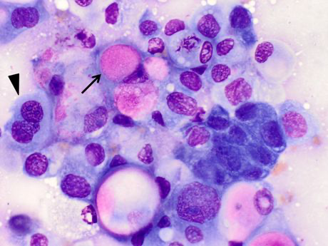
(round to polygonal cells - anisocytosis & anisokaryosis)
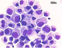
What techniques can be used to stage a TCC?
Haematology/ Biochemistry/Urine analysis (including culture):
May show neutrophilia, renal dysfunction, presence of UTI
Rule out any other problems and suitability for subsequent therapies
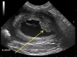
Abdominal ultrasound
Assess bladder wall, urethra, prostate and kidneys +/-metastasis in other organs.
FNA.
T&A radiography
Lung metastasis uncommon
Bone metastasis (especially lumbar vertebrae)
What are the treatment options for TCC?
Cystectomy (rarely poss but best survival)
External beam radiotherapy -(complications++)
Brachytherapy
Suitable for prostatic and urethral tumours with improved survival.
Medical treatment - commonly the only feasible option
NSAIDs and Mitoxantrone
Other chemotherapy drugs (Vinblastine, chlorambucil, carboplatin etc.)
Palliative care
Regular urine cultures and AB courses as appropriate
Cystotomy tubes/ Urethral stents (not good QoL)
What is the general prognosis of TCC?
Poor long term prognosis but quality of life can be maintained for several months.
Local disease most likely cause of death/euthanasia (MST 6-8 months).
Rapid deterioration due to renal failure or clinical signs associated with bone metastasis (pain) also possible.
Owner counselling and regular monitoring of quality of life.