Shoulder, Clavicle, AC Joints, & Scapula Positioning (RAD 1101)
 Call Kai
 Learn
 Practice Test
 Spaced Repetition
 Match
 Flashcards
 Knowt Play
1/235

There's no tags or description
Looks like no tags are added yet.
236 Terms

Shoulder - AP Projection
Explain how the patient should be positioned for an AP projection of the shoulder.
1. Supine or erect AP
Explain how the patient's body part should be positioned for an AP projection of the shoulder.
1. Rotate body slightly toward affected side to place scapula in contact (parallel) with IR
2. Abduct arm slightly

Shoulder - AP Projection in External Rotation
for non-trauma patients
Explain how the patient's part should be positioned for an AP projection in external rotation of the shoulder.
1. Supinate hand until humeral epicondyles are parallel to IR
2. Humerus is true AP (anatomical position)
For positioning an AP projection in external rotation of the shoulder, how is the hand positioned?
supine

For positioning an AP projection in external rotation of the shoulder, the humeral epicondyles are ________ to the IR when the hand is supinated
parallel

For positioning an AP projection in external rotation of the shoulder, how is the humerus positioned?
true AP (anatomical position)
The structures shown and criteria for evaluation of the shoulder in external rotation are...
- greater tubercle in profile
- the lesser tubercle is superimposed on the head of the humerus
- head of the humerus seen in profile
- greater tuberosity seen in profile on lateral aspect
- lesser tuberosity seen between greater tuberosity and head of the humerus

When meeting criteria for the AP projection of the shoulder in external rotation, what is seen in profile on the lateral aspect?
greater tuberosity
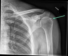
When meeting criteria for the AP projection of the shoulder in external rotation, what structure is superimposed on the head of the humerus and seen between the greater tuberosity and head?
the lesser tubercle/tuberosity
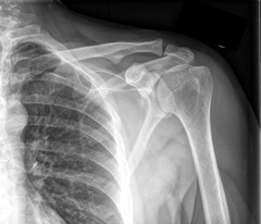
When meeting criteria for the AP projection of the shoulder in external rotation, what is seen in profile on the medial aspect?
head of the humerus

Is the AP projection in external rotation of the shoulder a non-trauma or trauma view?
non-trauma

Shoulder - Lateral Projection in Internal Rotation
for non-trauma patients
Explain how the patient's part should be positioned for a lateral projection in internal rotation of the shoulder.
1. Internally rotate hand until humeral epicondyles are perpendicular to the IR; back of hand on hip
2. Humerus is in lateral position
For positioning a lateral projection in internal rotation of the shoulder, how is the hand positioned?
internally rotated with the back of hand on hip

For positioning a lateral projection in internal rotation of the shoulder, the humeral epicondyles are _______ to the IR
perpendicular

For positioning a lateral projection in internal rotation of the shoulder, how is the humerus positioned?
lateral position
The structures shown and criteria for evaluation of the shoulder in internal rotation are...
- demonstrates lesser tubercle in profile on the medial aspect of the humeral head
- greater tuberosity superimposed on the head of the humerus
- more overlap of humerus and glenoid fossa than external rotation

When meeting criteria for the lateral projection of the shoulder in internal rotation, what is demonstrated in profile on the medial aspect of the humeral head?
lesser tubercle

When meeting criteria for the lateral projection of the shoulder in internal rotation, what is superimposed on the humeral head?
greater tuberosity

When meeting criteria for the lateral projection of the shoulder in internal rotation, what determines an internal rotation from the external rotation?
more overlap of humerus and glenoid fossa

Is the lateral projection in internal rotation of the shoulder a non-trauma or trauma view?
non-trauma

Shoulder - AP Projection in Neutral Rotation
for trauma patients
Explain how the patient's part should be positioned for an AP projection in neutral rotation of the shoulder.
1. Arm at side in neutral rotation (epicondyles are about 45-degrees to IR)
For positioning an AP projection in neutral rotation of the shoulder, the epicondyles are _________ to the IR when the hand is in neutral position at the side
about 45-degrees
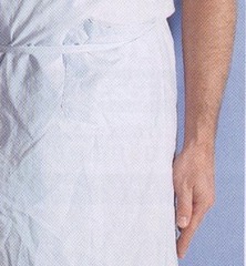
The structures shown and criteria for evaluation of the shoulder in neutral rotation are...
- proximal 1/3 of humerus
- upper scapula
- lateral 2/3 of scapula
- humeral head superimposing glenoid cavity
- greater and lesser tuberosities superimposed on head of humerus

When meeting criteria for the AP projection of the shoulder in neutral rotation, what part and how much of the humerus is shown?
proximal 1/3 of humerus

When meeting criteria for the AP projection of the shoulder in neutral rotation, what part and how much of the scapula is shown?
upper lateral 2/3 of scapula

When meeting criteria for the AP projection of the shoulder in neutral rotation, how is the humeral head positioned in terms of the glenoid cavity?
superimposed

When meeting criteria for the AP projection of the shoulder in neutral rotation, what is superimposed on the humeral head?
greater and lesser tuberosities

Is the AP projection in neutral rotation of the shoulder a non-trauma or trauma view?
trauma
When taking AP projections (in external and neutral rotation) and lateral projection (in internal rotation) of the shoulder, the CR is _____ to the IR
perpendicular
When taking AP projections (in external and neutral rotation) and lateral projection (in internal rotation) of the shoulder, the CR enters where?
1" inferior to coracoid process of 1 3/4" inferior to lateral portion of clavicle
The structures shown in all 3 positions of the AP projections (in external and neutral rotation) and lateral projection (in internal rotation) of the shoulder is...
- slight overlap of humeral head over glenoid cavity


What is the error in this internal rotation X-ray of the shoulder? How do you correct it?
Humerus is not in alignment. It is angled outward. To correct, the humerus should be placed parallel to the body if possible.
What device is used to absorb scattered radiation when taking a shoulder X-ray?
boomerang


Shoulder Joint - Inferosuperior Axial Projection - Lawrence Method (Axillary View)
for non-trauma patients
Explain how the patient should be positioned for an inferosuperior axial projection (Lawrence Method) of the shoulder joint.
1. Supine
2. Shoulder and entire arm elevated 2-3 inches
When should you not attempt an inferosuperior axial projection (Lawrence Method) of the shoulder joint?
if a fracture or dislocation is present
Explain how the patient's body part should be positioned for an inferosuperior axial projection (Lawrence Method) of the shoulder joint.
1. Abduct arm to right angle (90 degrees) to the body
2. Arm externally rotated with pad under entire arm
3. Turn head away from affected side
4. IR is vertical in holder against neck (parallel to joint)

For positioning an inferosuperior axial projection (Lawrence Method) of the shoulder joint, the arm is abducted how many degrees when the patient is supine?
90 degrees to the body

For positioning an inferosuperior axial projection (Lawrence Method) of the shoulder joint, how is the arm rotated when the patient is supine?
externally rotated
For positioning an inferosuperior axial projection (Lawrence Method) of the shoulder joint, how is the patient's head positioned if they are lying down?
turned away from the affected side
For positioning an inferosuperior axial projection (Lawrence Method) of the shoulder joint, how is the IR placed when the patient is supine?
vertical in a holder against the patient's neck (parallel to the joint)
When taking an inferosuperior axial projection (Lawrence Method) of the shoulder joint, the CR is horizontal through what?
the axilla (armpit)

When taking an inferosuperior axial projection (Lawrence Method) of the shoulder joint, the CR is perpendicular to what joint?
the shoulder joint

When taking an inferosuperior axial projection (Lawrence Method) of the shoulder joint, the CR is angled how?
medially 25-30 degrees

The structures shown in an inferosuperior axial projection (Lawrence Method) of the shoulder joint are the...
- scapulohumeral joint
- coracoid process in profile
- lesser tubercle in profile
- dislocation of proximal humerus
- AC joint

When meeting criteria for an inferosuperior axial projection (Lawrence Method) of the shoulder joint, what should be met?
- slight overlap of scapulohumeral joint
- coracoid process seen in profile and pointing anteriorly
- lesser tubercle seen in profile

When meeting criteria for an inferosuperior axial projection (Lawrence Method) of the shoulder joint, how is the scapulohumeral joint seen?
slight overlap

When meeting criteria for an inferosuperior axial projection (Lawrence Method) of the shoulder joint, which process is seen in profile and points where?
coracoid process seen in profile pointing anteriorly

When meeting criteria for an inferosuperior axial projection (Lawrence Method) of the shoulder joint, which tuberosity is seen in profile?
lesser tubercle


Shoulder Joint - Superior-Inferior Axial Lateral Projection
Explain how the patient should be positioned for a superior-inferior axial lateral projection of the shoulder joint.
1. Patient seated at end of x-ray table, leaning laterally over table
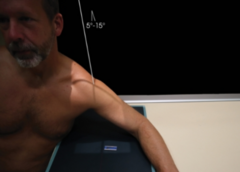
Explain how the patient's body part should be positioned for a superior-inferior axial lateral projection of the shoulder joint.
1. Elbow flexed 90 degrees, hand is pronated
2. Patient leans laterally over the IR
3. Abduct humerus as much as possible
4. Tilt the patient's head away from the shoulder

For positioning a superior-inferior axial lateral projection of the shoulder joint, the elbow is flexed how many degrees?
flexed 90 degrees
For positioning a superior-inferior axial lateral projection of the shoulder joint, how is the hand positioned?
pronated
For positioning a superior-inferior axial lateral projection of the shoulder joint, the patient is seated and how are they positioned over the IR?
leaning laterally
For positioning a superior-inferior axial lateral projection of the shoulder joint, the humerus is ________ as much as possible
abducted
For positioning a superior-inferior axial lateral projection of the shoulder joint, how is the patient's head positioned?
head is tilted away from the shoulder
When taking a superior-inferior axial lateral projection of the shoulder joint, the CR is angled how much in order to make it perpendicular to a line between the AC joint and the superior angle of the scapula?
5-15 degrees

The structures shown in a superior-inferior axial lateral projection of the shoulder joint are the...
- lateral view of proximal humerus
- glenohumeral articulation is visualized
- coracoid process is seen on end
- bursitis, shoulder impingement, osteoporosis, osteoarthritis and tendonitis
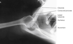

What is the error in this axillary shoulder projection? How do you correct this?
Humerus is not in same plane as shoulder. Humerus is foreshortened. To correct, place entire arm in the same plane to correctly align CR, part, and IR. Arm is foreshortened.

What is the error in this axillary shoulder projection? How do you correct this?
Central ray not directed correctly to axilla. To correct, correctly align CR, part, and IR. Arm is elongated.

What is the error in this axillary shoulder projection? How do you correct this?
Incorrect centering of IR, also quantum mottle and incorrect collimation. To correct, center to IR correctly, collimate, and increase the amount of radiation.

What is the error in this axillary shoulder projection?
Collimation is open too much.

Shoulder - PA Transaxillary Projection (Hobbs Modification)
Explain how the patient should be positioned for a PA transaxillary projection (Hobbs Modification) of the shoulder.
1. Prone or erect PA
Explain how the patient's body part should be positioned for a PA transaxillary projection (Hobbs Modification) of the shoulder.
1. The arm is raised superiorly as much as possible
2. The head is turned away from affected side

For positioning for a PA transaxillary projection (Hobbs Modification) of the shoulder, how is the arm positioned?
arm is raised superiorly as much as possible

For positioning for a PA transaxillary projection (Hobbs Modification) of the shoulder, how is the head positioned?
head is turned away from affected side
When taking a PA transaxillary projection (Hobbs Modification) of the shoulder, the CR is perpendicular to what?
the axilla and the humeral head passing through the glenohumeral joint
The structures shown and criteria for evaluation of the PA transaxillary projection (Hobbs Modification) of the shoulder are...
- lateral view of proximal humerus in relationship to glenohumeral articulation
- coracoid process is seen on end
- demonstrates bursitis, shoulder impingement, osteoarthritis, and tendonitis
The PA transaxillary projection (Hobbs Modification) of the shoulder shows the relationship between what?
the lateral view of proximal humerus to the glenohumeral articulation

When meeting criteria for evaluation for the PA transaxillary projection (Hobbs Modification) of the shoulder, what process is seen on the end?
coracoid process

This view demonstrates bursitis, shoulder impingement, osteoarthritis, and tendonitis:
PA transaxillary projection of the shoulder (Hobbs Modification)

What projections is considered a non-trauma series when looking at X-raying the shoulder?
1. AP projections: external and internal rotations
2. inferosuperior axial projection (axillary view) (Lawrence Method)
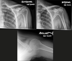
What projections are used for trauma patients when looking at X-raying the shoulder?
1. AP projection in neutral rotation
2. transthoracic lateral projection (Lawrence Method)
3. PA oblique projection (scapular Y method)

Glenoid Cavity - AP Oblique Projections - RPO & LPO Positions (Grashey Method)
for non-trauma patients
Explain how the patient should be positioned for an AP oblique projections in RPO & LPO positions (Grashey Method) of the glenoid cavity.
1. Supine or erect

Explain how the patient's body part should be positioned for AP oblique projections in RPO & LPO positions (Grashey Method) of the glenoid cavity.
1. Affected shoulder rotated 35-45 degrees toward the IR
2. Scapula is parallel to the IR
3. Arm slightly abducted in neutral rotation (or placed across the body)

For positioning the AP oblique projection (Grashey Method), the affected shoulder is rotated __________ degrees toward the IR
34-45 degrees

For positioning the AP oblique projection (Grashey Method), the scapula is _________ to the IR
parallel
For positioning the AP oblique projection (Grashey Method), how is the arm positioned?
the arm is slightly abducted in neutral rotation (or placed across the body)
When taking an AP oblique projections in RPO & LPO positions (Grashey Method) of the glenoid cavity, the CR is perpendicular to what?
the glenoid cavity

When taking AP oblique projections in RPO & LPO positions (Grashey Method) of the glenoid cavity, the CR is centered where?
the scapulohumeral joint

When taking AP Oblique Projections in RPO & LPO positions (Grashey Method) of the glenoid cavity, the CR is centered directly at the scapulohumeral joint where?
2" inferior and 2" medial to superio-lateral border of the shoulder

The structures shown in AP oblique projections in RPO & LPO positions (Grashey Method) of the glenoid cavity is the...
- glenoid cavity (scapulohumeral joint) in profile

When meeting criteria for evaluation for the AP oblique projections in RPO & LPO positions (Grashey Method) of the glenoid cavity, what should be met?
- anterior and posterior rim of glenoid fossa are superimposed (don't want to see the rim)
- glenoid cavity seen in profile
When meeting criteria for evaluation for the AP oblique projections (Grashey Method), what is superimposed?
anterior and posterior rim of the glenoid fossa

When meeting criteria for evaluation for the AP oblique projections (Grashey Method), what is seen in profile?
glenoid cavity


What is the error in this Grashey projection? How should it be corrected?
Patient under-rotated. Head of humerus overlaps glenoid fossa. To correct, rotate the patient more.

Shoulder - Transthoracic Lateral Projection (Lawrence Method)
for trauma patients
Explain how the patient should be positioned for a transthoracic lateral projection (Lawrence Method) of the shoulder.
1. Erect or supine

Explain how the patient's body part should be positioned for a transthoracic lateral projection (Lawrence Method) of the shoulder.
1. Lateral position
2. Affected shoulder against IR
3. Unaffected arm raised and hand placed on top of head
4. Affected arm in neutral position

For positioning the transthoracic lateral projection (Lawrence Method) of the shoulder, the affected side is where and in what position?
the affected side is against the IR in neutral position
For positioning the transthoracic lateral projection (Lawrence Method) of the shoulder, which arm is raised with the hand placed on top of the head?
unaffected arm is raised with hand placed on top of head
When taking a transthoracic lateral projection (Lawrence Method) of the shoulder, the CR is _________ to the IR
perpendicular
When taking a transthoracic lateral projection (Lawrence Method) of the shoulder, the CR is centered to what?
surgical neck of the humerus to IR