Lecture 3: Vertebral Joints + Ligaments, Spinal Cord
 Call Kai
 Learn
 Practice Test
 Spaced Repetition
 Match
 Flashcards
 Knowt Play
1/62

Earn XP
Description and Tags
meowowowowo
63 Terms
intervertebral joints
vertebral bones articulate with each other via intervertebral discs
fibrocartilage joints
zygapophysial joints
between superior and inferior articular facets of adjacent vertebrae
orientation of facets determines joint movement
synovial joints (plane)

costovertebral joints
thoracic vertebrae articulate with ribs
synovial joints (plane)

intervertebral discs
fibrocartilage disc between hyaline cartilage covered vertebral bodies
symphysis joints
consists of anulus fibrosus and nucleus pulposus
anulus fibrosus
rings of collagen fibers found in intervertebral discs
weight-bearing resiliency

nucleus pulposus
gel-like cartilage found within intervertebral discs
adds shock-absorption and height
becomes thinner and loses water content over time
accounts of ~25% of vertebral column height

intervertebral disc herniation
nucleus pulpous herniates through anulus fibrosus, impinging on spinal cord or spinal nerve roots leading to back pain and neuropathy
symptoms = pins and needles, pain, numbness, muscle weakness
vertebral ligaments
anterior longitudinal ligament
posterior longitudinal ligament
ligamentum flavum
intertransverse ligaments
interspinous ligaments
supraspinous ligament
nuchal ligament (cervical spine)
anterior longitudinal ligament
broad, flat sheet that covers the anterior and lateral sides of the vertebral bodies
extends from foramen magnum to the sacrum
prevents hyperextension of vertebral column

posterior longitudinal ligament
runs within vertebral canal along posterior side of the bodies
extends from C2 to the sacrum
prevents hyperflexion of the vertebral column
reinforces intervertebral discs posteriorly

ligamentum flavum
highly elastic ligament
extends from lamina of one vertebra to lamina of superior vertebra
makes up posterior wall of vertebral canal
resists hyperflexion

intertransverse ligaments
unites transverse processes
limits lateral flexion

interspinous ligaments
extend between spinous processes of each vertebra
limits flexion; well developed in Lumbar spine

supraspinous ligament
runs along very posterior surface of spinous processes
limits flexion; one tough, continuous ligament
extends from coccyx to C7
merges with nuchal ligament of cervical spine, extending to occipital protuberance

nuchal ligament
a strong and broad ligament located in the posterior region of the neck (nuchal region); a continuation of the supraspinous ligament in the cervical spine

spinal cord
major conduction pathway between brain and body
carries motor commands down to muscles
carries sensory information up from periphery
executes reflexes independently of the brain
extends from foramen magnum to L1-L2 and ends in conus medullaris
since shorter than vertebral column, inferior nerve roots progressively elongated + exit @ correct vertebral level, forming cauda equina
protected by vertebrae and vertebral column ligaments, meninges, CSF (cerebrospinal fluid)
conus medullaris
the tapered inferior-most part of the spinal cord that is attached to the filum terminale
where spinal cord terminates, at L1-L2 vertebral level
segment of spinal cord from which the sacral and coccygeal nerve roots arise
after the cord terminates, nerve roots descend within the spinal canal as individual rootlets, collectively referred to as the cauda equina
cauda equina
a bundle of spinal nerve roots at the base of the spinal cord that resemble a horse’s tail
a collection of peripheral nerve fibers that branch off the lower end of the spinal cord
contains a bundle of nerves that project distally within the enclosed cavity of the lumbar cistern from the spinal cord and conus medullaris toward the coccyx
lumbar cistern
an enlarged fluid-filled pocket of the subarachnoid space in the lower spine that extends from the end of the spinal cord to the second sacral vertebra
contains CSF, cauda equina, and filum terminale
area for spinal tap
spinal cord regions
cervical enlargement
thoracic
lumbar enlargement
sacral
cervical enlargement region of spinal cord
C3 to T1/T2
a widened region of the spinal cord that contains an increased volume of neurons to supply the upper limbs
houses extra motor and sensory neurons to control the upper limbs and form the brachial plexus
thoracic enlargement region of spinal cord
T1-T12
lumbar enlargement region of spinal cord
L1-L5
a widened section of the lower spinal cord that contains a high volume of neurons to supply sensory and motor signals to the lower limbs
sacral region of spinal cord
containins nerve roots (S1–S5) that manage sensation and movement in the lower body and control pelvic organs
s1-co
spinal cord enlargements
more fibers to innervate limbs
gray matter
central part of spinal cord
consists of dorsal horn, ventral horn, and lateral horn
dorsal horn
sensory information

ventral horn
motor information

ventral roots
the motor root of a spinal nerve, which carries motor information from the spinal cord to the rest of the body and leaves from the anterior side of the cord
ventral rootlets
small bundles of nerve fibers that emerge from the front part of the spinal cord and join together to form the ventral root
carry motor info OUT (autonomic and sensory)
lateral horn
autonomic information (thoracic)

white matter
peripheral part of spinal cord
consists of dorsal columns and laterocorticospinal tract
dorsal columns
sensory information
laterocorticospinal tract
the largest descending motor pathway in the central nervous system, responsible for controlling fine, voluntary movements of the contralateral (opposite side) limbs

spinal nerve
formed by dorsal and ventral roots of spinal cord
dorsal root and dorsal rootlets = sensory
dorsal root ganglion
ventral rootlets and ventral root = motor
therefore, this structure has mixed function

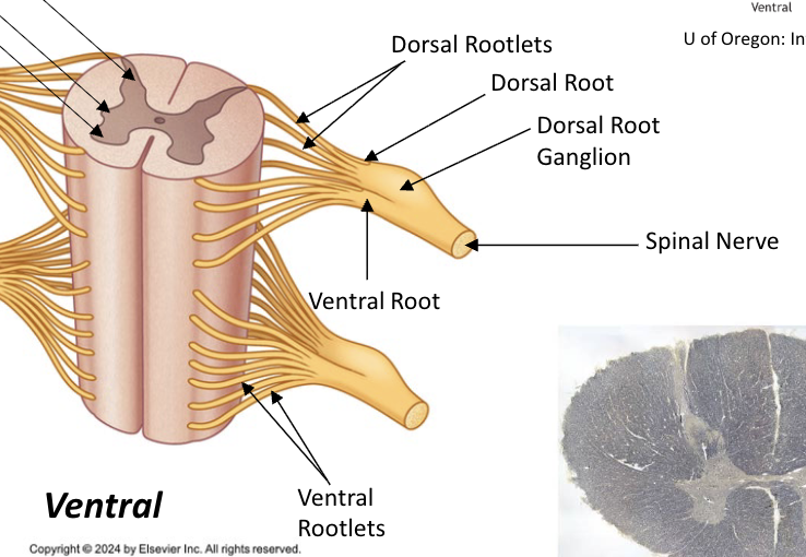
dorsal root ganglion
sensory cell bodies
emerges from the dorsal root of the spinal nerves
dorsal roots
carry sensory neural signals to CNS from PNS
carry sensory messages from various receptors (ie, pain and temperature) at the periphery toward the central nervous system for a response
dorsal rootlets
a small bundle of nerve fibers that emerges from the back of the spinal cord and joins other rootlets to form the sensory dorsal root of spinal nerve
meninges
dura mater
arachnoid mater
pia mater
dura mater
“tough mother”: outer covering of spinal cord inside vertebral canal
fibrous tissue (some elastic)
adheres to foramen magnum superiorly
anchored to coccyx via filum terminale
ensheathes dorsal and ventral roots and dorsal root ganglion, forming dural root sheaths
dural root sheaths
tubular extensions of the tough outer membrane surrounding the spinal cord, the dura mater, that covers a spinal nerve root as it exits the spinal canal
filum terminale
a delicate strand of fibrous connective tissue that anchors the bottom of the spinal cord to the tailbone
epidural space
between vertebral column and spinal cord dura mater
arachnoid mater
“spider mother”: lies against inner surface of dura
fibrous and elastic tissue
connected to pia mater via arachnoid trabeculae
arachnoid trabeculae
delicate connective tissue strands that connect arachnoid mater to pia mater
subarachnoid space
formed between arachnoid and pia, filled with CSF
forms lumbar cistern around cauda equina nerves
pia mater
“soft mother”: innermost covering of spinal cord and spinal roots
thin fibrous and elastic tissue
forms filum terminale
denticulate ligaments
fibrous ligaments that laterally attch pia to arachnoid/dura
protects cord from damage during movement
spinal nerves
31 pairs branch from the spinal cord
8 cervical
12 thoracic
5 lumbar
5 sacral
1 coccygeal
where C1 spinal nerve emerges
between occipital bone and atlas
where C1-7 spinal nerves exit
above same-named vertebrae, through intervertebral foramina
ie: C2 nerve emerges between C1 and C2 vertebrae
C8 nerve exits between C7 and T1 vertebrae (no C8 vertebrae)
how thoracic and coccygeal nerves exit
through intervertebral foramina or sacral foramina (angled inferiorly and posteriorly)
exit below same-named vertebrae
ie: T2 nerve emerges between T2 and T3 vertebrae
spinal nerve development
early in development, spinal cord fills vertebral canal → nerve roots exit horizontally at their own levels
spinal cord completes growth at birth long before vertebral column
lumber cistern forms as potential space
since each nerve’s exit is fixed at its segment of origin, elongating vertebral column stretches lower roots into progressively longer, more steeply inferiorly and posteriorly angled courses forming cauda equina below cord’s end
somatic innervation
consists of
dorsal ramus
ventral ramus
the nervous system network that supplies sensory input from the skin, joints, and limbs and controls voluntary contractions of skeletal muscles
dorsal ramus
mixed nerve fibers that go to deep back muscles and overlying skin

ventral ramus
mixed nerve fibers that go to remaining trunk and limbs


dermatome
band of skin supplied by sensory fibers of a single dorsal spinal nerve root
adjacent dermatomes overlap
nerve root injury → dermatome-patterned sensory loss

cervical dermatome
C2-C8 skin regions
thoracic dermatome
T1-T12 skin regions
lumbar dermatome
L1-L5 skin regions
sacral dermatome
S1-S5 skin regions
myotome
the muscle(s) supplied by a single ventral spinal nerve roots
muscles supplied by multiple segments → single-root injury weakens, rarely paralyzes
tested clinically as movements
root injury → movement-pattern weakness; peripheral nerve injury→complete paralysis of its muscle
nerve plexus
nerve network where ventral rami of several spinal nerves merge and redistribute fibers into named peripheral nerves