Adv Anat Unit 1
 Call Kai
 Learn
Learn Practice Test
 Spaced Repetition
 Match
Match Flashcards
 Knowt Play
1/67

There's no tags or description
Looks like no tags are added yet.
Name | Mastery | Learn | Test | Matching | Spaced | Call with Kai | Chat |
|---|
No analytics yet
Send a link to your students to track their progress
68 Terms
Sagittal Plane
divides the body into left and right sides
Frontal plane
divides the body into anterior and posterior portions
Transverse plane
divides the body into superior and inferior portions
Movement in the sagittal plane
rotates around the mediolateral axis
Movement in the frontal plane
rotates around the anteroposterior axis
Movement in the transverse plane
rotates around the longitudinal axis
Movement and planes
To confirm what plane a certain movement occurs in, imagine that plane is made of two pieces of glass and placed around your body. Can you make the certain movement between the two pieces of glass? If you can, then you know you are in the right plane for that movement!
Rotational movement occurs
In a plane rotating about an axis
Mediolateral axis
Runs side to side or medial/lateral. (Imagine an arrow going through one arm and pointing out the other)
Anteroposterior axis
runs front to back or anteroposteriorly. (Imagine an arrow going through the stomach and coming out through the back)
Longitudinal axis
Runs top to bottom or longitudinally (Imagine an arrow going through the top of the head and coming out through the foot)
Rotational movements
feature moving points of the object that do not all travel equal distance or velocity. (proximal elbow travels less distance than the distal hand during elbow flexion)
Translational movements
uniformly move. (walking forward or a skier sliding down a mountain)
Deformities in the Frontal Plane
The bone or bones will abnormally be more lateral or medial. Valgus is when the distal end of the bone is more lateral than usual relative to the midline. Varus is when the distal end of a bone is more medial than usual relative to the midline.

Deformities in the Sagittal plane
Includes recurvation which is excessive bowing posteriorly and antecurvation which is excessive bowing anteriorly.
Deformities in the Horizontal plane
rotational deformities or “twists” in bones. Retroversion is rotated posteriorly and anteversion is rotated anteriorly.
Kyphoses
Vertebral curvature that is concave anteriorly. Includes thoracic and sacral curves
Lordoses
Vertebral curvature that is concave posteriorly. Includes cervical and lumbar curves
Scoliosis
Excessive vertebral curvature from side to side
Differences between the Male and Female pelvis
The female ilia are laterally flared to provide a wider pelvis, the female pelvis inlets are wider and oval while the male inlets are somewhat heart shaped, the subpubic angle in the female is wider (greater than 90 degrees)
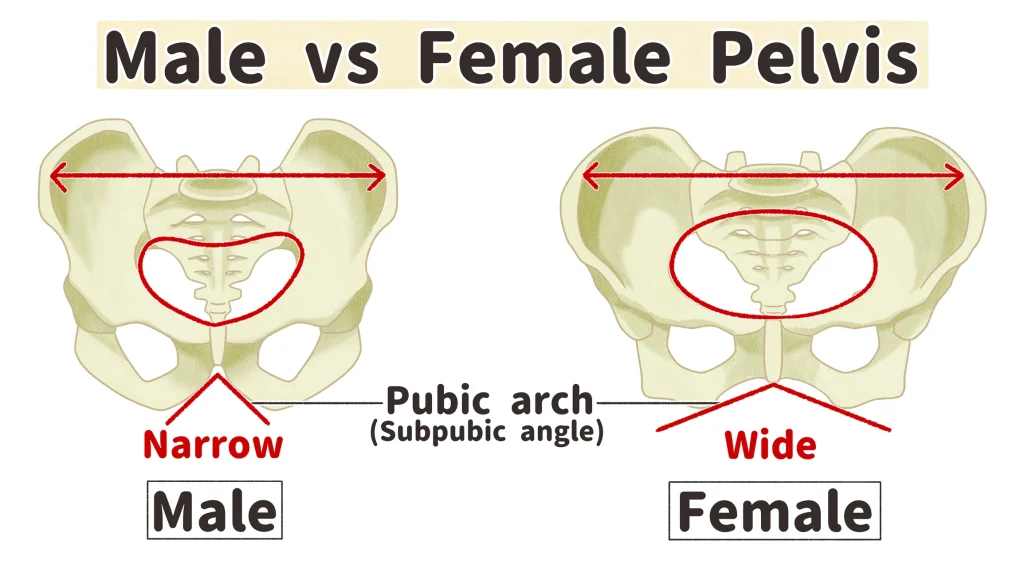
Pelvic Inlet
opening in the pelvis whose physical boundaries are defined by the pelvic prim

Pelvic Outlet
inferior to the pelvic inlet, boundaries are defined by the coccyx, ischial tuberosities, and the inferior border of the pubic symphysis
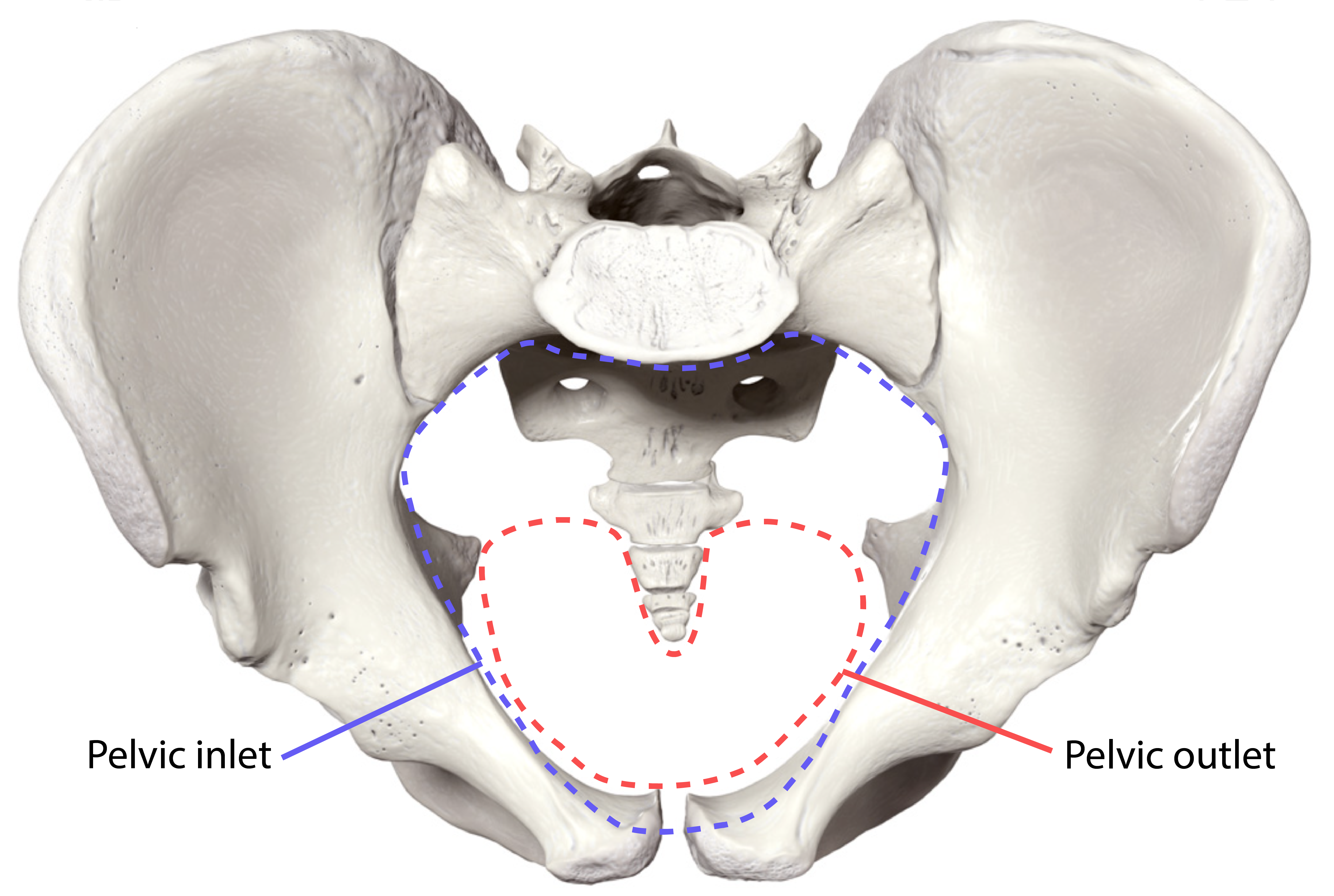
Subpubic angle

Femoral Angles
Angle of Inclination, Q Angle, Torsion Angle
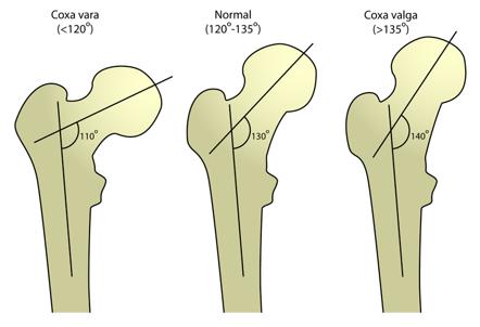
Angle of Inclination
Is the angle of the head/neck of the femur in relation to the shaft. Is normally between 120-130 degrees. This angle is greatest at birth and diminishes with age. Angle is less in females due to the increased width of hips and increased Q angle of the femur.
Q Angle
Angle between the vertical line through the midpoint of the patella and the oblique line connecting the midpoint of the patella to the anterior superior iliac spine. Is normally 8-10 degrees for males and up to 15 degrees for females. This angle is not always the same on both sides. Excessive Q angles are likely to increase risks of patellofemoral pathology.

Angle of Torsion
The twist of the femur. The head and neck of the femur are not perfectly aligned with the condyles, they are at an angle to each other. Normal angle is between 7-15 degrees.

Excessive anteversion
Angle of torsion greater than 15 degrees. result in toe gait
Retroversion
angle of torsion is less than 7 degrees. results in toe our gait
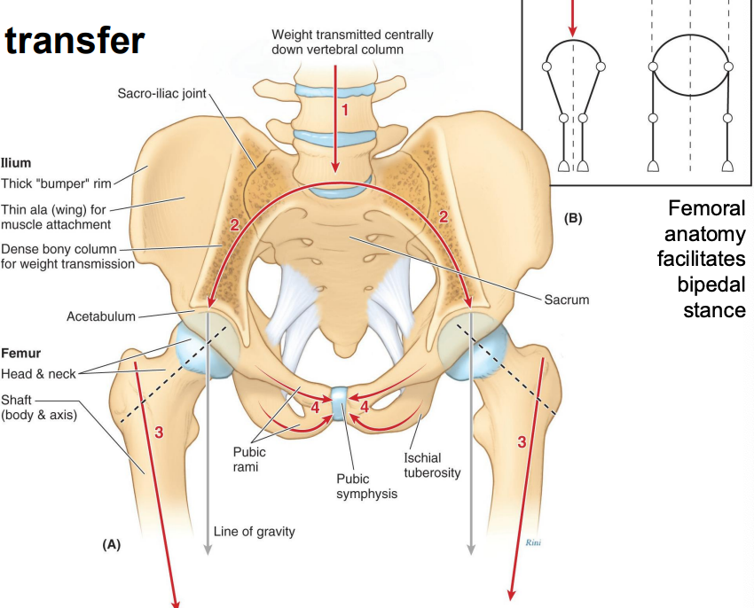
Pelvic girdle weight transfer
Weight is transferred down the vertebral column 2. At the sacrum, the weight is split and transferred down the arch to the femurs. 3. The neck of the femur transfers the weight down the main body of the femur. 4. The pubic bone acts as a strut to brace the pelvis and support the arch. 5. Femoral anatomy allows for bipedal stance
Valgus
the distal element of the bone is more lateral than normal
Varus
the distal element of the bone is more medial than normal
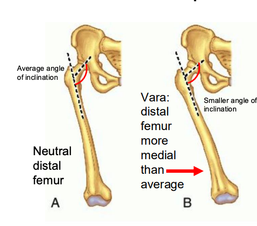
Coxa Vara
distal femur is more medial than average, femoral angle of inclination is decreased (less than 120 degrees)
Coxa Valga
the distal femur is more lateral than average, femoral angle of inclination is increased (greater than 140 degrees)

Genu varum
distal tibia is more medial than average, this is normal until 18-24 months of age
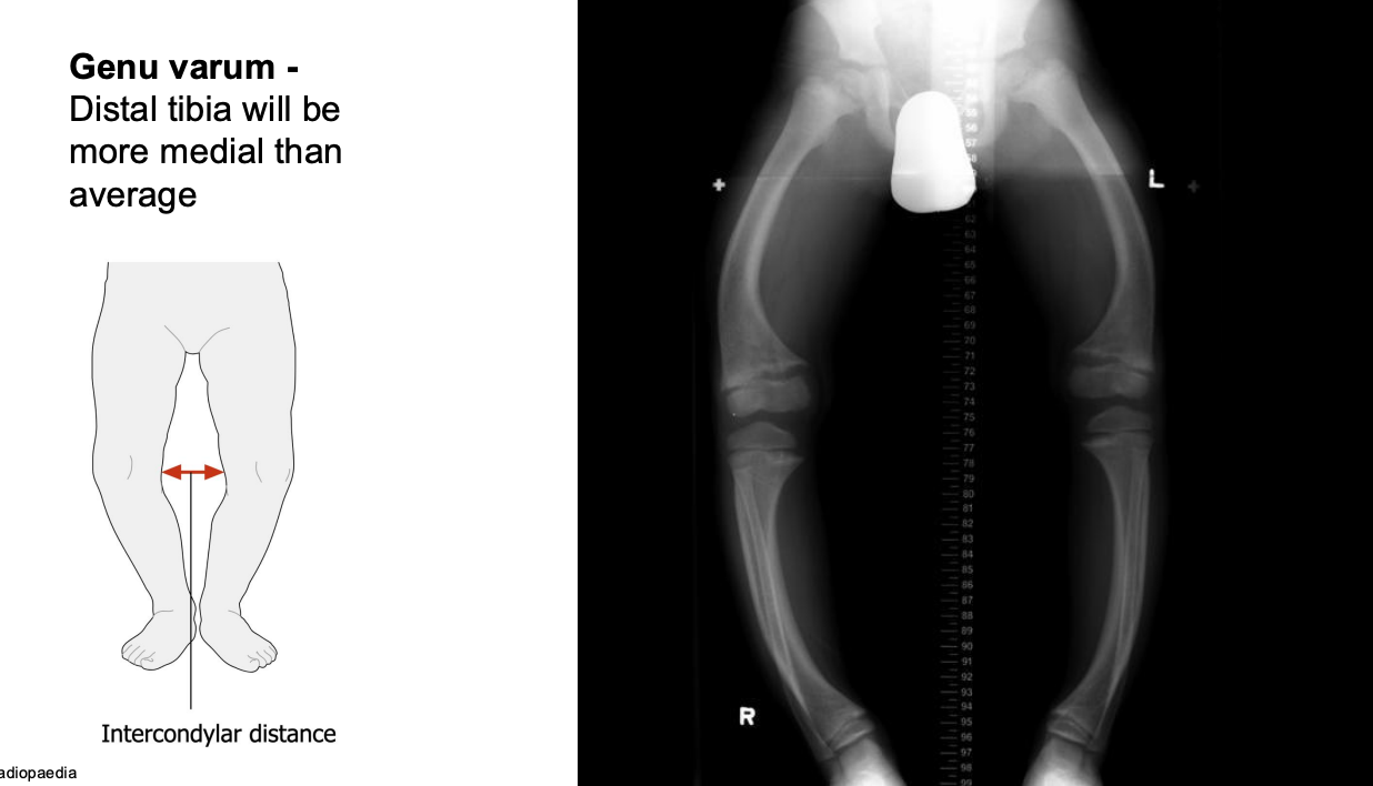
Genu valgum
distal tibial is more lateral than average, common from 2-11 years old

Humeral Angles
Inclination, Torsion
Humeral Inclination
The angle between the axis of the head/neck of the humerus and the anatomical axis of the humeral shaft. Is usually around 135 degrees

Humeral Torsion
The twist of the humerus. The angle between the axis of the head/neck of the humerus and the inter-epicondylar axis. Averages around 20 degrees so it is considered retroversion. Allows the head to of humerus to better align with the scapula that sits on a slanted thoracic cage. At birth its as high as 60 degrees but decreases during childhood. Overloading the shoulder in youth can reduce the normal rotation during development (ex: elite baseball players)

Upper Limb Carrying Anlge
Formed by the interception of the dashed lines shown running through the humerus and ulna as the elbow is fully extended. Normal ranges are between 10-15 degrees. The carrying angle influences how objects are held by individuals. Those with a more extreme carrying angle may be more likely to pronate the forearm when holding objects in the hand to keep the elbow closer to the body

Relationship between Joint Mobility and Joint stability
The more mobile a join it, the less stable. The more stable a joint it, the less mobile
Classification of joints
Joints are classified by structure (the connective tissue involved in the binding of the 2 bones) and function (the relative degree of mobility between the 2 bones). Structure determines function
What types of joints are classified by structure?
Fibrous, Cartilaginous, synovial
Fibrous
Bones held together by dense regular connective tissue
Cartilaginous
bones held together by cartilage
Synovial
bones separated by a fluid-filled cavity which is enclosed by a capsule and stabilized by ligaments MOST COMMON AND FOCUS IN THIS CLASS
What Joins are classified by function?
synarthrosis, amphiarthrosis, diarthrosis
Synarthrosis
an immobile joint
Amphiarthorsis
a slightly mobile joint
Diarthrosis
a freely mobile joint MOST COMMON AND OUR FOCUS IN THIS CLASS
Articular Capsule of synovial joint
double layer that surrounds the entire joint
Synovial Membrane of Synovial Joint
Lines the inner capsule
Synovial Plicae of synovial joint
folds in the synovial membranes of large capsules like the knee and elbow that increase the surface area and allow for full joint motion without stressing the capsule
Joint cavity of synovial joints
a physical space separating the two articulating bones filled with synovial fluid
Synovial fluid of Synovial Joint
fluid inside the joint cavity
Articular cartilages of Synovial Joints
hyaline cartilage lining the appositional surfaces of the articulating bones
Synovial Joint

Ligament
connect bone to bone to maintain integrity of joint within normal range of motion. arranged in a variety of direction to withstand forces in multiple directions. strengthens most synovial membranes
Bursae
Fluid filled sacs that cushion and reduce friction in tight spaces
Tendon Sheaths
Fluid filled sacs that cushion and reduce friction in tight spaces
Types of Synovial Joints
Uniaxial, biaxial, multiaxial
Uniaxial
bone moves in one plane. (plane, hinge, pivot)
Biaxial
bone moves in two planes (condylar, saddle)
Mutliaxial
bone moves in more than two places (ball and socket)
Gliding Joint
Uniaxial. Movement in plane surface. Glide happens in two directions. (Intercarpals, intertarsals, acromioclavicular)
Pivot Joint
Uniaxial. Rotation around longitudinal axis. (Atlanto-axial joint, radiohumeral)
Hinge Joint
Uniaxial. Flexion and extension only (humeroulnar, interphalengeal)
Modified Hinge/bicondylar Joint
Uniaxial and Biaxial. Flexion, extension