L46 - Principles of Radiology
 Call Kai
 Learn
Learn Practice Test
 Spaced Repetition
 Match
Match Flashcards
 Knowt Play
1/63

There's no tags or description
Looks like no tags are added yet.
Name | Mastery | Learn | Test | Matching | Spaced | Call with Kai | Chat |
|---|
No analytics yet
Send a link to your students to track their progress
64 Terms
What is the difference between X-rays and a Radiograph?
X-rays = Electromagnetic energy produced by an X-ray generator
Radiograph = Image produced after X-rays pass through the patient and reach the detector
What are the basic Steps in Producing a Radiograph?
X-ray generator produces X-rays
X-rays pass through the patient
Tissues block different amounts of X-rays
Remaining X-rays reach the detector
Radiographic image is created

What determines whether a structure appears BLACK or WHITE on a radiograph?
The number of X-rays that reach the detector
What happens when MORE X-rays are blocked by a structure?
Fewer X-rays reach the detector → Structure appears WHITE (radiopaque)
What happens when MORE X-rays pass through a structure?
More X-rays reach the detector → Structure appears BLACK (radiolucent)
Why do different tissues produce different shades of gray?
Different tissues block different amounts of X-rays due to differences in their composition and density
Why is radiography described as the "science of shadows"?
A radiograph is a 2D silhouette of a 3D structure created when tissues block X-rays, similar to how an object blocks light to create a shadow

What is the major limitation of a single radiograph?
A 3D patient is compressed into a 2D image, resulting in loss of depth information
What spatial relationships may be difficult to determine from one radiograph?
Above vs. Below
Left vs. Right
Inside vs. Outside another structure
What are Orthogonal Radiographic Projections?
Two radiographic projections taken 90° apart

Why should at least TWO orthogonal views be obtained?
To determine the LOCATION and SHAPE of structures by providing information about the missing third dimension

How do orthogonal views help determine the location of a structure?
First projection provides information in two dimensions
Second projection provides the missing dimension
Together, they allow mental reconstruction of the structure's 3D location
Why can the shape of a structure appear different between orthogonal views?
A 3D structure can have different silhouettes when viewed from different directions


Clinical application: A pin appears over a dog's nasal region on one radiograph. Why is a second orthogonal projection necessary?
The pin could be in the mouth, nasal cavity, surrounding tissues, or outside the patient
A second projection helps determine its actual location

How does CT use the same principle as orthogonal radiographs?
CT obtains many X-ray projections from different angles and uses computer reconstruction to create detailed images that can be viewed in different planes
What causes Magnification on a radiograph?
X-rays diverge outward from their source → so an object's distance from the detector affects its apparent size
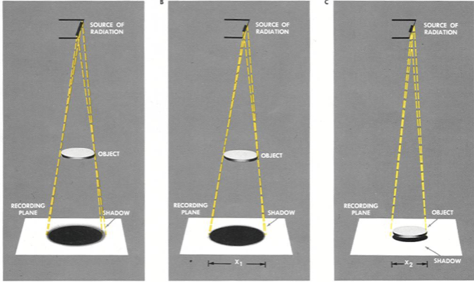
What happens when an object is positioned CLOSER to the detector?
Less magnification
Image size is closer to the object's true size

What happens when an object is positioned FARTHER from the detector?
More magnification
Object appears larger with less distinct edges
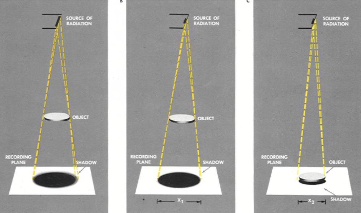
How can Magnification be minimized when accurate measurements are needed?
Position the structure of interest as close to the detector as possible
Why is minimizing magnification particularly important in orthopedic radiography?
Accurate measurements of bone size and anatomy are important for orthopedic assessment

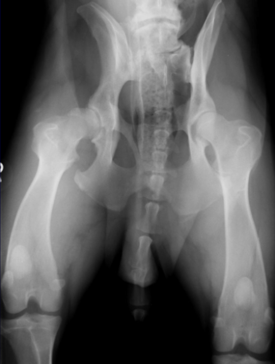
Clinical application: Two femurs appear different in size on a pelvic radiograph. Which is farther from the detector?
Larger-appearing femur = Farther from detector
Smaller-appearing femur = Closer to detector

What is Radiographic Distortion?
Alteration of a structure's original geometry due to variable magnification along the object
How can Distortion change the apparent shape of a structure?
The structure may falsely appear shorter, wider, uneven, or abnormally shaped
How can Radiographic Distortion be prevented?
Proper patient positioning:
Keep limbs straight
Keep spine straight
Keep structure of interest as parallel to the detector as possible
What is the difference between Magnification and Distortion?
Magnification = Object appears larger than its true size
Distortion = Object's shape or proportions are altered by unequal magnification
What are the THREE Major Steps in Radiographic Interpretation?
1. Determine whether an abnormality exists.
2. Describe the lesion using the 6 Roentgen signs
3. Generate a list of differential diagnoses
Abnormalities can only be recognized by comparing them with normal anatomy
Normal anatomy varies among species, breeds, body sizes, and individual patients
What is the importance of pattern recognition in radiographic interpretation?
Consistent positioning, image orientation, and systematic evaluation make abnormalities easier to identify
What is the Hanging Protocol?
A standardized method of displaying radiographs in the same orientation:
Head → Left
Tail → Right
Dorsal aspect → Top

Why should a lesion be described before assigning a diagnosis?
Objective description using Roentgen signs helps identify possible causes without prematurely assuming a diagnosis
What are three common pitfalls of radiographic interpretation?
Normal species and breed variations
Prior bias or tunnel vision
Failure to evaluate the entire radiograph
What are the SIX Roentgen signs?
1. Location
2. Number
3. Shape
4. Size
5. Opacity
6. Margination

What does LOCATION describe?
Where the lesion is located relative to surrounding anatomical structures
What does NUMBER describe?
Whether lesions are single, multiple, or multifocal
What does SHAPE describe?
The lesion's geometric appearance, such as round, linear, or amorphous
What does SIZE describe?
The lesion's dimensions, determined by direct measurement or comparison with another anatomical structure.
What does OPACITY describe?
The brightness or darkness of a structure on a radiograph

What does MARGINATION describe?
The visibility and appearance of a structure's borders

What determines how clearly the margins of a structure can be seen?
The relative density and opacity of the surrounding material

How should the Roentgen signs be used when evaluating an abnormality?
Systematically describe its location, number, shape, size, opacity, and margination before generating differential diagnoses
What is Radiopacity?
The degree to which a structure BLOCKS X-rays
Radiopaque structures appear WHITE
Example: Bone

What is Radiolucency?
The ability of a structure to ALLOW X-rays to pass through
Radiolucent structures appear BLACK
Example: Gas
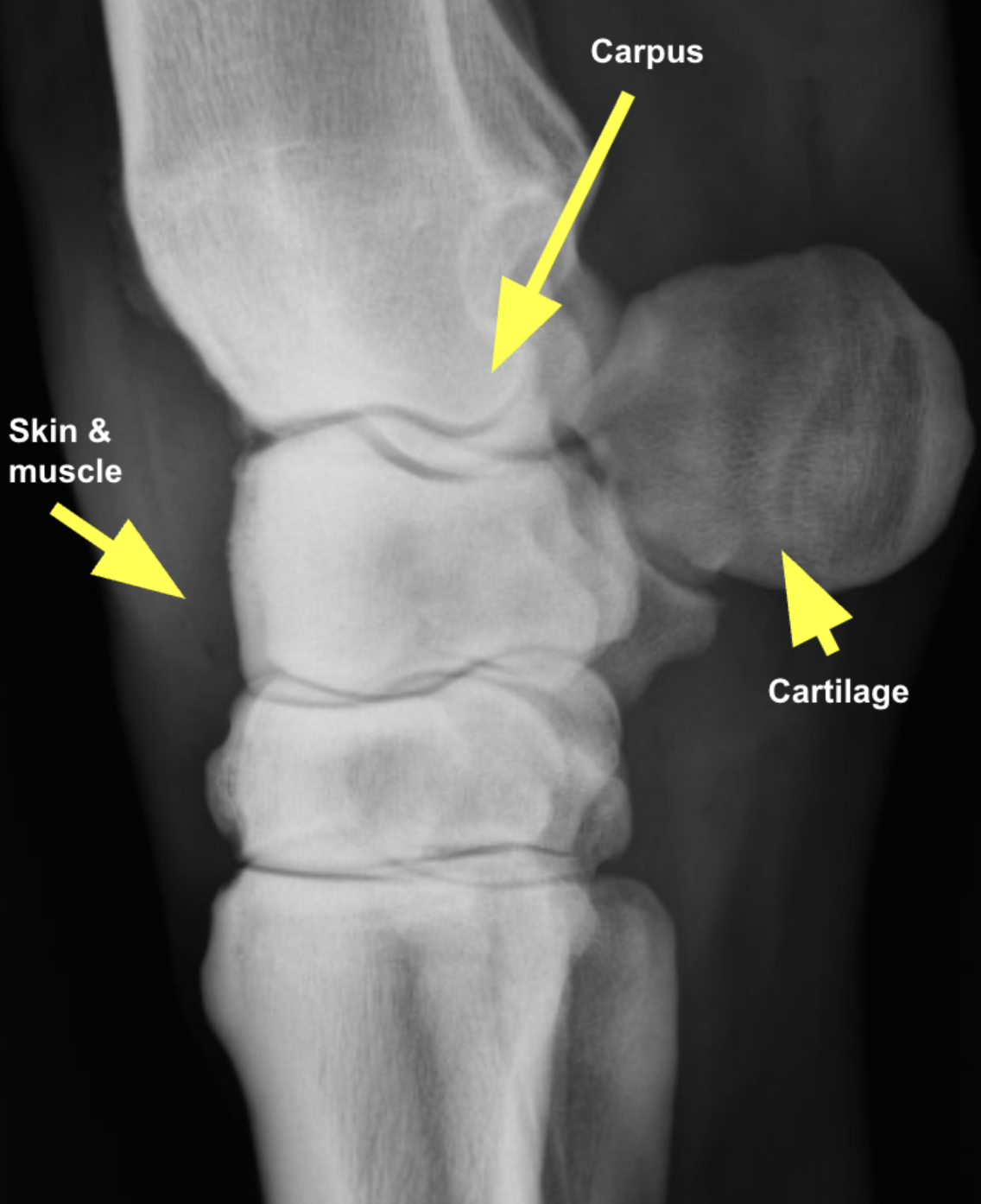
List the FIVE basic radiographic opacities from BLACKEST to WHITEST.
Gas → Fat → Soft tissue/Fluid → Mineral → Metal
Which radiographic opacity is the BLACKEST (least opaque)?
Gas/air
Examples:
Lung
Tracheal gas
Which radiographic opacity appears DARK GRAY?
Fat
Example
Falciform fat
Which radiographic opacity appears GRAY?
Soft tissue/fluid
Examples:
Liver
Spleen
Muscle
Blood
Water
Which radiographic opacity appears LIGHT GRAY to WHITE?
Mineral
Examples:
Bone
Mineralized material
Which radiographic opacity is the WHITEST (most opaque)?
Metal
Examples:
Metallic pins
Microchips
Metallic foreign bodies
Can soft tissue and fluid generally be differentiated based on radiographic opacity alone?
No
Soft tissue and fluid have approximately the same opacity
Why are structures with different opacities easier to distinguish when adjacent?
Differences in opacity create contrast, making the borders easier to identify
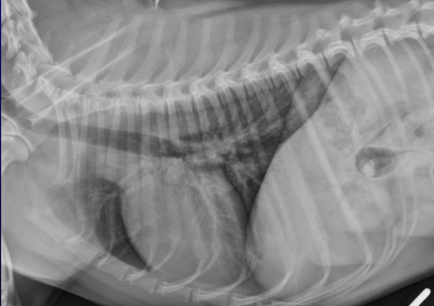
What is the Silhouette sign (Silhouetting)?
When two structures of equal radiographic opacity are in contact → their SHARED borders become indistinct
EX: Ascites (fluid-filled abdomen)
EX: Mass DIRECTLY contacts the heart

What is Border effacement?
Loss of the visible border between two structures of similar opacity that are in direct contact
What is Summation?
When two structures are superimposed along the X-ray beam →their densities have an ADDITIVE effect, increasing the overall opacity
EX: Mass OVERLAPS the heart on the X-ray beam

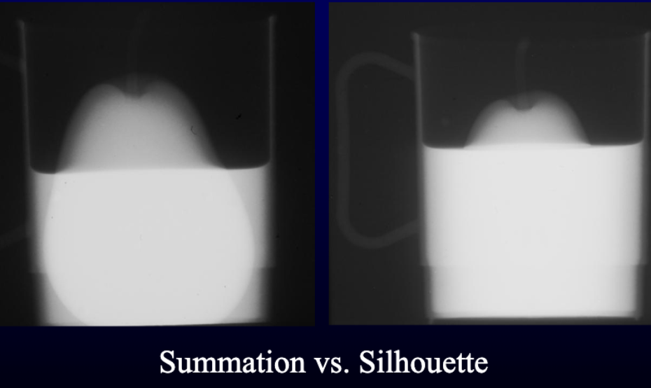
What is the Key difference between Silhouette and Summation?
Silhouette = Similar opacity + direct contact → Loss of visible border
EX: Pear inside the water
Summation = Superimposed structures → Increased overall opacity
EX: Pear outside the cup, overlapping it in your view
Radiopaque vs. Radiolucent SUMMARY
Feature | Radiopaque | Radiolucent |
X-rays blocked | More | Fewer |
X-rays reaching detector | Fewer | More |
Appearance | White | Black |
Example | Bone | Gas |
Key concept | Blocks X-rays | Allows X-rays through |
Remember:
More X-rays reach detector → BLACK
Fewer X-rays reach detector → WHITE
Orthogonal Projections SUMMARY
Feature | Single Projection | Orthogonal Projections |
Number of views | 1 | At least 2 |
Angle | One direction | 90° apart |
Depth information | Limited | Additional dimension |
Location | Difficult to determine | Easier to determine |
Shape | May be misleading | More accurately assessed |
Main limitation | 3D → 2D | Helps recover spatial information |
Remember: Orthogonal views = LOCATION + SHAPE
Magnification vs. Distortion SUMMARY
Feature | Magnification | Distortion |
Definition | Apparent increase in size | Alteration of original geometry |
Cause | Object farther from detector | Unequal magnification |
Appearance | Larger, less distinct edges | Shorter, wider, uneven, abnormal shape |
Prevention | Object close to detector | Proper positioning |
Clinical importance | Accurate orthopedic measurements | Accurate assessment of shape |
Remember:
Farther from detector → More magnification
Poor positioning → Distortion
The 6 Roentgen Signs SUMMARY
Roentgen Sign | What It Describes | Examples |
Location | Where is the lesion? | Relative to anatomical structures |
Number | How many lesions? | Single, multiple, multifocal |
Shape | What shape? | Round, linear, amorphous |
Size | How large? | Measured or compared with anatomy |
Opacity | How black or white? | Radiopaque, radiolucent |
Margination | How visible are the borders? | Distinct vs. indistinct |
Remember: Describe the lesion BEFORE generating differential diagnoses.
Five Radiographic Opacities SUMMARY
Opacity | Appearance | Examples |
Gas | Blackest | Lung, tracheal gas |
Fat | Dark gray | Falciform fat |
Soft tissue/fluid | Gray | Liver, spleen, muscle, blood, water |
Mineral | Light gray/white | Bone, rocks/mineral |
Metal | Whitest | Pins, microchips, metallic FB |
Remember: Gas → Fat → Soft tissue/Fluid → Mineral → Metal.
Silhouette vs. Summation SUMMARY
Feature | Silhouette | Summation |
Definition | Two structures of equal opacity contact | Two structures overlap |
Relationship | Direct contact | Superimposed |
Main effect | Border disappears | Opacity increases |
Mechanism | Similar opacities cause border effacement | Combined thickness blocks more X-rays |
Key phrase | Loss of border | Additive opacity |
Remember:
Silhouette → Same opacity + touching = Border disappears
Summation → Overlapping structures = Increased opacity
Radiographic Interpretation Pitfalls SUMMARY
Pitfall | Why It Matters |
Species/breed variation | Normal anatomy may appear different |
Prior bias/tunnel vision | May overlook alternative explanations |
Failure to evaluate entire radiograph | May miss additional abnormalities |
Inconsistent image orientation | Makes pattern recognition more difficult |
How a Radiograph Is Produced SUMMARY
X-ray generator produces X-rays
X-rays travel through patient
Tissues block different amounts of X-rays
Remaining X-rays reach detector
Detector produces radiographic image
Radiographic Interpretation Steps SUMMARY
1. Identify abnormality
Compare with normal anatomy
2. Describe using 6 Roentgen signs
Location • Number • Shape • Size • Opacity • Margination
3. Generate differential diagnoses
Determine possible causes of the appearance