Autonomic Nervous UNIT 1
 Call Kai
 Learn
Learn Practice Test
 Spaced Repetition
 Match
Match Flashcards
 Knowt Play
1/13

There's no tags or description
Looks like no tags are added yet.
Name | Mastery | Learn | Test | Matching | Spaced | Call with Kai | Chat |
|---|
No analytics yet
Send a link to your students to track their progress
14 Terms
Autonomic nervous system: Neurons
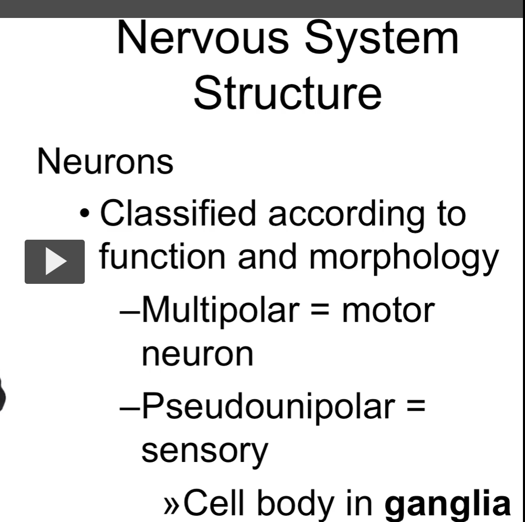
multipolar neurons are mainly motor neurons
pseudounipolar neurons are mainly sensory neurons
picture of the multipolar neuron, cell body is in the CNS, axon terminals to to the effector organ
picture of the pseudounipolar neuron, one axon goes to the receptor organ, the other axon goes to the cns (spinal cord), then the spinal cord sends the information to the brain.


The other type of cell in the nervous system is the neuroglia. The neuroglia has the function to support, insulate, and nourish neurons.
Neurons communicate with each other by the synapse.


Organization of the Nervous System
The central nervous system is the brain and spinal cord
knowledge gap: the peripheral nervous system is the cranial nerves and spinal nerves.
The nerves exit through the intervertebral foramen in the spinal cord.
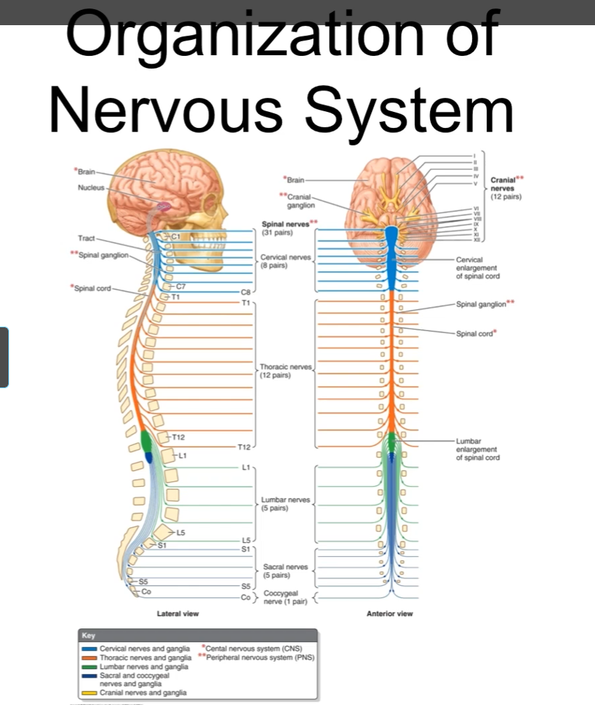
This image shows the distribution of the organization of the nervous system.
31 pairs of spinal nerves
The spinal nerves are classified according to where they exit the spinal cord.
C8, T12, L5, S5
All of them are exiting both sets of the spinal cord in the region called the intravertebral foramen.
The cervical and lumbar regions are enlarged because they supply the upper and lower limbs.
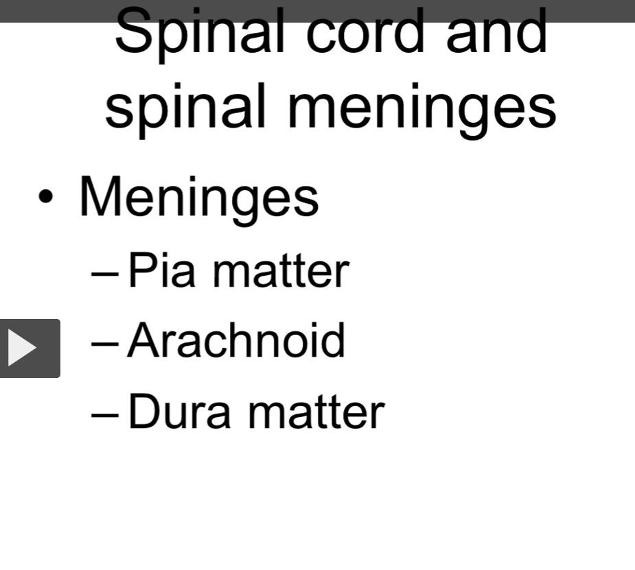
The meninges “membranes” of the brain are the
dura matter
arachnoid matter
pia matter
The meninges “membranes” of the central nervous system are the
dura matter
arachnoid matter
pia matter
knowledge gap: the meninges covers both the spinal cord and the brain.


-Spinal nerves arise from the spinal cord gray matter
cell bodies are located in the H-shaped inner gray matter
myelinated axons constitute the outer white matter
knowledge gaps: the spinal nerves have rootlets that converge to form the anterior nerve roots and anterior nerve roots.
Spinal Nerves
Posterior/dorsal: sensory neurons or afferent neurons
Anterior/ventral: motor neurons or efferent neurons
spinal nerves are mixed nerves
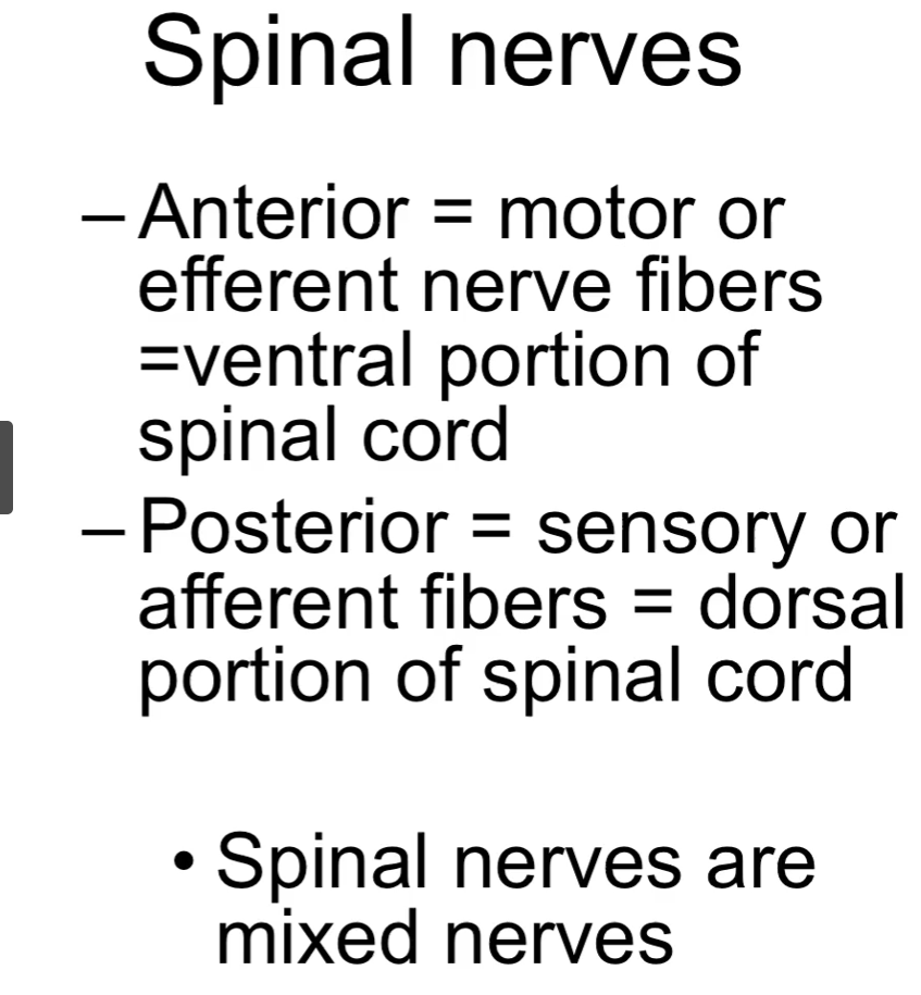

“spinal nerves are mixed nerves” meaning.


dorsal and ventral rootlets
spinal nerves converge
spinal nerves again divide into posterior rami (to supply back muscles and skin) and anterior rami (to supply remaining anterolateral wall and limbs).
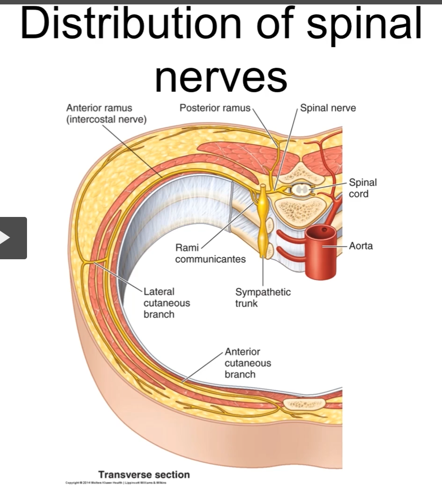
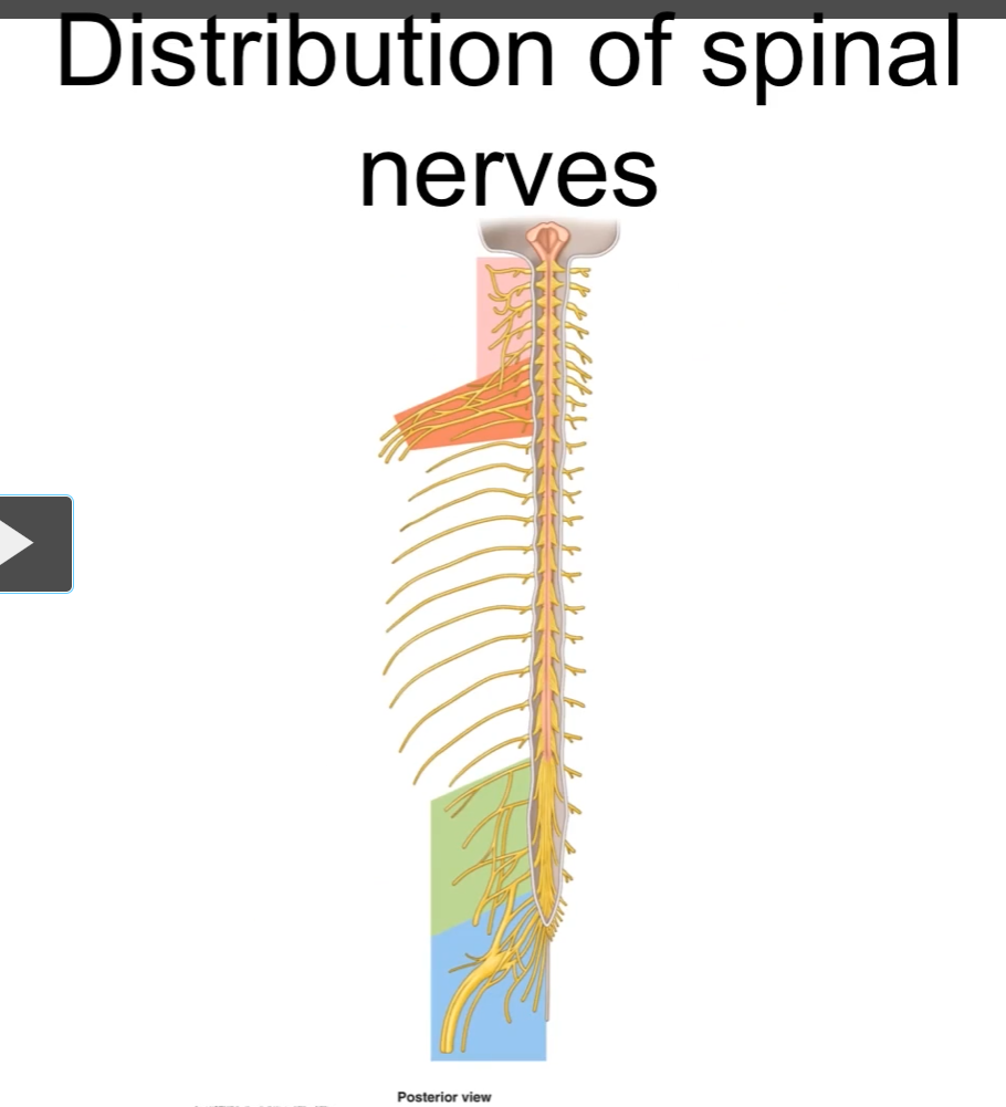
The “plexi” are the areas where the spinal nerves mix with the other nerves
Plexus Formation
Most anterior rami contribute to plexus formation for limb innervation
The posterior rami innervate the back and skin.
There is no plexus in the area of the trunk.


Plexus Formation
Plexus formation leads to the formation of multisegmental peripheral nerves.
As you can see, from a cell in the C5 region of the spinal cord, we can have the contribution to various nerves. One spinal nerve is contributing to motor fibers to many different peripheral nerves.

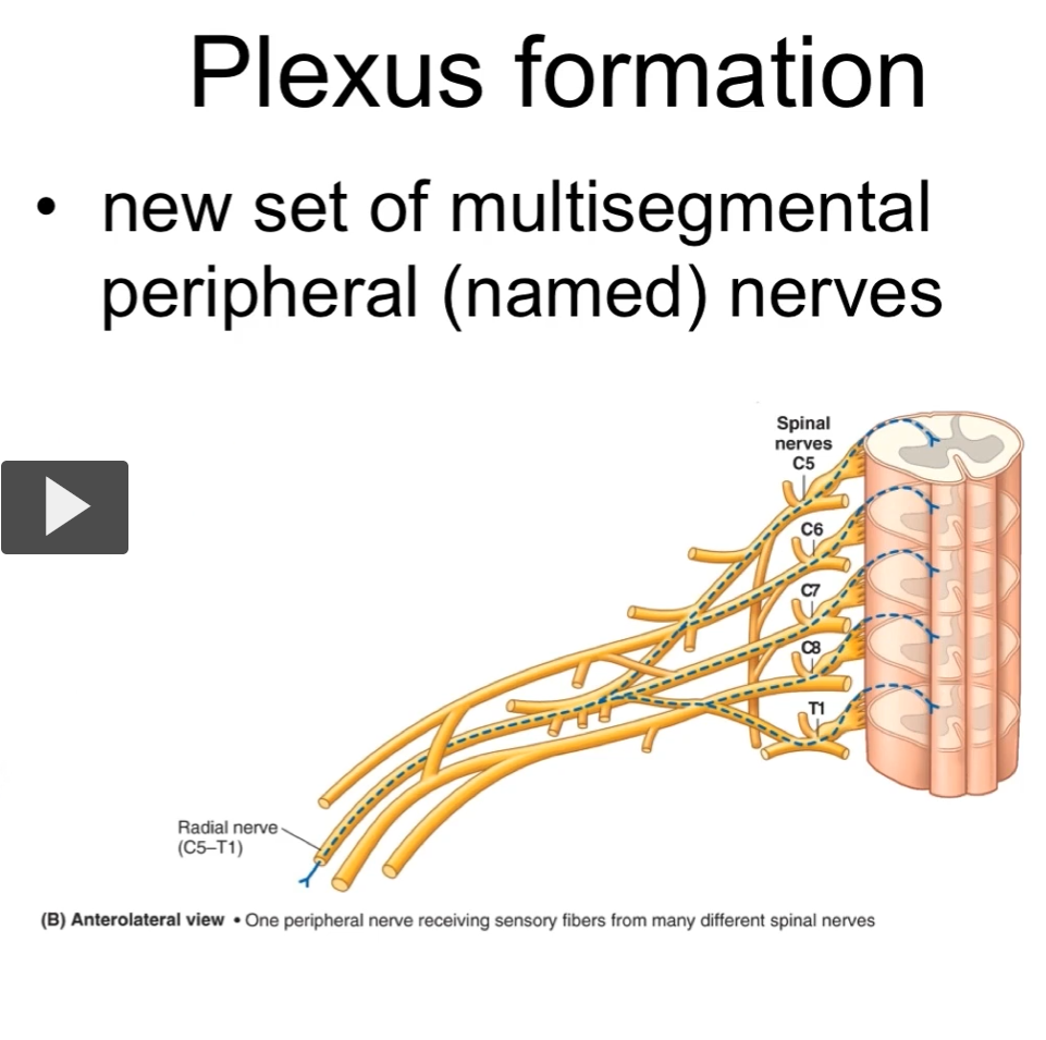
In this other example, we have one peripheral nerve receiving the sensory fibers from many different spinal nerves
The nervous system is divided into the a somatic nervous system and autonomic nervous system. The somatic nervous system is mainly a motor system that permits the voluntary and reflexive movements by contracting the skeletal muscle.


Somatic Fibers
GSA: general somatic afferent
GSE: general somatic efferent
Visceral Fibers
GVA: general visceral afferent
GVE: general visceral efferent
Pathway
touch a hot iron
sensory receptors in skin send signal to peripheral axon of pseudounipolar neuron
sensory pseudounipolar neuron cell body is in the dorsal root ganglion
remember a ganglion is a collection of neuron cell bodies outside the central nervous system.
heat signal reaches the spinal cord via the central axon of the pseudounipolar neuron entering via the posterior (dorsal) rootlets.
central axon of the pseudounipolar neuron synapses on interneuron in gray matter of the spinal cord
interneuron synapses on motor neuron in the ventral grey region of the spinal cord
axon of motor neuron exits the spinal cord through the anterior rootlets
anterior rootlets exit the spinal cord through the anterior rami
anterior rami reaches the muscle that allows the body to remove the hand from the source of the heat.
this is a reflex in this case.


There are different types of neurons in the peripheral nervous system.
Sensory neurons receive the stimuli and reach to the spinal cord
Motor neurons reach target organs
Autonomic motor neurons control movement in muscles that are not skeletal muscle (smooth muscle and cardiac muscle). It is also going to innervate glands to cause it’s contraction.
This is what was talked about in the above flashcard
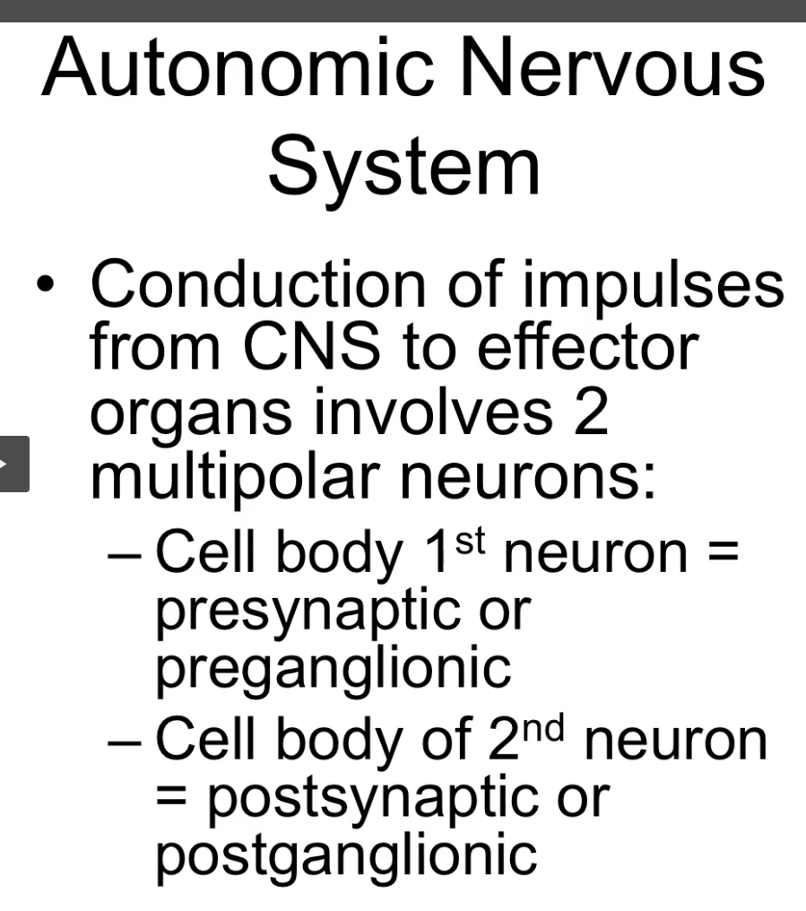
Note: Autonomic motor pathways use a two-neuron chain (preganglionic and postganglionic) connected by a ganglion to amplify, distribute, and finely control signals sent to involuntary organs.


The autonomic nervous system innervates smooth muscle, cardiac muscle and glands

The autonomic efferent fibers are organized into sympathetic and parasympathetic divisions.
The cells of the sympathetic division come from the thorax and lumbar divisions of the spinal cord
The cells of the parasympathetic division come from the cranio and sacral divisions of the spinal cord
The presynaptic neuron is always located in the spinal cord
The postganglionic neuron is always located outside the spinal cord
stopped at 21:34