Muscles
 Call Kai
 Learn
Learn Practice Test
 Spaced Repetition
 Match
Match Flashcards
 Knowt Play
1/81

There's no tags or description
Looks like no tags are added yet.
Name | Mastery | Learn | Test | Matching | Spaced | Call with Kai | Chat |
|---|
No analytics yet
Send a link to your students to track their progress
82 Terms
Epicranius
Moves the scalp; elevates the eyebrows

Frontalis (frontal belly)
Elevates the eyebrows and wrinkles the forehead

Occipitalis (occipital belly)
Retracts (pulls posteriorly) the scalp

Orbicularis Oculi
closes the eyelids (blinking, squinting)

Orbicularis Oris
Closes and protrudes the lips

Buccinator
Compresses the cheek against the teeth

Zygomaticus Major
Elevates the corners of the mouth (smiling)

Masseter
Elevates the mandible

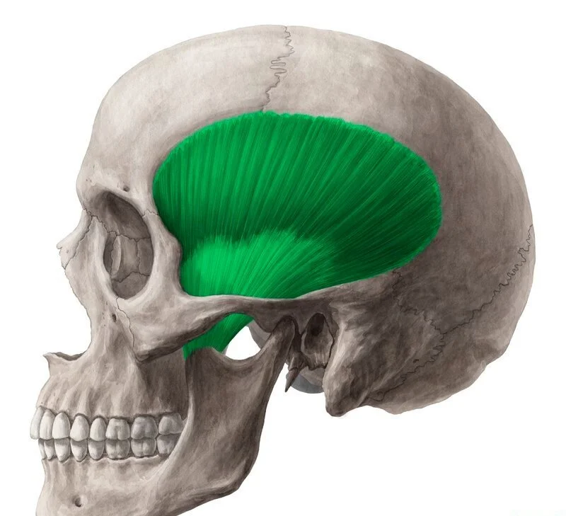
Temporalis
Elevates and retracts the mandible
Depressor Labii Inferioris
Depresses the lower lip

Sternocleidomastoid
Both sides: flex the neck; one side: laterally flexes the head to the same side and rotates it to the opposite side

Pectoralis Major
Flexes, adducts, and medially rotates the arm

Pectoralis Minor
Depresses and protracts the scapula

Serratus Anterior
rotracts and upwardly rotates the scapula

Diaphragm
Flattens to increase thoracic volume (inspiration)

Central Tendon (diaphragm)
Central tendon where the diaphragm's fibers converge and pull during contraction

External Intercostals
Elevate the ribs

Internal Intercostals
Depress the ribs

External Oblique
Flexes the trunk, laterally flexes it to the same side, and rotates it to the opposite side; compresses the abdomen

Internal Oblique
Flexes the trunk, laterally flexes it to the same side, and rotates it to the same side; compresses the abdomen

Transversus Abdominis
Compresses the abdomen

Rectus Abdominis
Flexes the trunk (vertebral column) and compresses the abdomen

CT: Linea Alba
Fibrous midline band separating the right and left rectus abdominis; anchors the abdominal muscles

CT: Aponeurosis of External Oblique
Broad tendon sheet transmitting the pull of the external oblique (trunk flexion and rotation) to the linea alba

CT: Tendinous Intersections
Fibrous bands dividing the rectus abdominis into segments; give the muscle strength during trunk flexion

Trapezius
Elevates (upper fibers), retracts (middle fibers), and depresses (lower fibers) the scapula; extends the neck

Latissimus Dorsi
Extends, adducts, and medially rotates the arm

Rhomboid Major
Retracts and downwardly rotates the scapula

Rhomboid Minor
Retracts and downwardly rotates the scapula

Levator Scapulae
Elevates the scapula

Teres Major
Extends, adducts, and medially rotates the arm

Teres Minor
Laterally rotates the arm (Part of rotator cuff)

Infraspinatus
Laterally rotates the arm (Part of rotator cuff)

Supraspinatus
Abducts the arm; initiates abduction (part of rotator cuff)

Subscapularis
Medially rotates the arm (part of rotator cuff

Erector Spinae
Extends the vertebral column; laterally flexes it when one side contracts

Deltoid
Abducts the arm; anterior fibers flex and medially rotate it; posterior fibers extend and laterally rotate it

Triceps Brachii
Extends the forearm at the elbow; long head also extends and adducts the arm

Long, Medial, and Lateral Heads (triceps brachii)
extends the forearm at the elbow; the long head also extends and adducts the arm at the shoulder

Biceps Brachii
Flexes the forearm at the elbow and supinates the forearm; weakly flexes the arm at the shoulder

Long and Short Heads (biceps brachii)
Both flex the forearm at the elbow and supinate the forearm; also weakly flex the arm at the shoulder

Brachialis
Flexes the forearm at the elbow

Brachioradialis
Flexes the forearm at the elbow

Extensor Digitorum
Extends the fingers (digits 2-5) and the wrist

Extensor Carpi Ulnaris
Extends and adducts the hand at the wrist

Extensor Carpi Radialis Longus
Extends and abducts the hand at the wrist

Flexor Carpi Radialis
Flexes and abducts the hand at the wrist

Palmaris Longus
Weakly flexes the hand at the wrist

Flexor Carpi Ulnaris
Flexes and adducts the hand at the wrist

Sulcus Carpi (= carpal tunnel)
Passageway on the anterior wrist through which the tendons of the forearm flexors (wrist and finger flexion)

CT: Inguinal Ligament
ligament along the lower edge of the external oblique aponeurosis from the anterior superior iliac spine to the pubic tubercle; separates the abdomen from the thigh

Iliopsoas
Flexes the thigh at the hip and flexes the trunk

Psoas Major
Flexes the thigh at the hip and flexes the trunk

Iliacus
Flexes the thigh at the hip

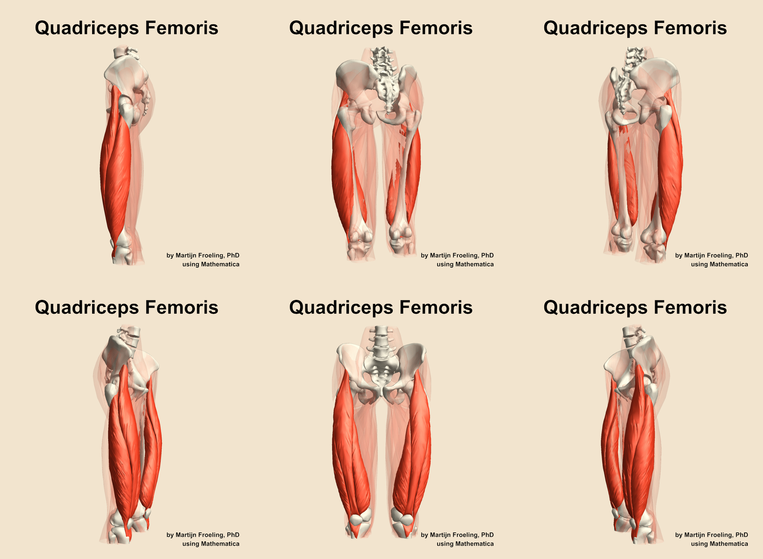
Quadriceps Femoris
Extends the leg at the knee
Rectus Femoris
Extends the leg at the knee and flexes the thigh at the hip

Vastus Lateralis
Extends the leg at the knee

Vastus Medialis
Extends the leg at the knee

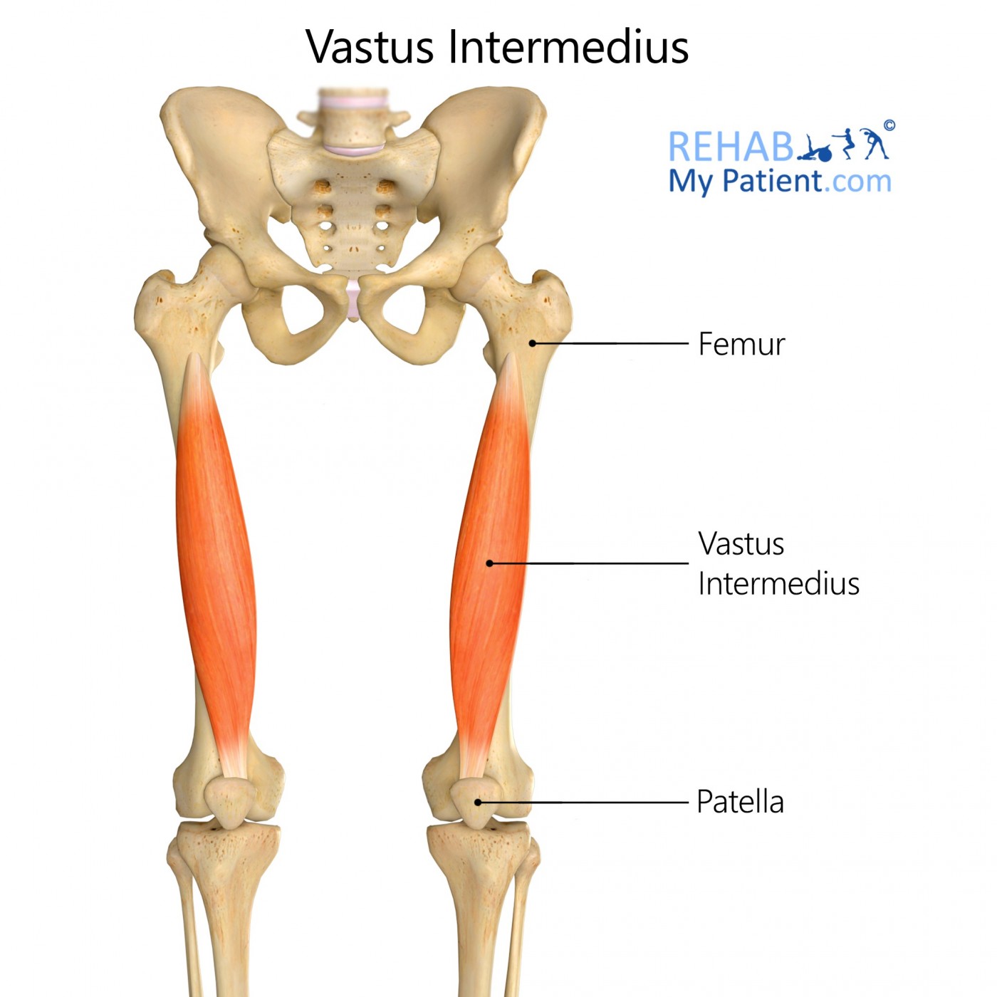
Vastus Intermedius
Extends the leg at the knee
Pectineus
Flexes and adducts the thigh at the hip

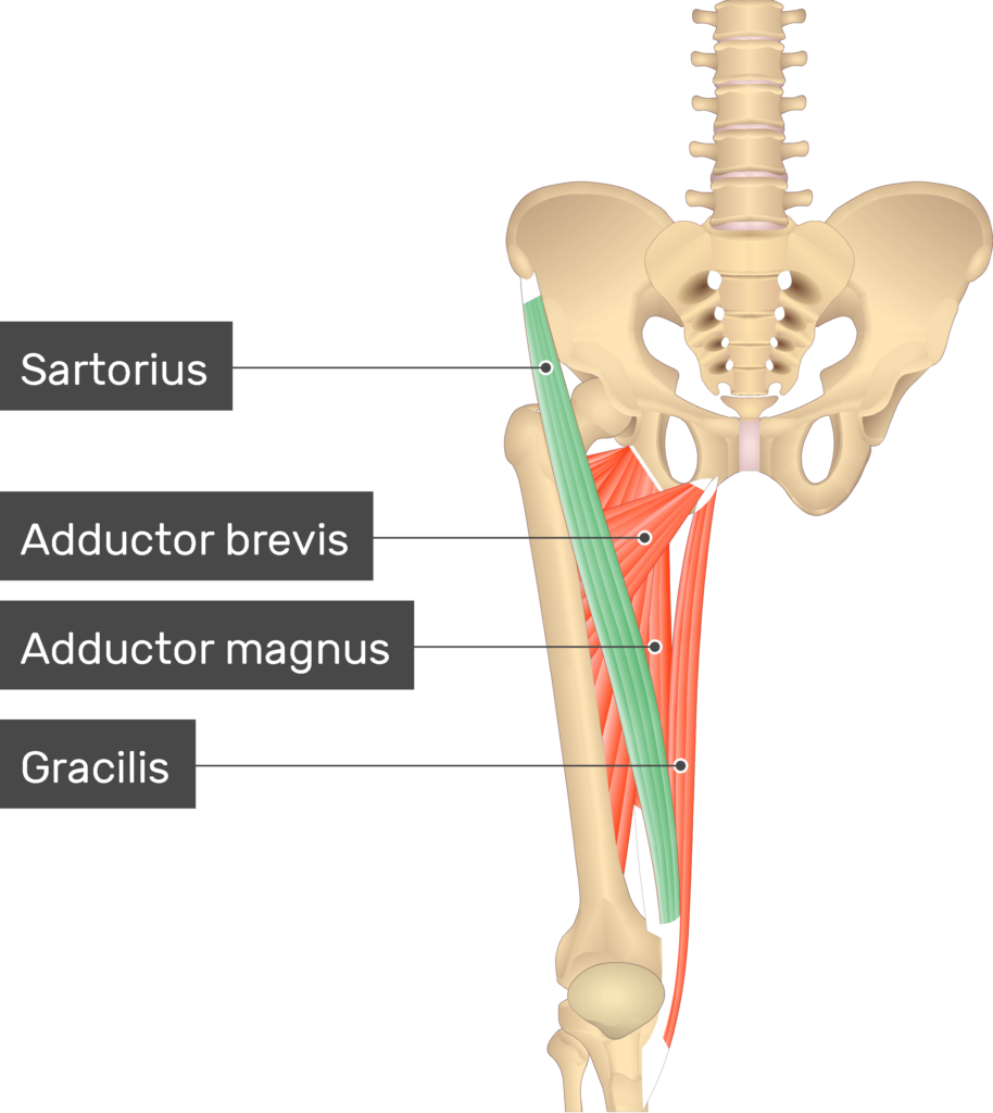
Gracilis
Adducts the thigh at the hip; flexes and medially rotates the leg at the knee

Sartorius
Flexes, abducts, and laterally rotates the thigh at the hip; flexes the leg at the knee
CT: Patellar Ligament
Ligament from the patella to the tibial tuberosity; transmits the quadriceps pull to extend the leg at the knee

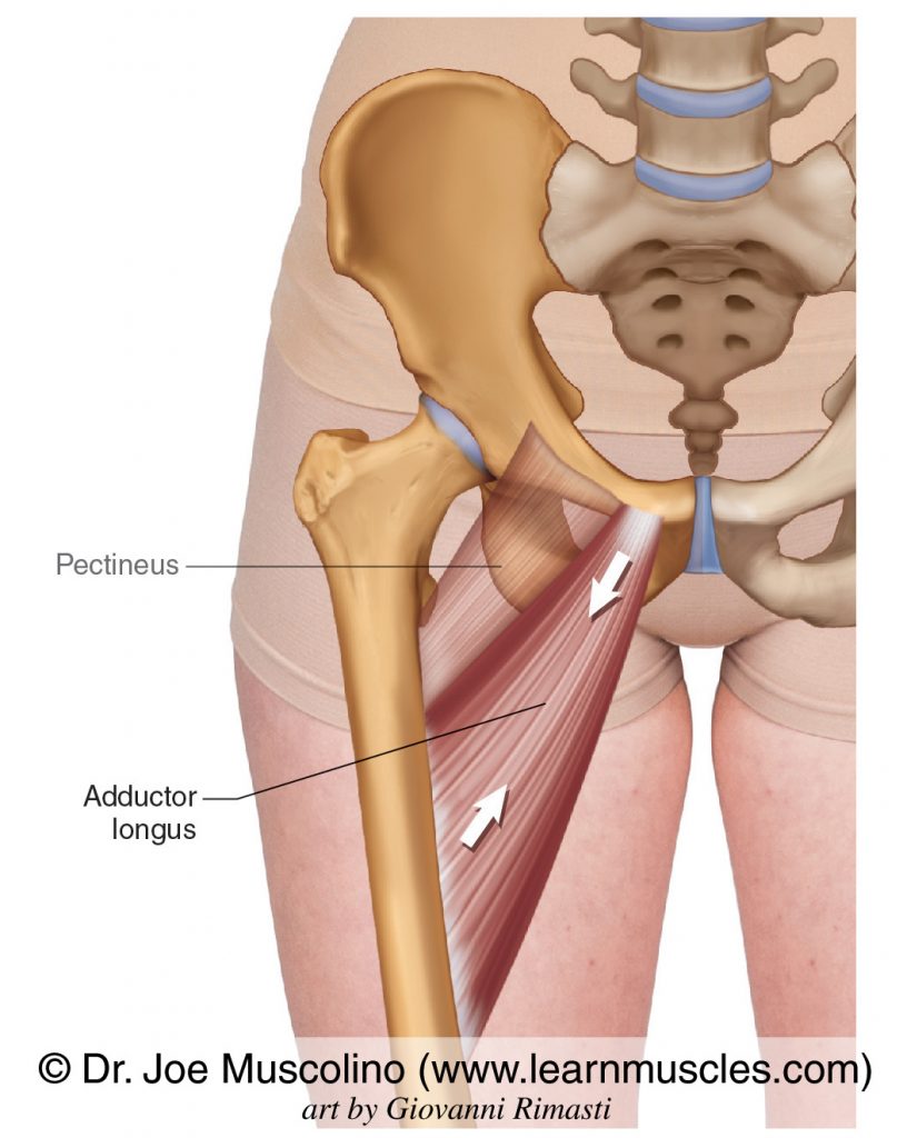
Adductor Longus
Adducts, flexes, and medially rotates the thigh at the hip
Adductor Magnus
Adducts the thigh; anterior part flexes the thigh, posterior part extends it

Tensor Fasciae Latae
Flexes and abducts the thigh at the hip; stabilizes the knee via the iliotibial tract

CT: Iliotibial Tract (Band)
Band of fascia along the lateral thigh that transmits the pull of the tensor fasciae latae and gluteus maximus; stabilizes the hip and knee

Gluteus Maximus
xtends and laterally rotates the thigh at the hip

Gluteus Medius
Abducts and medially rotates the thigh at the hip

Gluteus Minimus
abducts and medially rotates the thigh at the hip

Piriformis
Laterally rotates and abducts the thigh at the hip

Hamstrings
Extend the thigh at the hip and flex the leg at the knee

Semitendinosus
Extends the thigh at the hip; flexes and medially rotates the leg at the knee

Semimembranosus
Extends the thigh at the hip; flexes and medially rotates the leg at the knee

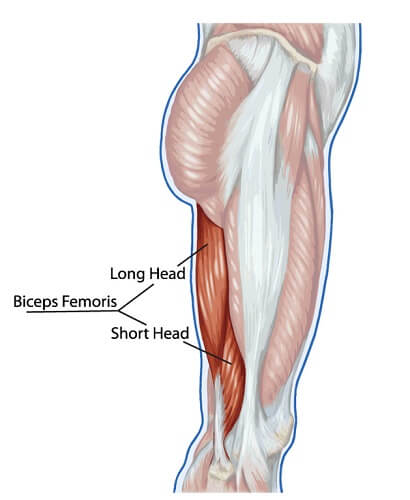
Biceps Femoris
Extends the thigh at the hip; flexes and laterally rotates the leg at the knee

Long Head (biceps femoris)
Extends the thigh at the hip and flexes the leg at the knee

Short Head (biceps femoris)
Flexes the leg at the knee
Gastrocnemius
Plantar flexes the foot at the ankle and flexes the leg at the knee

Soleus
Plantar flexes the foot at the ankle

CT: Calcaneal (Achilles) Tendon
transmits their pull for plantar flexion of the foot

Tibialis Anterior
Dorsiflexes and inverts the foot
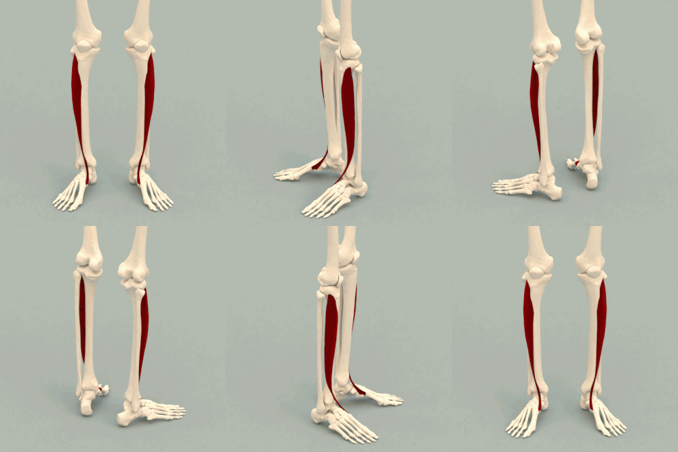
Fibularis Longus
Plantar flexes and everts the foot
