Human Body Systems
 Call Kai
 Learn
Learn Practice Test
 Spaced Repetition
 Match
Match Flashcards
 Knowt Play
1/203

There's no tags or description
Looks like no tags are added yet.
Name | Mastery | Learn | Test | Matching | Spaced | Call with Kai | Chat |
|---|
No analytics yet
Send a link to your students to track their progress
204 Terms

Parietal Bone
one of two bones that form the upper sides and roof of the skull, protecting the brain.

Frontal Bone
bone that forms the forehead and the upper part of the eye sockets, protecting the front of the brain.

Occipital Bone
forms the back and base of the skull, protecting the back of the brain.
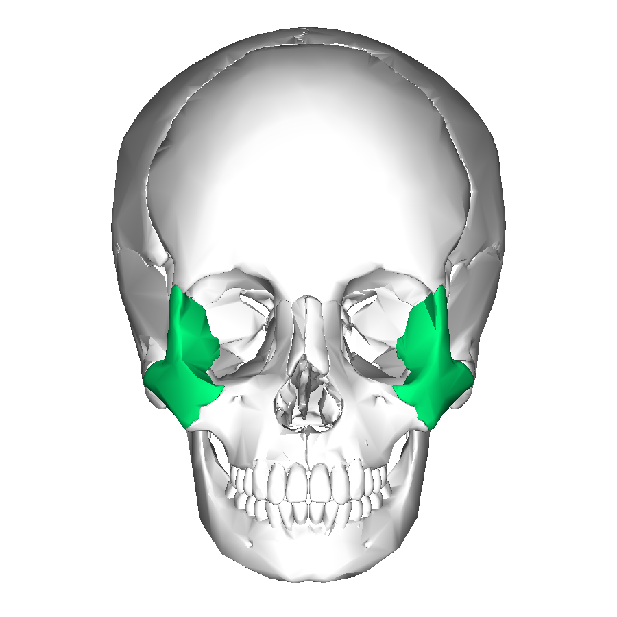
Zygomatic Bone
forms the cheekbones and helps create the outer edges of the eye sockets.

Temporal Bone
forms the lower sides of the skull, houses the structures of the inner and middle ear, and helps protect the brain.

Maxilla
upper jawbone that holds the upper teeth and forms part of the nose and eye sockets.

Clavicle
connects the sternum (breastbone) to the scapula (shoulder blade) and helps support the shoulder.

Scapula
flat, triangular bone in the upper back that connects the upper arm bone to the clavicle and helps with shoulder movement.

Sterum
flat bone in the center of the chest that connects to the ribs and helps protect the heart and lungs.

Humerous
long bone in the upper arm that connects the shoulder to the elbow.

Ribcage
framework of ribs, the sternum, and thoracic vertebrae that protects the heart and lungs.

Vertebral column
series of bones called vertebrae that supports the body, protects the spinal cord, and allows movement and flexibility.

Radius
forearm bone on the thumb side that helps the wrist and forearm rotate.

Pelvic girdle
group of bones that connects the legs to the trunk, supports the body's weight, and protects the pelvic organs.
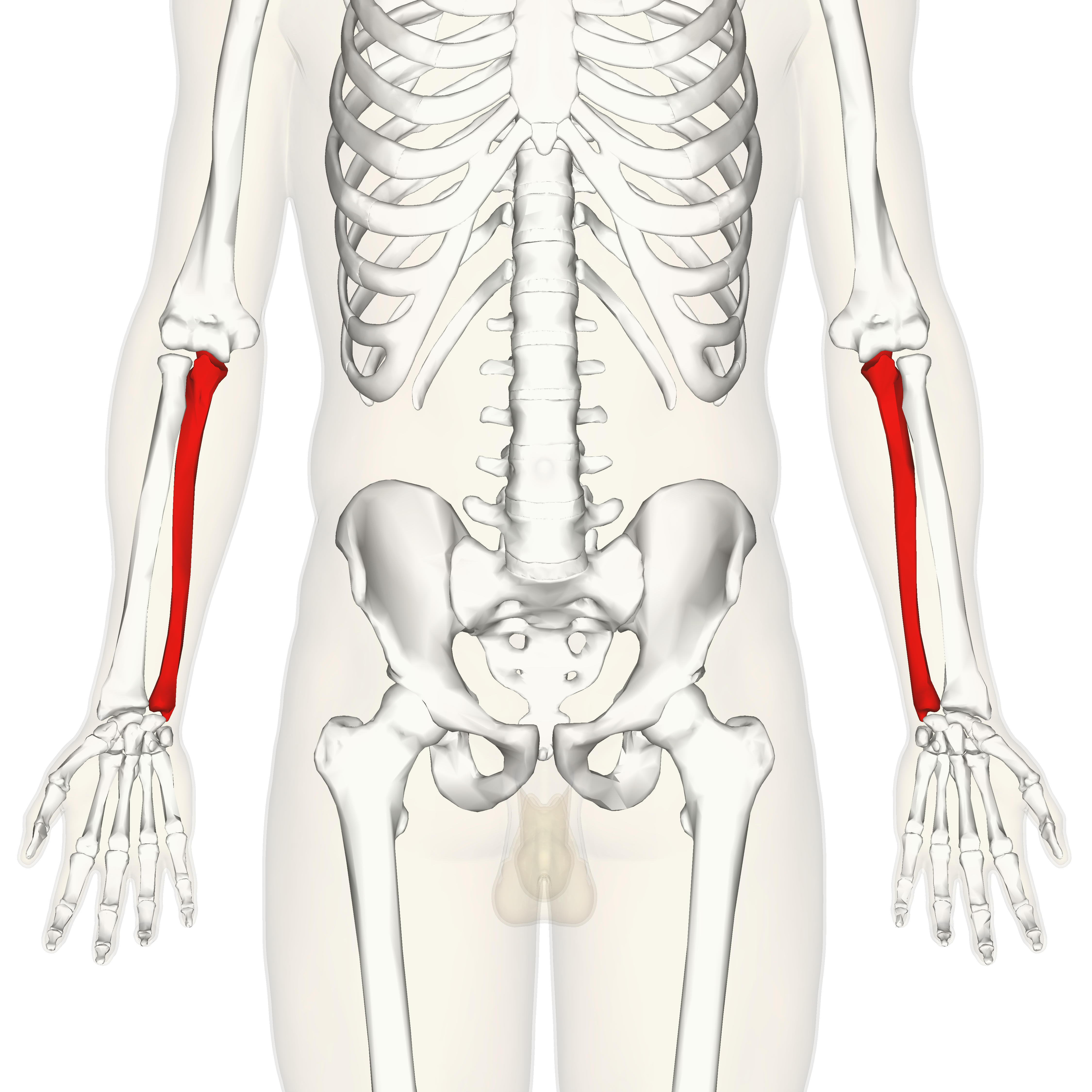
Ulna
forearm bone on the pinky-finger side that helps form the elbow joint.

Carpals
eight small bones in each wrist that connect the forearm to the hand and allow wrist movement.

Metacarpals
five bones in each hand that form the palm and connect the wrist bones to the finger bones.

Phalanges
bones in the fingers and toes that allow them to bend and move.

Femur
thigh bone and the longest, strongest bone in the human body, connecting the hip to the knee.

Patella
small bone at the front of the knee that protects the knee joint and helps the leg straighten.

Tibia
larger, weight-bearing bone in the lower leg that connects the knee to the ankle.

Fibula
smaller bone on the outer side of the lower leg that helps stabilize the ankle and support the leg.

Tarsals
seven bones in each ankle that connect the lower leg to the foot and help support body weight and movement.

Metatarsals
five long bones in each foot that form the middle of the foot and connect the tarsals to the toe bones.

Cervical curvature
C1-C7 (7 vertebrae)

Thoracic curvature
T1-T12 (12 vertebrae)

Lumbar curvature
L1-L5 (5 vertebrae)

Sacrum
5 fused vertebrae

Coccyx
4 fused vertebrae

True ribs
Ribs 1-7 connected to the sternum

False ribs
Ribs 8-12 not directly connected to sternum

Long bone
bone that is longer than it is wide and helps support the body and allow movement, such as the femur or humerus.

Short bone
bone that is roughly cube-shaped and provides stability and support with limited movement, such as the carpals in the wrist and tarsals in the ankle.

Flat bone
thin, often slightly curved bone that protects internal organs and provides a surface for muscle attachment, such as the sternum, ribs, and skull bones.

Irregular bone
bone with a complex shape that doesn’t fit into other bone categories, such as the vertebrae in the spine.

Extra fact: Hyoid bone
only bone in the body that is not connected to another bone (used as the airway motor control between jaw and airway)

Extra fact: Femur
largest bone int he body

Extra fact: Stapes
smallest bone in the body (ear bone)
Extra fact: 270-300
how many bones are you born with
Extra fact: 206
how many bones do you have as an adult
Extra fact: 27
bones in one hand/wrist

Extra fact: connective tissue
what kind of tissue is bone

Coronal suture
immovable joint that connects the frontal bone to the two parietal bones across the top of the skull.

Nasal bone
two small bones that form the bridge of the nose and support its structure.

Vomer
thin, flat bone in the skull that forms the lower part of the nasal septum, separating the left and right nasal cavities.
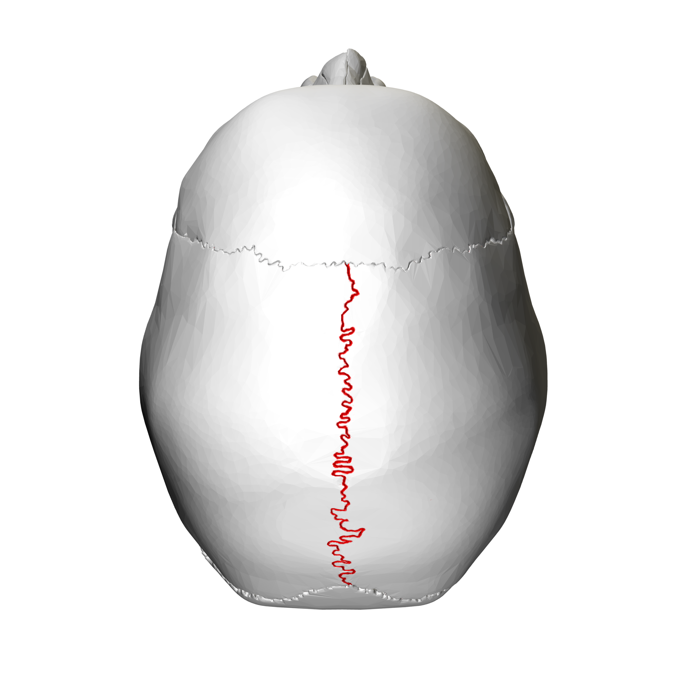
Sagittal suture
immovable joint that connects the two parietal bones along the midline at the top of the skull.

Squamous suture
immovable joint that connects each parietal bone to the temporal bone on the sides of the skull.

Frontal bone
forehead and the upper part of the eye sockets, protecting the front of the brain.
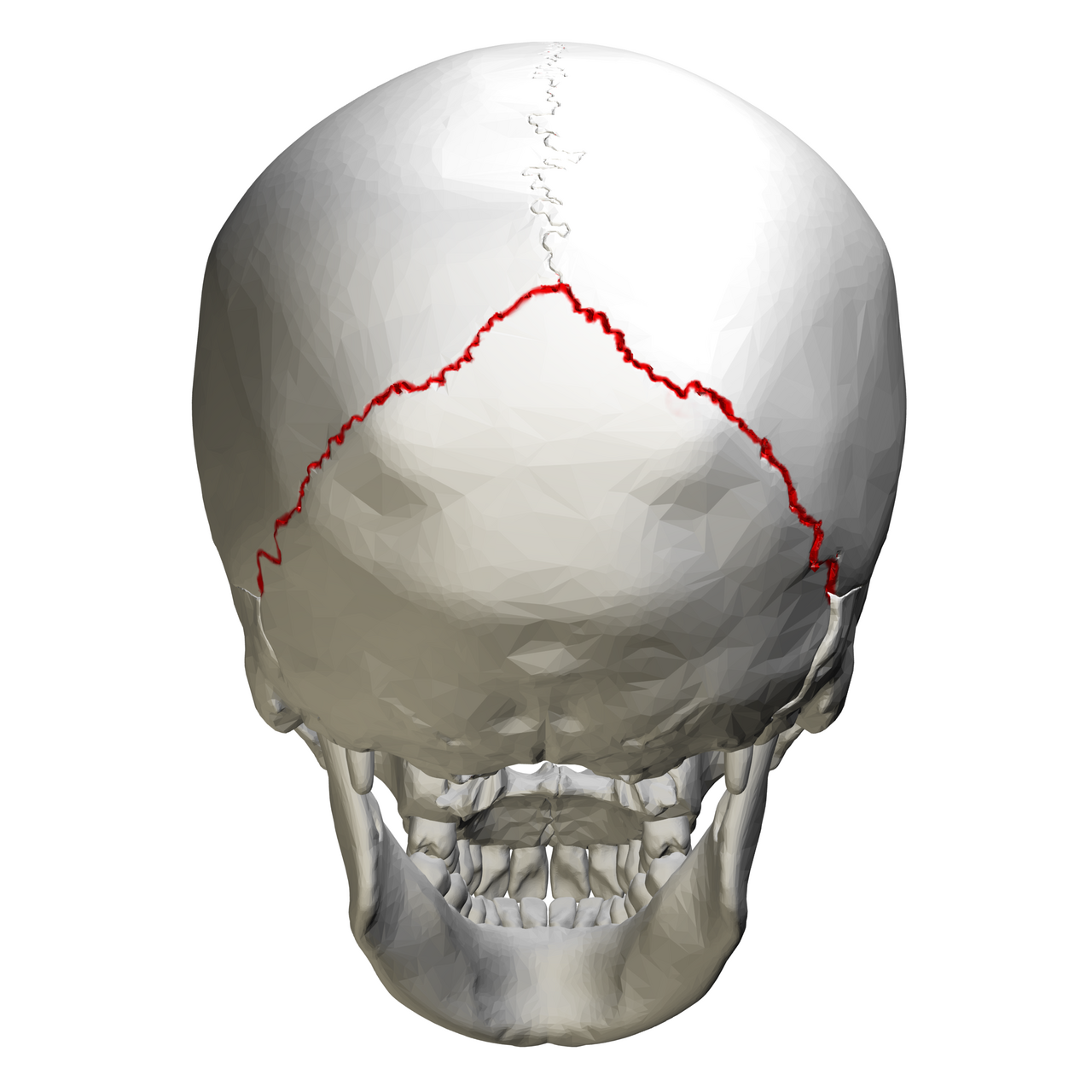
Lambdoidal suture
immovable joint that connects the occipital bone to the two parietal bones at the back of the skull.

Mastoid process
rounded projection of the temporal bone behind the ear that serves as an attachment point for neck muscles.

Styloid process
thin, pointed projection of the temporal bone that serves as an attachment point for muscles and ligaments of the tongue and throat.

Zygomatic process (arch)
bony arch on each side of the skull formed by parts of the zygomatic and temporal bones, creating the prominence of the cheek.

Sphenoid
butterfly-shaped bone at the base of the skull that helps form the eye sockets and supports the brain.

Ethmoid
small, delicate bone between the eyes that forms part of the nasal cavity, the nasal septum, and the eye sockets.

Occipital bone
forms the back and base of the skull, protecting the back of the brain.

Foramen magnum
large opening at the base of the skull where the spinal cord passes through to connect with the brain.

Occipital condyles
two rounded projections on the occipital bone that connect the skull to the first cervical vertebra (atlas), allowing the head to nod up and down.

Manubrium
upper portion of the sternum (breastbone) that connects to the clavicles and first ribs.

Body (of sternum)
long, middle portion of the breastbone that connects to the ribs and forms most of the sternum.

Xiphoid process
small, pointed bottom part of the sternum that serves as an attachment point for muscles and ligaments.

Superior border (scapula)
shortest upper edge of the shoulder blade, located near the base of the neck.

Spine of scapula
prominent ridge on the back of the shoulder blade that provides attachment for muscles.

Acromion process
bony projection at the top of the scapula that connects with the clavicle to form the acromioclavicular joint.

Scapula
flat, triangular shoulder blade that connects with the clavicle and humerus and provides attachment points for shoulder muscles.

Glenoid cavity
shallow socket on the scapula that connects with the head of the humerus to form the shoulder joint.

Axillary border (scapula)
thick outer edge of the shoulder blade closest to the armpit, providing attachment points for muscles.
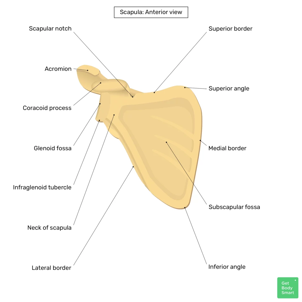
Medial border (scapula)
long inner edge of the shoulder blade closest to the spine.
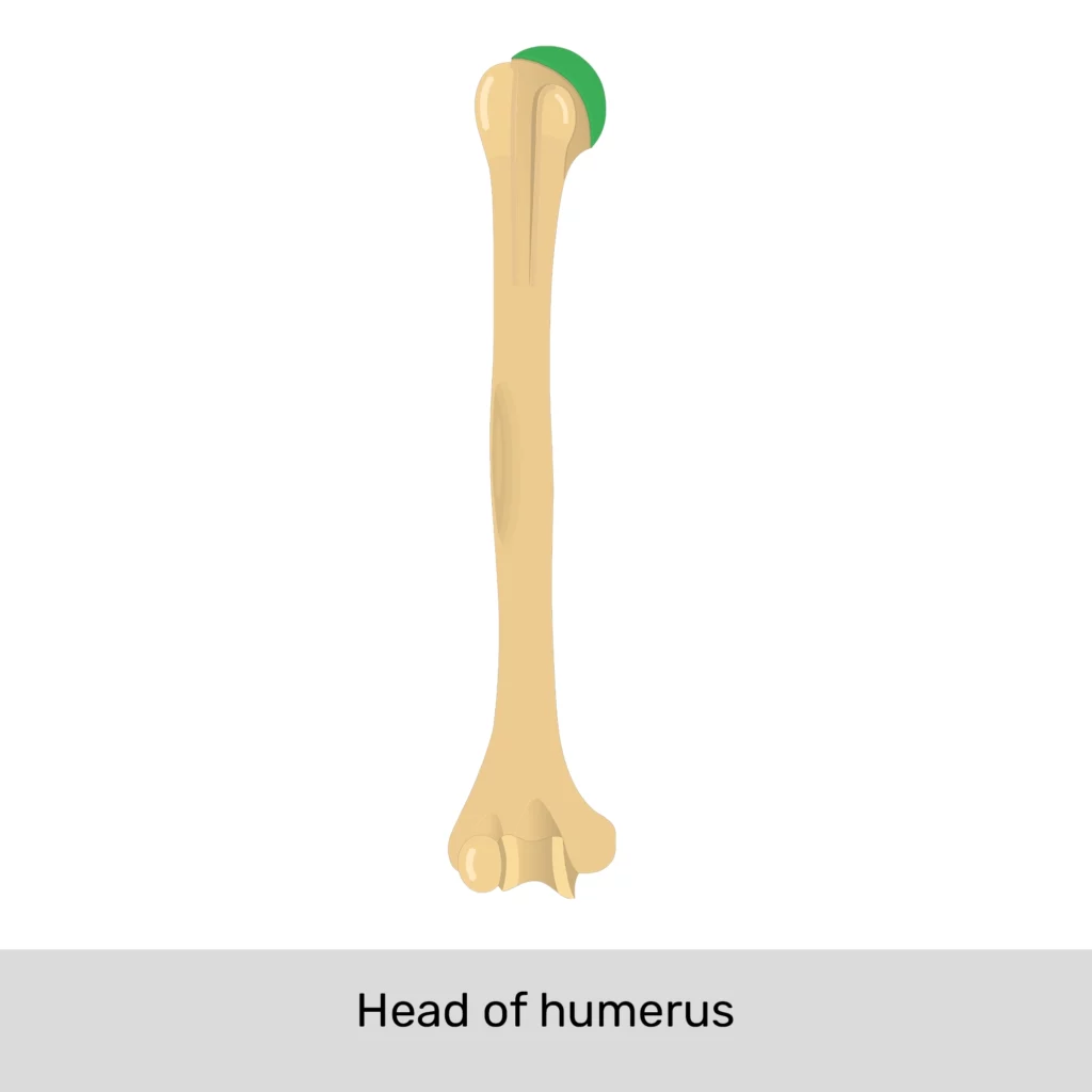
Head of humerous
rounded upper end of the upper arm bone that fits into the glenoid cavity of the scapula to form the shoulder joint.

Greater tubercle
large bony projection near the top of the humerus that serves as an attachment point for rotator cuff muscles.
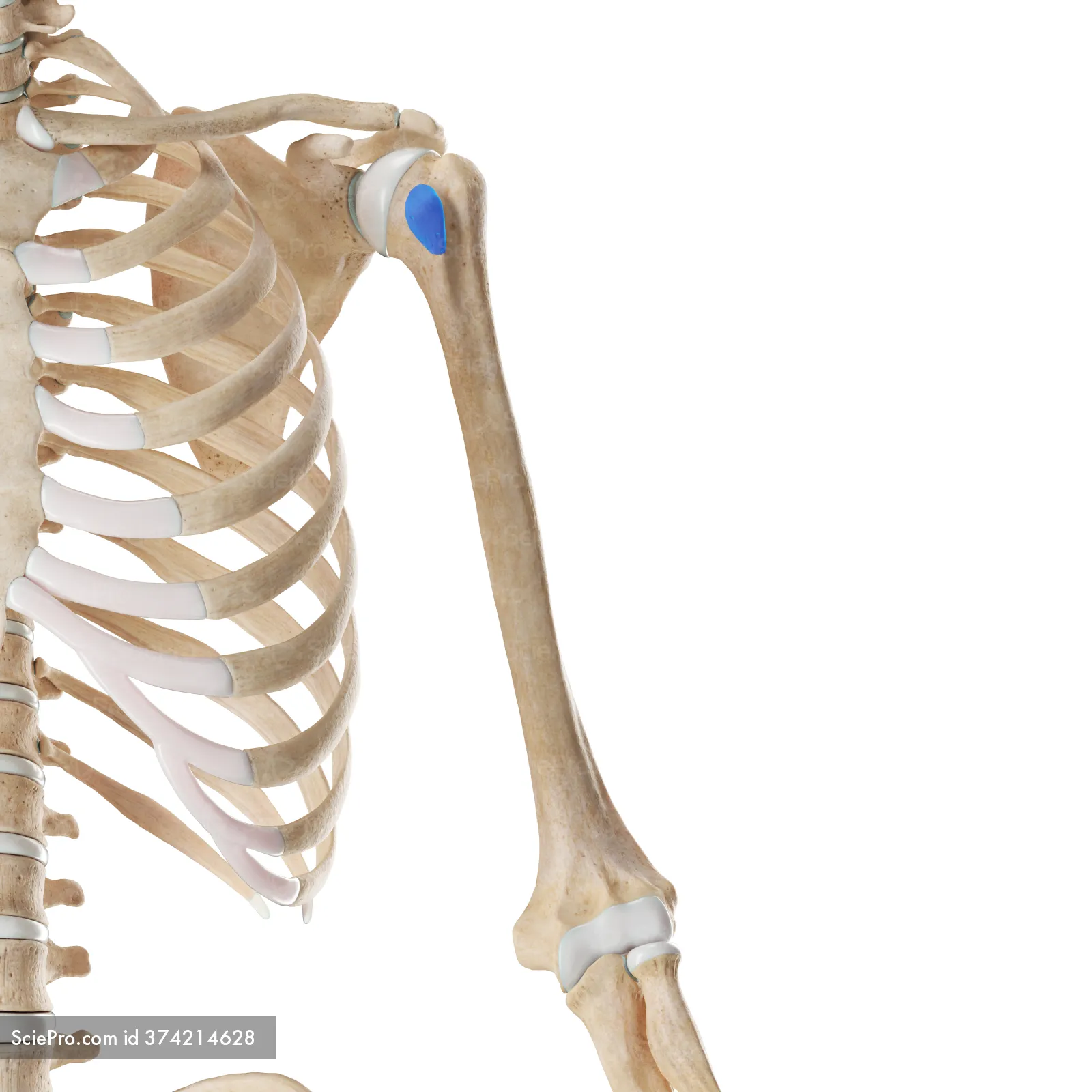
Lesser tubercle
small bony projection near the top of the humerus that serves as an attachment point for the subscapularis muscle.

Bicipital groove
narrow groove on the humerus that holds the tendon of the long head of the biceps muscle.
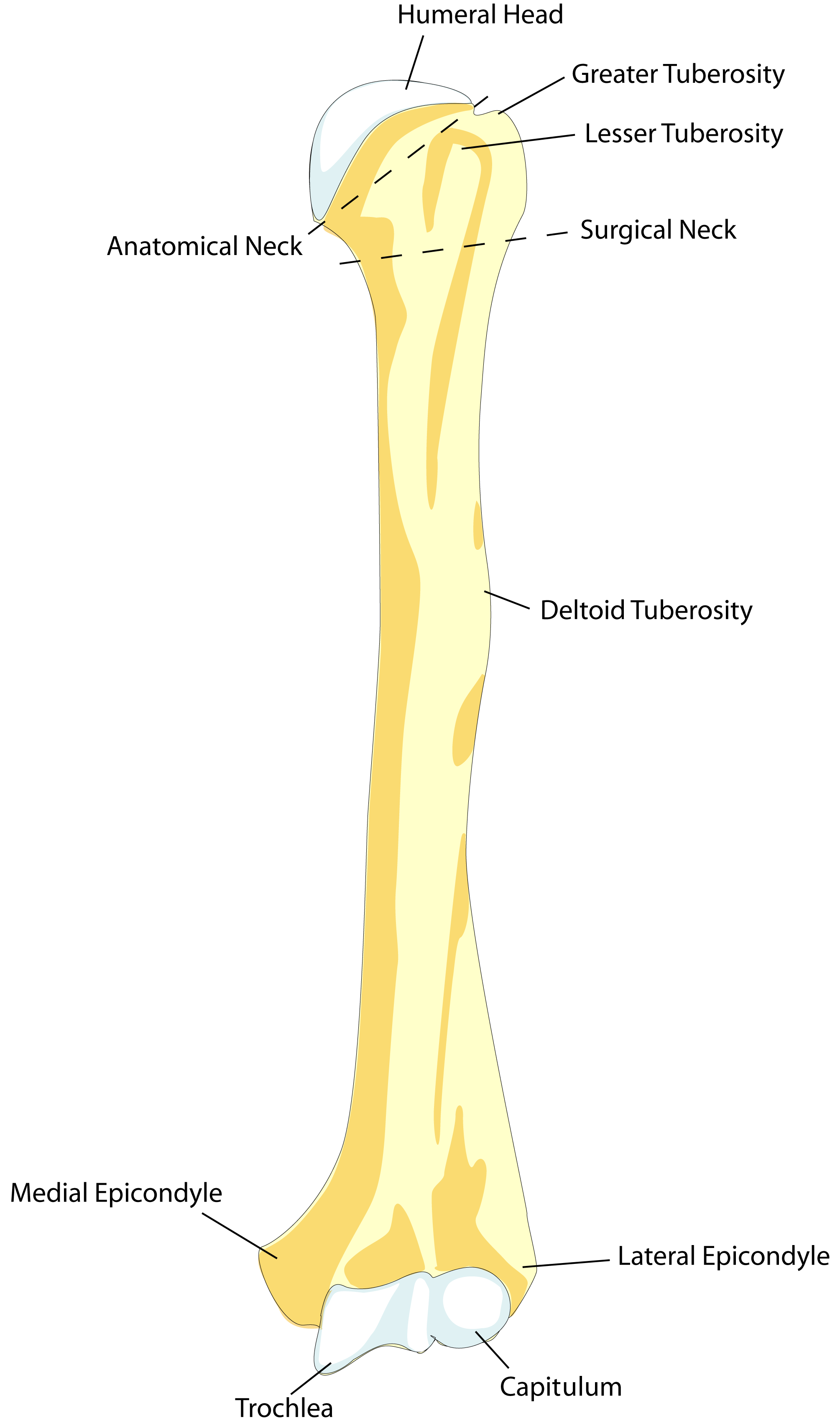
Deltoid tuberosity
roughened area on the humerus where the deltoid muscle attaches to help lift and move the arm.

Humerous bone
long bone of the upper arm that connects the shoulder to the elbow.

Capitulum
rounded projection at the lower end of the humerus that connects with the radius bone at the elbow joint.

Trochlea
spool-shaped structure at the lower end of the humerus that connects with the ulna to form part of the elbow joint.

Olecranon process
prominent projection at the upper end of the ulna that forms the point of the elbow and helps stabilize the elbow joint.

Trochlear notch
deep, curved notch at the upper end of the ulna that fits around the trochlea of the humerus to form the elbow joint.

Radial tuberosity
roughened projection on the radius bone where the biceps muscle attaches to help bend the elbow.

Radius bone
forearm bone on the thumb side that helps rotate the forearm and connects with the wrist and elbow.

Ulna bone
forearm bone on the pinky-finger side that forms part of the elbow joint and helps stabilize the forearm.

Head of ulna
rounded lower end of the ulna that connects with the radius and helps form the wrist joint.
Styloid process
pointed bony projection at the end of the radius or ulna that provides attachment for ligaments and helps stabilize the wrist.

Distal phalange
bone at the tip of each finger or thumb that supports the fingertip and nail.

Medial/middle phalange
middle bone of each finger between the proximal and distal phalanges; the thumb does not have a middle phalanx.

Proximal phalange
finger bone closest to the hand that connects to the metacarpal bones.

Metacarpals
five long bones in each hand that form the palm and connect the wrist bones to the finger bones.

SLTP, TTCH
First row: “So Long To Pullman”, Second row: “Time To Come Home”

Scaphoid
small, boat-shaped carpal bone on the thumb side of the wrist that connects the hand to the forearm.

Lunate
crescent-shaped carpal bone in the wrist that connects with the radius and helps allow wrist movement.

Triquetrum
small, pyramid-shaped carpal bone on the pinky-finger side of the wrist that helps stabilize the wrist joint.
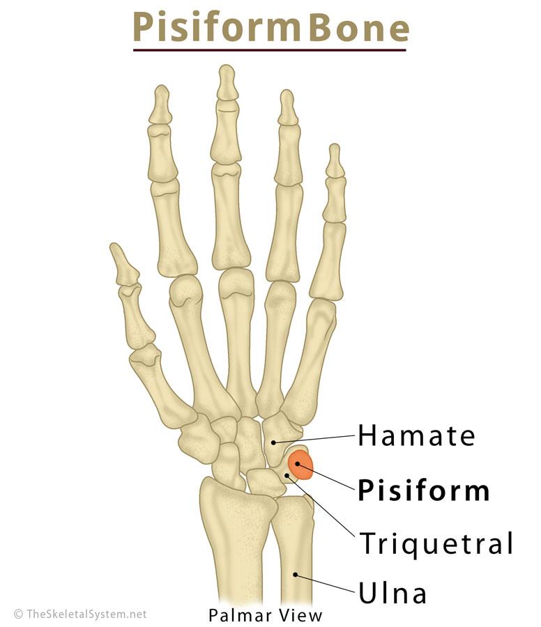
Pisiform
small, pea-shaped carpal bone on the pinky-finger side of the wrist that serves as an attachment point for tendons and helps wrist movement.
Trapezoid
small carpal bone in the wrist that connects with the index finger’s metacarpal and helps stabilize the hand.

Trapezium
carpal bone on the thumb side of the wrist that connects with the thumb’s metacarpal and allows a wide range of thumb movements.
Capitate
largest carpal bone, located in the center of the wrist, that connects with several hand bones and helps stabilize the wrist.
Hamate
hook-shaped carpal bone on the pinky-finger side of the wrist that helps support the hand and provides attachment points for ligaments and tendons.
Sacrum
triangular bone at the base of the spine formed by five fused vertebrae that connects the spine to the pelvis and supports body weight.
Ilium
largest, upper part of the hip bone that supports the body’s weight and provides attachment points for muscles.
Anterior superior iliac spine (ASIS)
bony projection at the front of the ilium that serves as an attachment point for muscles and ligaments of the hip and thigh.
Acetabulum
cup-shaped socket in the pelvis where the head of the femur fits to form the hip joint.
Pubic bone
front portion of the pelvis that joins with the other pubic bone to form the pubic symphysis and help support the pelvic organs.